Photonic Forensics
How a laser beam and a little gold can identify body fluids at crime scenes
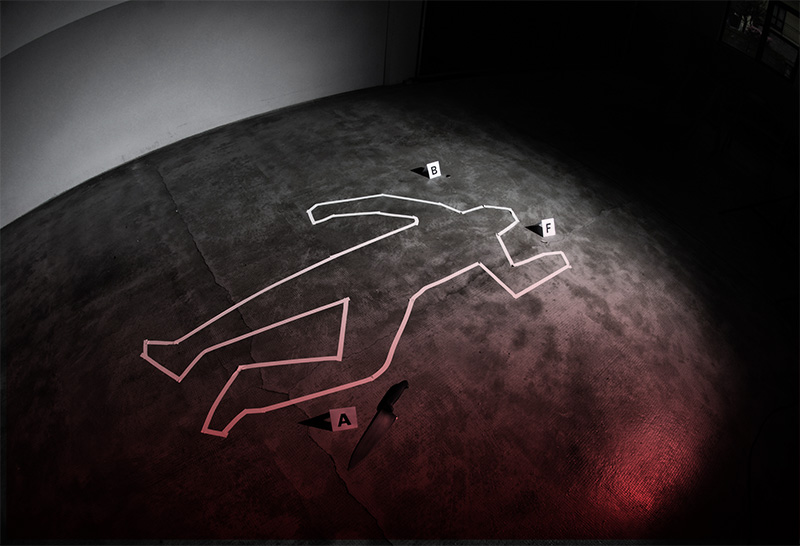
Fade in: A motel room, morning. The bed is made. The towels are folded. The room looks pristine, but caution tape hangs from the door. A forensic team sprays the scene with the chemical reagent Luminol, revealing a blue Pollock-like splatter invisible to the naked eye. The team documents the stain and collects a sample. By the time the forensic scientist back in the lab confirms the stain is blood, two men are crossing into Mexico with a body in the trunk.
It’s a scene from a typical crime show, but it won’t be long before investigators—both fictional and real-life—will be able to get results well before the bad guys reach the border. Lawrence Ziegler, a professor of chemistry at Boston University’s College of Arts & Sciences, is developing a machine that will identify body fluids at crime scenes precisely, on site, and in under an hour.
The instrument—a Portable Raman Microscope—is powered by what’s called surface enhanced Raman spectroscopy (SERS), a marriage of laser spectroscopy and nanotechnology. And it will be relatively easy to use. The forensic scientist will place a fluid sample in the machine, which will then compare the substance to those in a pre-programmed library. The sample is then identified as blood, saliva, semen, or one of many other fluids that could be present at a crime scene.
The critical component of the instrument is a glass chip painted with a metal—typically gold. The paint is nanostructured, or composed of coarse particles that generate enhanced optical fields. “That’s where the black magic is,” says Ziegler, who is developing the machine with the help of a two-year grant from the National Institute of Justice and BU’s Biomedical Forensic Sciences program.
To illustrate how the machine works, Ziegler places a drop of blood on the gold-covered chip and, in a process called “Raman scattering,” targets a laser at it. The laser interacts with the chip where the blood’s molecules touch the gold. Each type of molecule present in the blood scatters light from the surface of the chip at a different wavelength along a spectrum; the spectrometer separates these wavelengths to produce a frequency spectrum. Every type of body fluid generates a unique spectrum, or fingerprint.
The technique is so precise that it can differentiate human blood from that of other animals, as well as determine the age of some samples, suggesting the approximate time frame in which a crime occurred. It can also classify the components of a mixture of body fluids, such as picking up a trace of semen in vaginal fluid, a distinction that could improve forensic accuracy in rape cases.
Ziegler’s research in this area developed in the wake of the September 11 attacks, when the US Department of Defense awarded BU funding to create an instrument for detecting Bacillus anthracis, the bacterium that causes the disease anthrax. Ziegler has also received funding from the National Institutes of Health to apply SERS technology to diagnosing blood infections, enabling physicians to more quickly and accurately prescribe antibiotics. Other possible applications include rapid diagnostics for STDs and urinary tract infections, and the detection of cancer cells.
“My life prior to this experience was exclusively in chemical physics, and when my wife’s friends would ask me what I’m interested in, I’d try to convince them that the ‘first measurements of the fifth order susceptibility’ really is important,” Ziegler says with a laugh, as he recalls past difficulties describing the relevance of his research. Now, this additional line of research gives him a different kind of satisfaction. “It addresses long-standing problems and challenges in society that are broadly recognized and profound.”