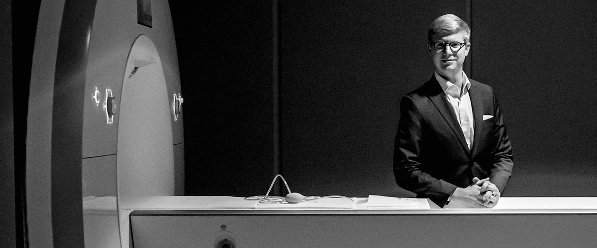
Tyler Perrachione uses BU’s new Siemens Prisma 3 Tesla MRI machine to scan the human brain. Photo by Mira Whiting
TYLER PERRACHIONE walked out of his office, crossed Commonwealth Avenue, and entered the Rajen Kilachand Center for Integrated Life Sciences & Engineering’s Cognitive Neuroimaging Center (CNC). There, he spent two hours scanning a human’s brain for a study using BU’s new Siemens Prisma 3 Tesla MRI machine. It was the first time Perrachione, director of the Communication Neuroscience Research Laboratory, didn’t have to schlep across the river to MIT’s brain imaging center. The nine-story, 170,000-square-foot Kilachand Center opened in 2017 thanks to a record $115 million gift from BU Trustee Rajen Kilachand (Questrom’74, Hon.’14).
“Everything went swimmingly,” says Perrachione, an assistant professor of speech, language, and hearing sciences, whose enthusiasm is echoed by his colleague Jason Bohland, the center’s associate director. The MRI machine—which supplies higher quality scans than older machines in record time—is “especially an advantage for people like Perrachione who are developing new technology or trying something that’s a little out of the ordinary,” says Bohland. “Just being able to come over here quickly is a huge advantage.”
The MRI machine (see “Scanning for Speech,” below) has been a game changer not just for neuroscientists, but for Sargent researchers working in a range of disciplines; Assistant Professor Deepak Kumar scans his subjects’ knees and hips, for example. The machine is just one of many BU technologies that facilitates Sargent’s pioneering research.
Here’s how Sargent faculty are using tech, from decoding speech to tracking walking patterns.
Scanning for Speech
This MRI machine is six million times more powerful than the Earth’s magnetic field.
“The new MRI machine lets us get higher quality images of the brain [than older machines] in a smaller amount of time, which is important when we are looking at things that the brain has to do very fast, like understand speech,” says Perrachione. “Being able to take high quality pictures faster is also important when doing studies with children, who sometimes don’t like to be in the machine for a long time. We are using these new technologies to study which parts of the brain change as you listen to different people talk, how these changes help you understand speech more efficiently, and how this plasticity might be reduced in developmental communication disorders like dyslexia.”
Walk this way

The lab’s treadmill and track feature 21 muscle-monitoring sensors, 6 force plates, and 18 optical motion cameras.
At the Neuromotor Recovery Laboratory, “We use optical and inertial motion trackers in combination with physiological sensors to study how people who have neurological conditions, such as a stroke, walk” and to help them improve their walking ability, says Louis N. Awad, an assistant professor of physical therapy and athletic training and director of the lab. The image above features the lab’s treadmill and track with 21 sensors to monitor muscle activity, 6 force plates to measure gait, 18 optical motion cameras to triangulate a subject’s position in 3-D, and a projector and screen that process the data.
Detecting brain activation

The fNIRS technology uses near-infrared light to detect changes in blood flow as humans think, speak, read, and write.
Swathi Kiran, director of the Aphasia Research Laboratory, typically uses brain imaging techniques (fMRI) to examine how the damaged brain recovers language. Even small head movements can distort the data, however, so in recent studies Kiran has turned to functional near-infrared spectroscopy (fNIRS) to study activation in the brain. The fNIRS technology “is easier to set up and study, and with a clinical population like people who have had a stroke, this is a huge advantage,” says Kiran, associate dean for research and a professor of speech, language, and hearing sciences. The technology relies on near-infrared light that, when targeted on the scalp and brain, can detect changes in blood flow as humans think, speak, read, or write.
Capturing motion

Deepak Kumar, an assistant professor of physical therapy and athletic training, uses a 3-D motion capture system at the Movement & Applied Imaging Lab to “assess movement patterns in people who have osteoarthritis or who may be at risk for developing it,” he says. His goal is to “understand how their movement patterns may be interwoven with their disease—and how we can change their movements to help them feel better.” Above, a subject drops from a step onto a force platform, then jumps straight up. Data from sensors worn on the body are represented as red dots, and the arrows indicate the force he exerts on the ground.
Zooming into the brain

Researchers use a diamond knife to slice brain tissue into slivers 2,000 times thinner than a piece of paper, then magnify it up to 500,000 times.
Sargent researchers are systematically looking at the developing human brain at a higher resolution than ever before. In a 2018 study published in PLoS Biology, assistant professor Vasileios Zikopoulos and his health sciences colleagues used the GeminiSEM 300 electron microscope to—for the first time—study the axons, or cables, that connect neurons and facilitate their communication across different areas of the brain. Housed at the Human Systems Neuroscience Laboratory, the microscope enabled researchers to study the organization of various areas of the brain and explore what goes wrong in the connections between them in disorders like autism.
Zikopoulos found that axons are tangled, which scrambles neural signals. This miscommunication causes problems commonly seen in autism, such as extreme focus and inability to shift attention. The researchers are now exploring changes in the brain’s organization in schizophrenia, depression, and other disorders.