")
Back to Journals » Orthopedic Research and Reviews » Volume 17
β-Caryophyllene and Statins in Bone Fracture Healing – A Narrative Review
Authors Marom H, Khan MA, Darvish N, Tornetta III P, Khoury A, Weil YA, Skelley NW, Allison DC, Meiron S , Ehrmann Barr T
Received 13 November 2024
Accepted for publication 9 January 2025
Published 23 January 2025 Volume 2025:17 Pages 31—42
DOI https://doi.org/10.2147/ORR.S506427
Checked for plagiarism Yes
Review by Single anonymous peer review
Peer reviewer comments 2
Editor who approved publication: Professor Clark Hung
Hilik Marom,1,* Mansoor A Khan,1,* Nissim Darvish,2,* Paul Tornetta III,3,* Amal Khoury,4,* Yoram A Weil,5,* Nathan WM Skelley,6,* Daniel C Allison,7,* Sahar Meiron,2,* Tami Ehrmann Barr1,*
1R&D, OrthoTreat Ltd, Tel Aviv-Jaffa, Israel; 2Corporate Office, OrthoTreat Ltd, Tel Aviv-Jaffa, Israel; 3Department of Orthopaedic Surgery and Orthopaedic Trauma, Chobanian and Avedisian School of Medicine, Boston Medical Center, Boston, MA, USA; 4Division of Orthopaedic Surgery, Orthopaedic, and Reconstructive Trauma Surgery, Tel Aviv Sourasky Medical Center, Tel Aviv, Israel; 5Department of Orthopaedics, Hadassah Hebrew University Hospital, Jerusalem, Israel; 6Department of Orthopaedics, Sanford Health, Sioux Falls, SD, USA; 7Department of Orthopaedic Oncology, Cedars-Sinai Medical Center, Los Angeles, CA, USA
*These authors contributed equally to this work
Correspondence: Tami Ehrmann Barr, Email [email protected]
Abstract: Bone fractures are a leading cause of morbidity and healthcare expenditure globally. The complex healing process involves inflammation, cartilage formation, mineralization, and bone remodeling. Current treatments like immobilization, surgery, and bone grafting, though effective, pose significant challenges, such as prolonged recovery and high costs. Emerging therapies such as biological agents, pharmacological treatments, and physical stimulation techniques are also associated with high costs, side effects, and practical applicability limitations. There is a critical need for alternative therapies that are cost-effective, safe, and easy to use. Recent studies suggest the potential of β-caryophyllene (BCP) and statins in promoting bone healing. BCP, a naturally occurring anti-inflammatory and antioxidant compound found in essential oils, enhances osteoblast activity and inhibits osteoclastogenesis. Statins, known for their cholesterol-lowering effects, also promote bone formation and reduce bone resorption through multiple biochemical pathways. Both BCP and statins have shown promising results in preclinical studies, enhancing bone density and promoting fracture healing. This review explores the individual and potential synergistic effects of BCP and statins on bone fracture healing. It highlights the complementary mechanisms of these agents: BCP’s anti-inflammatory and osteogenic properties and statins’ ability to inhibit osteoclast activity and promote angiogenesis. Combining BCP and statins could offer a multifaceted approach to enhance fracture healing, reduce complications, and improve patient outcomes. While individual effects are supported preclinically, further studies investigating synergies, formulations, and clinical translation are needed to develop this promising novel therapeutic approach for improving fracture repair outcomes.
Keywords: bone regeneration, fracture repair, osteoblast, osteogenesis, piezoelectric effect
Introduction
Bone fractures are among the most prevalent musculoskeletal injuries globally, contributing significantly to healthcare costs, with an estimated 178 million new cases annually.1 Fracture healing is a complex process involving, inter alia, inflammation, cartilage formation, mineralization, and bone remodeling.2–4 Impairments in this process can lead to delayed healing, nonunion, or malunion, resulting in morbidity, disability, and reduced quality of life.5 Standard treatments for fracture healing include immobilization, surgical interventions, and bone grafting; however, these methods face challenges such as prolonged immobilization, surgical risks, donor site morbidity, and high costs.6,7 Additional treatments, such as biological therapies, pharmacological treatments, shock wave therapy, low-intensity pulsed ultrasound (LIPUS), and electrical bone growth stimulation, offer various benefits but also present challenges like high costs, side effects, and the need for long-term commitment and specialized equipment.6
Biological therapies, such as growth factors, stem cells, or osteoinductive agents, enhance healing at the cellular level but are expensive, complex to administer, and have uncertain long-term outcomes.8 Often used off-label, pharmacological treatments pose risks of side effects, long-term adverse effects, and potential contraindications with other medications.9 These include gastrointestinal issues, jaw osteonecrosis, femur fractures, renal toxicity, flu-like symptoms with bisphosphonates; hypercalcemia, dizziness, leg cramps, unclear cancer risk with teriparatide; hypocalcemia, infections, jaw osteonecrosis with denosumab; renal and gastrointestinal anomalies, cardiovascular risks, delayed bone healing with non-steroidal anti-inflammatory drugs; osteoporosis, infections, hyperglycemia, wound healing issues with corticosteroids; and hepatic toxicity, cardiovascular problems, mood swings, hormone suppression with anabolic steroids.9–11
Hormone replacement therapy is associated with an increased risk of breast cancer, thromboembolic events, cardiovascular disease, and stroke, and is contraindicated in patients with a history of hormone-sensitive cancers.12 Calcitonin nasal sprays tend to cause nausea, flushing, injection reactions, cancer risk, and allergies.11 Shock wave therapy can be uncomfortable and variably effective, depending on the fracture type.13 LIPUS offers a non-invasive option but requires daily use for several months, and its effectiveness is debated.14 Electrical bone growth stimulation, while useful, can cause skin irritation, is inconvenient for continuous use, and is unsuitable for patients with electronic medical devices like pacemakers.15 These considerations must be carefully weighed for clinical decision-making. Certain populations, including the elderly and individuals with comorbidities like diabetes and osteoporosis, are at higher risk for impaired fracture healing.16 Thus, there is an urgent need for alternative or adjuvant therapies that are cost-effective, easy to use, patient-friendly, safe, have a long shelf-life, and can enhance and accelerate the fracture healing process, reduce complications, and improve patient outcomes.
Recent studies highlight the possibilities of β-caryophyllene (BCP) and statins in promoting bone formation and fracture healing.17,18 BCP is a naturally occurring bicyclic sesquiterpene found in various essential oils, spices, and plants, known for its anti-inflammatory and antioxidant properties.19 Statins, on the other hand, are widely used cholesterol-lowering drugs that have been found to exert pleiotropic effects, including bone anabolic actions.17,20 BCP has been shown to protect against bone defects caused by Vitamin D deficiency through the upregulation of klotho expression.21 It also demonstrates anticancer potential by inducing apoptosis in osteosarcoma cells via ROS and the JAK1/STAT3 pathway,22 and enhances wound healing when incorporated into a hydrogel with nano-emulsified BCP.23 Meanwhile, statins play a significant role in bone metabolism by promoting osteoblast activity and reducing bone resorption, making them effective for treating bone diseases.24,25 Local delivery methods further improve their efficacy.26 Together, both BCP and statins hold promise for advancing bone health treatments. This review aims to provide a comprehensive overview of the current literature on the potential roles and mechanisms of action of BCP and statins in bone fracture healing, highlighting their therapeutic potential and future research directions, which could ultimately lead to improved outcomes for patients suffering from fractures and other conditions that compromise bone integrity.
A thorough literature search for scholarly articles published in English was conducted in PubMed and Google Scholar databases, using combinations of the medical subject heading (MeSH) terms “β-caryophyllene”, “statins”, “bone fracture”, “fracture healing”, “osteoblasts”, “osteoclasts”, “bone formation”, and “bone resorption”, separated by suitable Boolean operators. Relevant articles published between the years 2000 and 2022 were included. Studies before 2000, review articles, case reports, and articles not available in English were excluded (Figure 1).
 |
Figure 1 Flow diagram depicting literature search and study selection. |
Overview of BCP and Statins
BCP is a bicyclic sesquiterpene with the chemical formula alpha-zingiberene (C15H24). It occurs naturally in two isomeric forms: α‐caryophyllene (α‐humulene) and iso‐caryophyllene (Z‐BCP), often in a mixture with its oxidation product, BCP oxide.27 Widely distributed in the plant kingdom, BCP is found in various essential oils, spices, and herbs, including clove oil (Syzygium aromaticum), black pepper (Piper nigrum), and cannabis (Cannabis sativa).27 BCP is known for its volatile nature and distinctive woody, spicy aroma, contributing to the characteristic scent of numerous plants and spices. BCP exhibits various biological activities, including anti-inflammatory, antioxidant, antimicrobial, and analgesic properties (Figure 2).28 In terms of bone metabolism, BCP promotes osteoblast differentiation and mineralization while inhibiting osteoclast formation and bone resorption, potentially through modulation of the Wnt/β-catenin pathway.18,29 Beyond its effects on bone metabolism, BCP protects against seizures.30 It also shows promise in preventing the development of neurodegenerative diseases like Alzheimer’s.31 Additionally, BCP inhibits cytochrome P450 isoforms, particularly cytochrome P450 3A4 (CYP3A4), mitigating adverse effects from excessive drug levels.32
 |
Figure 2 Effect of BCP on bone fracture healing. BCP offers multiple benefits, including significant anti-inflammatory, immunomodulatory, and antioxidant effects, and enhanced osteoblastic mineralization, crucial for fracture healing. Additionally, BCP’s analgesic and antimicrobial activities reduce pain perception and risk of infection. |
Numerous studies have demonstrated BCP’s low toxicity profile, with no significant adverse effects observed in animal models even at relatively high doses.33 Additionally, BCP is widely present in many foods and food products and has been granted Generally Recognized as Safe status by the US Food and Drug Administration.34 BCP has exhibited diverse therapeutic effects, such as neuroprotective properties against cerebral ischemic injury through the reduction of inflammation and oxidative stress,35 protective effects against hepatic fibrosis and apoptosis in animal models,36 and the potential to promote osteoblastic mineralization while inhibiting osteoclastogenesis and adipogenesis, suggesting its utility in enhancing bone formation.18 Furthermore, BCP has been shown to enhance cell migration, angiogenesis, and collagen deposition, contributing to wound healing.37 It has also demonstrated neuroprotective effects against cerebral ischemia-reperfusion injury by regulating necroptotic neuronal death and inflammation (Table 1).38
 |
Table 1 Summary of the Studies on BCP and Bone Fracture Healing |
Statins, widely known as cholesterol-lowering medications, have garnered significant interest due to their pleiotropic effects beyond lipid regulation, particularly in promoting bone regeneration and enhancing fracture healing.39 These effects are mediated through multiple mechanisms that modulate osteoblast and osteoclast activity, angiogenesis, and inflammatory responses.40 At the molecular level, statins inhibit 3-hydroxy-3-methylglutaryl coenzyme A (HMG-CoA) reductase, a key enzyme in the mevalonate pathway.41 This inhibition reduces downstream metabolites, such as farnesyl pyrophosphate (FPP) and geranylgeranyl pyrophosphate (GGPP), consequently impairing protein prenylation of small GTPases. Disruption of these signaling pathways affects cytoskeletal organization and impedes osteoclast formation and function.17,40,41
Role of BCP in Bone Fracture Healing
After a bone fracture, the body initiates an inflammatory response characterized by hematoma formation and the recruitment of polymorphonuclear neutrophils to clear debris.42 Chemokines such as monocyte chemoattractant protein-1 and interleukin-6 (IL-6) attract macrophages crucial for subsequent bone healing. Additionally, macrophages and osteoMacs play a pivotal role in the adaptive immune response initiated by lymphocytes, ultimately promoting bone regeneration.42 Disruption in this inflammatory phase can hinder proper bone healing and increase the risk of non-union. Due to its potential therapeutic effects, BCP is gaining attention, including its anti-inflammatory and bone-healing properties.36,43 Macrophages also play a crucial role in osteogenesis, with both M1 and M2 macrophages being essential for ensuring proper formation and recovery during the inflammatory and remodeling phases.44 Acting as a selective agonist of cannabinoid receptor type 2 (CB2), BCP exhibits potent anti-inflammatory properties by inhibiting various cytokines and factors involved in inflammation, such as IL-1β and IL-6.36 Its engagement with peroxisome proliferator-activated receptors alpha and gamma contributes to its anti-arthritic effects.45
Several preclinical investigations have shed light on the potential benefits of BCP in bone fracture healing. BCP stimulates osteoblastic mineralization while suppressing adipogenesis and osteoclastogenesis. By selectively activating CB2 receptors, BCP promotes bone formation and inhibits bone resorption, enhancing bone mineral density.46,47 The distinctive expression patterns of CB2 in osteoclasts from menopausal women highlight its role in inhibiting osteoclast activity, with reductions in CB2 signaling efficacy associated with lower bone density and the onset of osteoporosis.47,48 Despite its widespread use and generally recognized safety, interactions with other compounds or medications, as well as potential effects in specific populations, warrant further investigation and consideration.
Statins and Bone Fracture Healing
Statins stimulate osteoblast differentiation by enhancing the expression of bone morphogenetic protein-2 (BMP-2) and osteocalcin while suppressing the activation of Rho and Rho kinase.40,49 This dual action promotes bone formation and mineralization. Moreover, statins activate Smad3, a critical mediator of transforming growth factor-beta (TGF-β) signaling, pivotal for maintaining bone mass by inhibiting osteoblast apoptosis and fostering bone formation.50,51 Additionally, statins modulate the osteoprotegerin (OPG)/receptor activator of nuclear factor-kappa beta ligand (RANKL)/RANK system, a key regulator of osteoclastogenesis, by upregulating OPG expression and downregulating RANKL expression, consequently inhibiting osteoclast formation and activity.52 This anti-osteoclastogenic effect is complemented by the promotion of osteoblastogenesis through the induction of estrogen receptor-alpha (ER-α) expression, leading to reduced RANKL expression and further hindering osteoclast formation.53
Statins also exhibit pro-angiogenic properties, promoting neovascularization and enhancing collateral blood flow.54 This angiogenic activity is mediated through the Akt/phosphatidylinositol 3-kinase (Akt/PI3K) pathway, resulting in increased endothelial cell proliferation and secretion of angiogenic factors such as vascular endothelial growth factor (VEGF).55 Additionally, statins possess anti-inflammatory properties, contributing to their beneficial role in fracture healing. They promote the polarization of macrophages toward the anti-inflammatory M2 phenotype and upregulate the expression of anti-inflammatory cytokines like interleukin-13 (IL-13) and growth factors.56 These foster an environment conducive to tissue repair and regeneration (Figure 3).
 |
Figure 3 Statins exhibit pro-angiogenic properties. They modulate various pathways. They enhance estrogen receptor expression, inhibit osteoclast differentiation by modulating the OPG/RANKL/RANK signaling pathway, and promote bone formation (depicted in red). They inhibit actin depolymerization and enhance osteoblast differentiation leading to bone formation via the FPP/GGPP/Rho/BMP-2/P13-K pathway and VEGF (depicted in blue). They also activate TGF-β signaling by activating Smad3, essential for maintaining bone mass. Deletion of Smad3 decreases bone formation due to increased osteoblast apoptosis. Statins induce Smad3 expression, promoting bone formation (depicted in green). |
Preclinical investigations strongly support the beneficial impact of statins on fracture healing. Early research showed that compactin inhibited bone resorption by blocking the fusion of osteoclasts and disrupting their actin ring.41 Subsequent studies suggested that statins differentially regulated VEGF synthesis in endothelial and vascular smooth muscle cells.57 In vitro studies demonstrated that simvastatin induced the expression of ER-α in murine bone marrow stromal cells and suppressed apoptosis in osteoblastic cells through the TGF-β Smad3 pathway.58,59 Simvastatin also induces ER-α expression in bone, restoring bone loss and reducing ER-α expression and uterine wet weight in ovariectomized rats.60 Additionally, inhibiting actin depolymerization enhances osteoblast differentiation and bone formation in human stromal stem cells.61 Further exploration revealed that accumulation of FPP inhibited calvarial osteoblast differentiation, while direct inhibition of GGPP synthase affected osteoblast differentiation.62 Also, GGPP synthase was found to be downregulated during osteoblast differentiation,49 while another study indicated that pitavastatin enhanced BMP-2 and osteocalcin expression in human osteoblasts by inhibiting Rho-associated kinase.40 Animal model studies have further substantiated these findings, showing that statin administration enhances callus formation, increases bone density, and improves the biomechanical properties of the healing bone (Table 2).17,63
 |
Table 2 Summary of the Studies on Statins and Bone Fracture Healing |
Studies also demonstrate that topical simvastatin accelerates wound healing in diabetes by enhancing angiogenesis and lymphangiogenesis.64 Recently, an exploration into the potential of simvastatin-loaded lyophilized wafers as a dressing for chronic wounds yielded positive outcomes.65 An in vitro study has illustrated that simvastatin activates Akt and promotes angiogenesis in normocholesterolemic animals.55 Ex vivo studies have further supported these findings suggesting that statins promote angiogenesis (Table 3).66
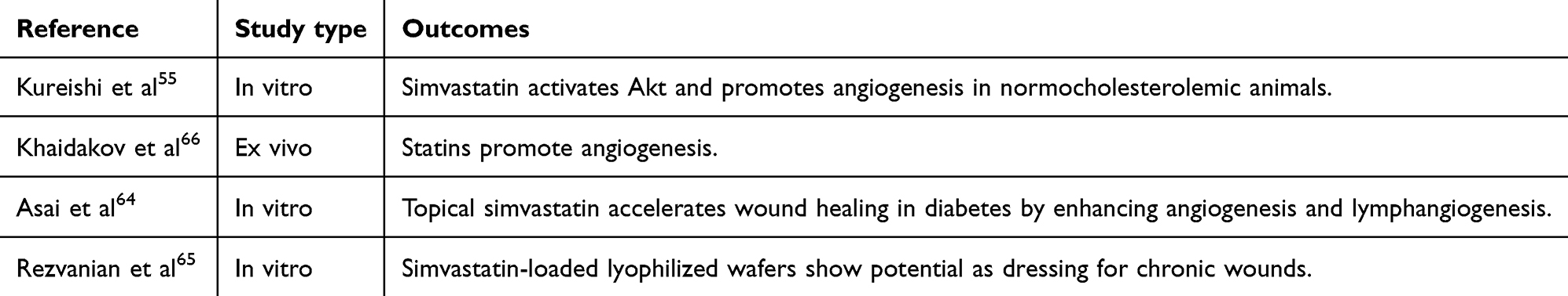 |
Table 3 Effect of Statins on Bone Regeneration |
The Promise of Combinatorial Therapy
Combinatorial therapies offer an advanced approach to bone fracture healing, leveraging multiple biological pathways. Utilizing therapeutic agents with complementary mechanisms offers several key advantages, such as reducing the likelihood of resistance, lowering individual drug doses, and minimizing potential side effects.67 Combining statins and BCP may enhance the therapeutic impact beyond their individual capabilities. Statins inhibit osteoclast differentiation via the RANK/RANKL pathway,52,68 while BCP promotes osteoblast differentiation and exerts anti-inflammatory effects.27,69,70 This complementary targeting fosters an environment conducive to accelerated fracture repair. Statins and BCP also modulate inflammation by inhibiting cytochrome P450 isoforms like CYP3A4, thereby balancing inflammation and cytotoxicity.36,71 The combination can also mimic the piezoelectric effect in bone, where mechanical loading generates electrical potentials that govern bone remodeling.72,73 Statins reduce osteoclastogenesis, favoring bone formation,52 while BCP promotes osteoblast differentiation.69 This duality echoes bone’s natural response to mechanical strains and electrical signals.74–76 A controlled-release formulation of statins and BCP could provide a multi-faceted approach to bone repair, enhancing therapeutic efficacy (Figure 4).
 |
Figure 4 Potential combined effect of BCP and statins on bone fracture healing. Bone remodeling involves osteoblast and osteoclast activity regulated by cytokines and the piezoelectric effect, with mechanical deformation generating negative charges in compressed areas and positive charges in tension areas. These changes influence osteocyte activity, leading to osteogenesis and osteoclastogenesis via NF-κB signaling, NFATc1, and osteocalcin mRNA expressions. Statins and BCP together counteract tensile stress by inhibiting RANK/RANKL binding and NF-κB activation (depicted in red blunt arrow), while enhancing compression stress effects by increasing osteocalcin production and inhibiting NF-κB (depicted in green sharp arrow and red blunt arrow). |
While preclinical studies specifically investigating the combinatorial effects of BCP and statins on bone fracture healing are limited, several studies have examined the individual effects of these compounds in animal models. Research findings indicate that the administration of BCP can promote callus formation in rodent fracture healing models.37,77 Similarly, numerous studies have reported that statin administration can accelerate fracture healing, improve callus formation, and enhance the biomechanical properties of the healing bone in various animal models, including rodents.17,63 While these studies provide valuable insights into the individual effects of BCP and statins on bone fracture healing, further preclinical investigations are needed to evaluate their potential synergistic effects when combined.
The synergy between BCP and statins, particularly lovastatin, can prevent RANKL from binding to its receptor (RANK) and inhibit RANKL release through BCP, a selective CB2 receptor agonist. Statin-induced OPG via ER-α activation further inhibits RANKL binding. BCP also mitigates the inflammatory response from high statin levels, while statins reduce BCP’s cytotoxicity.78 Understanding the synergistic effects of BCP and statins on bone’s piezoelectric properties could further improve fracture healing and bone health. Bone remodeling involves cellular responses of osteoblasts and osteoclasts to several key cytokines and an electrochemical process due to piezoelectric dipoles generated by bone tissue deformation. This dual process influences osteocyte activity, regulating bone resorption and formation through cytokine production. The piezoelectric effect plays a vital role in bone healing and repair. During a fracture, mechanical loading can stimulate the piezoelectric effect, enhancing the healing process. Compressive pressures generate negative potentials, while tensile stresses produce positive potentials, influencing the electric charges and amplitudes generated during activities such as walking. The piezoelectric potential produced by bone tissue deformation influences osteocyte activity within the bone matrix. Osteocytes sense mechanical force via their processes in the canaliculi and then produce several cytokines to regulate osteoclast-mediated bone resorption and osteoblast-mediated bone formation.79 Further preclinical and clinical studies are needed to explore these effects and optimize dosing and administration strategies for the best therapeutic outcomes. Research on the combinatorial use of BCP and statins could lead to more effective and personalized fracture treatments, ultimately improving patient outcomes.
Strengths, Limitations, and Future Perspectives
This article comprehensively reviews the potential roles and mechanisms of action of BCP and statins in bone fracture healing, covering both preclinical and clinical evidence. It offers valuable insights into potential therapeutic strategies for enhancing bone fracture healing. However, limitations include a primary focus on mechanistic aspects with limited discussion on clinical outcomes and patient perspectives. While the manuscript discusses novel treatment approaches, such as combinatorial therapy using BCP and statins, and in particular lovastatin, further exploration of clinical trial data and translational implications would enhance its relevance to clinical practice. Further preclinical investigations to assess the synergistic effects of combining BCP and statins on bone fracture healing could delve into optimal dosing regimens, formulations, and delivery methods to maximize therapeutic efficacy while minimizing potential adverse effects. Mechanistic studies elucidating the underlying pathways involved in the synergistic action of BCP and statins on bone regeneration could provide valuable insights for future drug development and personalized treatment approaches in bone fracture care.
Clinical Implications and Translational Opportunities
The possible combinatorial use of BCP and statins holds significant clinical implications and translational opportunities. When proven effective and safe in clinical trials, this approach can transform fracture treatment approaches, particularly in complex cases or for patients at high risk, such as the elderly, individuals with comorbidities, and smokers. This strategy may offer substantial benefits for managing osteoporosis and other bone defects, potentially improving outcomes for patients with compromised bone health. Potential applications include developing localized delivery systems, such as implants, scaffolds, or injectable formulations, for controlled and sustained release at the fracture site, thereby reducing the risk of systemic side effects associated with oral administration of statins. Furthermore, the anti-inflammatory properties and potential modulation of the immune response could have implications beyond fracture healing, finding applications in other musculoskeletal conditions involving inflammation and tissue repair. The development of controlled-release formulations for localized and sustained delivery to the fracture site represents a critical area for future investigation.
Conclusion
The combination of BCP and statins holds promise for enhancing bone fracture healing. BCP, with its anti-inflammatory and bone-promoting properties, stimulates osteoblast activity and inhibits osteoclast formation, while statins promote osteoblast differentiation, inhibit osteoclasts, and improve angiogenesis. Together, they target complementary pathways, potentially accelerating healing and improving bone density. Although preclinical results are promising, further research is needed to optimize dosing, delivery methods, and long-term safety. Successful validation in clinical trials could lead to more effective, patient-friendly fracture treatments, especially for high-risk populations, with potential applications in other musculoskeletal conditions.
Abbreviations
Akt/PI3K, Akt/phosphatidylinositol 3-kinase; BCP, β-caryophyllene; BMP-2, Bone morphogenetic protein-2; CB2, Cannabinoid receptor type 2; CYP3A4, Cytochrome P450 3A4; ER-α, Estrogen receptor-alpha; FPP, Farnesyl pyrophosphate; GGPP, Geranylgeranyl pyrophosphate; HMG-CoA, 3-hydroxy-3-methylglutaryl coenzyme A; IL-1β, Interleukin-1 beta; IL-6, Interleukin-6; IL-13, Interleukin-13; LIPUS, Low-intensity pulsed ultrasound; MeSH, Medical subject heading; OPG, Osteoprotegerin; RANKL, Receptor activator of nuclear factor-kappa beta ligand; TGF-β, Transforming growth factor-beta; VEGF, Vascular endothelial growth factor.
Acknowledgments
Manuscript writing assistance, editorial, and publication support were provided by Dr. Harsha SK and Dr. Priyanka Biswas Karmakar from Turacoz Group (www.turacoz.com).
Author Contributions
All authors made a significant contribution to the work reported, whether that is in the conception, study design, execution, acquisition of data, analysis, and interpretation, or in all these areas; took part in drafting, revising, or critically reviewing the article; gave final approval of the version to be published; have agreed on the journal to which the article has been submitted; and agree to be accountable for all aspects of the work.
Funding
This research was fully funded by OrthoTreat Ltd., which provided the financial resources necessary to execute the review.
Disclosure
All authors are employees, managers, and advisors of OrthoTreat Ltd., who analyzed and submitted the article. Dr Nathan Skelley reports personal fees for consultancy from Conmed corporation, outside the submitted work. Dr Tami Ehrmann Barr has a patent WO2023/021514A1 pending to OrthoTreat Ltd. The authors declare that they have no other competing interests in this work.
References
1. Wu AM, Bisignano C, James SL. et al. Global, regional, and national burden of bone fractures in 204 countries and territories, 1990–2019: a systematic analysis from the Global Burden of Disease Study 2019. Lancet Healthy Longev. 2021;2(9):e580–e592. doi:10.1016/S2666-7568(21)00172-0
2. Einhorn TA, Gerstenfeld LC. Fracture healing: mechanisms and interventions. Nat Rev Rheumatol. 2015;11(1):45–54. doi:10.1038/nrrheum.2014.164
3. Claes L, Recknagel S, Ignatius A. Fracture healing under healthy and inflammatory conditions. Nat Rev Rheumatol. 2012;8(3):133–143. doi:10.1038/nrrheum.2012.1
4. Gibon E, Lu LY, Nathan K, Goodman SB. Inflammation, ageing, and bone regeneration. J Orthop Translat. 2017;10:28–35. doi:10.1016/j.jot.2017.04.002
5. Zura R, Xiong Z, Einhorn T, et al. Epidemiology of fracture nonunion in 18 human bones. JAMA Surg. 2016;151(e162775):e162775. doi:10.1001/jamasurg.2016.2775
6. Schlickewei C, Kleinertz H, Thiesen DM, et al. Current and future concepts for the treatment of impaired fracture healing. Int J Mol Sci. 2019;20(22):5805. doi:10.3390/ijms20225805
7. Kim T, See CW, Li X, Zhu D. Orthopedic implants and devices for bone fractures and defects: past, present and perspective. Eng Regen. 2020;1:6–18. doi:10.1016/j.engreg.2020.05.003
8. Virk M, Lieberman J. Biologic adjuvants for fracture healing. Arthritis Res Ther. 2012;14(5):225. doi:10.1186/ar4053
9. Tu KN, Lie JD, Wan CK, et al. Osteoporosis: a review of treatment options. P & T. 2018;43:92.
10. Li -S-S, He S-H, Xie PY, et al. Recent progresses in the treatment of osteoporosis. Front Pharmacol. 2021;12:717065. doi:10.3389/fphar.2021.717065
11. Tabatabaei-Malazy O, Salari P, Khashayar P, Larijani B. New horizons in treatment of osteoporosis. DARU. 2017;25(1):1–16. doi:10.1186/s40199-017-0167-z
12. Lowe G. Hormone replacement therapy and cardiovascular disease: increased risks of venous thromboembolism and stroke, and no protection from coronary heart disease. J Intern Med. 2004;256(5):361–374. doi:10.1111/j.1365-2796.2004.01400.x
13. Petrisor B, Lisson S, Sprague S. Extracorporeal shockwave therapy: a systematic review of its use in fracture management. Indian J Orthop. 2009;43(2):161–167. doi:10.4103/0019-5413.50851
14. Lou S, Lv H, Li Z, Zhang L, Tang P. The effects of low-intensity pulsed ultrasound on fresh fracture: a meta-analysis. Medicine (Baltimore). 2017;
15. Sandy R. ELECTROPHYSICAL AGENTS - contraindications and precautions: an evidence-based approach to clinical decision making in physical therapy. Physiother Can. 2020;62:1–80. doi:10.3138/ptc.62.5
16. Hernandez R, Do TP, Critchlow CW, Dent RE, Jick SS. Patient-related risk factors for fracture-healing complications in the United Kingdom general practice research database. Acta Orthop. 2012;83(6):653–660. doi:10.3109/17453674.2012.747054
17. Mundy G, Garrett R, Harris S, et al. Stimulation of bone formation in vitro and in rodents by statins. Science. 1999;286(5446):1946–1949. doi:10.1126/science.286.5446.1946
18. Yamaguchi M, Levy RM. β-Caryophyllene promotes osteoblastic mineralization, and suppresses osteoclastogenesis and adipogenesis in mouse bone marrow cultures in vitro. Exp Ther Med. 2016;12(6):3602–3606. doi:10.3892/etm.2016.3818
19. Dahham S, Tabana YM, Iqbal MA, et al. The anticancer, antioxidant and antimicrobial properties of the sesquiterpene β-caryophyllene from the essential oil of aquilaria crassna. Molecules. 2015;20(7):11808–11829. doi:10.3390/molecules200711808
20. Kavalipati N, Shah J, Ramakrishan A, Vasnawala H. Pleiotropic effects of statins. Indian J Endocrinol Metab. 2015;19(5):554–562. doi:10.4103/2230-8210.163106
21. Dong W, Postlethwaite BC, Wheller PA, et al. Beta-caryophyllene prevents the defects in trabecular bone caused by Vitamin D deficiency through pathways instated by increased expression of klotho. Bone Joint Res. 2022;11(8):528–540. doi:10.1302/2046-3758.118.BJR-2021-0392.R1
22. Annamalai V, Kotakonda M, Periyannan V. JAK1/STAT3 regulatory effect of β-caryophyllene on MG-63 osteosarcoma cells via ROS-induced apoptotic mitochondrial pathway by DNA fragmentation. J Biochem Mol Toxicol. 2020;34(e22514). doi:10.1002/jbt.22514
23. Parisotto-Peterle J, Bidone J, Lucca LG, et al. Healing activity of hydrogel containing nanoemulsified β-caryophyllene. Eur J Pharm Sci. 2020;148:105318. doi:10.1016/j.ejps.2020.105318
24. Granat MM, Eifler-Zydel J, Kolmas J. Statins-their role in bone tissue metabolism and local applications with different carriers. Int J Mol Sci. 2024;26(1):25. doi:10.3390/ijms25042378
25. Morse LR, Coker J, Battaglino RA. STATINS AND BONE HEALTH: a MINI REVIEW. Actual osteol. 2018;14(1):31–35.
26. Zhang Y, Bradley AD, Wang D, Reinhardt RA. Statins, bone metabolism and treatment of bone catabolic diseases. Pharmacol Res. 2014;88:53–61. doi:10.1016/j.phrs.2013.12.009
27. Gertsch J, Leonti M, Raduner S, et al. Beta-caryophyllene is a dietary cannabinoid. Proc Natl Acad Sci U S A. 2008;105(26):9099–9104. doi:10.1073/pnas.0803601105
28. Fidyt K, Fiedorowicz A, Strządała L, Szumny A. β -caryophyllene and β -caryophyllene oxide—natural compounds of anticancer and analgesic properties. Cancer Med. 2016;5(10):3007–3017. doi:10.1002/cam4.816
29. Chamani S, Liberale L, Mobasheri L, et al. The role of statins in the differentiation and function of bone cells. EJCI. 2021;
30. Zimmer A, Zimmer AM, Hohmann AG, Herkenham M, Bonner TI. Increased mortality, hypoactivity, and hypoalgesia in cannabinoid CB1 receptor knockout mice. Proc Natl Acad Sci U S A. 1999;96(10):5780–5785. doi:10.1073/pnas.96.10.5780
31. Shah SR, Werlang CA, Kasper FK, Mikos AG. Novel applications of statins for bone regeneration. Natl Sci Rev. 2015;2(1):85–99. doi:10.1093/nsr/nwu028
32. Ghazwani M, Hani U, Alqarni MH, Alam A. Beta caryophyllene-loaded nanostructured lipid carriers for topical management of skin disorders: statistical optimization, in vitro and dermatokinetic evaluation. Gels. 2023;9(7):550. doi:10.3390/gels9070550
33. Francomano F, Caruso A, Barbarossa A, et al. β-caryophyllene: a sesquiterpene with countless biological properties. Appl Sci. 2019;9(24):5420. doi:10.3390/app9245420
34. Viveros-Paredes J, González-Castañeda RE, Gertsch J, et al. Neuroprotective Effects of β-caryophyllene against dopaminergic neuron injury in a murine model of parkinson’s disease induced by MPTP. Pharmaceuticals. 2017;10(3):60. doi:10.3390/ph10030060
35. Chang H, Kim JM, Lee JC, Kim WK, Chun HS. Protective effect of β-caryophyllene, a natural bicyclic sesquiterpene, against cerebral ischemic injury. J Med Food. 2013;16(6):471–480. doi:10.1089/jmf.2012.2283
36. Mahmoud M, El Swefy S, Hasan RA, Ibrahim A. Role of cannabinoid receptors in hepatic fibrosis and apoptosis associated with bile duct ligation in rats. Eur J Pharmacol. 2014;742:118–124. doi:10.1016/j.ejphar.2014.08.021
37. Koyama S, Purk A, Kaur M, et al. Beta-caryophyllene enhances wound healing through multiple routes. PLoS One. 2019;14(e0216104):e0216104. doi:10.1371/journal.pone.0216104
38. Yang M, Lv Y, Tian X, et al. Neuroprotective Effect of β-caryophyllene on cerebral ischemia-reperfusion injury via regulation of necroptotic neuronal death and inflammation: in vivo and in vitro. Front Neurosci. 2017;11:583. doi:10.3389/fnins.2017.00583
39. Liao JK, Laufs U. Pleiotropic effects of statins. Annu Rev Pharmacol Toxicol. 2005;45(1):89–118. doi:10.1146/annurev.pharmtox.45.120403.095748
40. Ohnaka K, Shimoda S, Nawata H, et al. Pitavastatin enhanced BMP-2 and osteocalcin expression by inhibition of ho-associated kinase in human osteoblasts. Biochem Biophys Res Commun. 2001;287(2):337–342. doi:10.1006/bbrc.2001.5597
41. Woo J, Kasai S, Stern PH, Nagai K. Compactin suppresses bone resorption by inhibiting the fusion of prefusion osteoclasts and disrupting the actin ring in osteoclasts. J Bone Miner Res. 2000;15(4):650–662. doi:10.1359/jbmr.2000.15.4.650
42. Loi F, Córdova LA, Pajarinen J, Lin TH, Yao Z, Goodman SB. Inflammation, fracture and bone repair. Bone. 2016;86:119–130. doi:10.1016/j.bone.2016.02.020
43. Kogan N, Melamed E, Wasserman E, et al. Cannabidiol, a major non-psychotropic cannabis constituent enhances fracture healing and stimulates lysyl hydroxylase activity in osteoblasts. J Bone Miner Res. 2015;30(10):1905–1913. doi:10.1002/jbmr.2513
44. Orvalho JM, Fernandes JC, Moraes Castilho R, Fernandes GV. The macrophage’s role on bone remodeling and osteogenesis: a systematic review. Clin Rev Bone Miner Metab. 2023;21(1–4):1–13. doi:10.1007/s12018-023-09286-9
45. Irrera N, D’ascola A, Pallio G, et al. β-Caryophyllene Mitigates Collagen Antibody Induced Arthritis (CAIA) in mice through a cross-talk between CB2 and PPAR-γ receptors. Biomolecules. 2019;9(8):326. doi:10.3390/biom9080326
46. Idris A, Ralston SH. Role of cannabinoids in the regulation of bone remodeling. Front Endocrinol. 2012;3:136. doi:10.3389/fendo.2012.00136
47. Idris A. The promise and dilemma of cannabinoid therapy: lessons from animal studies of bone disease. Bonekey Rep. 2012;1:1. doi:10.1038/bonekey.2012.224
48. Rossi F, Siniscalco D, Luongo L, et al. The endovanilloid/endocannabinoid system in human osteoclasts: possible involvement in bone formation and resorption. Bone. 2009;44(3):476–484. doi:10.1016/j.bone.2008.10.056
49. Yoshida T, Asanuma M, Grossmann L, et al. Geranylgeranyl-pyrophosphate (GGPP) synthase is down-regulated during differentiation of osteoblastic cell line MC3T3-E1. FEBS Lett. 2006;580(22):5203–5207. doi:10.1016/j.febslet.2006.08.060
50. Borton AJ, Frederick JP, Datto MB, Wang XF, Weinstein RS. The loss of Smad3 results in a lower rate of bone formation and osteopenia through dysregulation of osteoblast differentiation and apoptosis. J Bone Miner Res. 2001;16(10):1754–1764. doi:10.1359/jbmr.2001.16.10.1754
51. Rodriguez-Vita J, Sanchez-Galan E, Santamaria B, et al. Essential role of TGF-beta/Smad pathway on statin dependent vascular smooth muscle cell regulation. PLoS One. 2008;
52. Kaji H, Kanatani M, Sugimoto T, Chihara K. Statins modulate the levels of osteoprotegerin/receptor activator of NFkappaB ligand mRNAin mouse bone-cell cultures. Horm Metab Res. 2005;37(10):589–592. doi:10.1055/s-2005-870538
53. Syed F, Khosla S. Mechanisms of sex steroid effects on bone. Biochem Biophys Res Commun. 2005;328(3):688–696. doi:10.1016/j.bbrc.2004.11.097
54. Weis M, Heeschen C, Glassford AJ, Cooke JP. Statins have biphasic effects on angiogenesis. Circulation. 2002;105(6):739–745. doi:10.1161/hc0602.103393
55. Kureishi Y, Luo Z, Shiojimam I, et al. The HMG-CoA reductase inhibitor simvastatin activates the protein kinase akt and promotes angiogenesis in normocholesterolemic animals. Nat Med. 2000;6(9):1004–1010. doi:10.1038/79510
56. Alkakhan W, Farrar N, Sikora V, et al. Statins modulate microenvironmental cues driving macrophage polarization in simulated periodontal inflammation. Cells. 2023;12(15):1961. doi:10.3390/cells12151961
57. Frick M, Dulak J, Cisowski J, et al. Statins differentially regulate vascular endothelial growth factor synthesis in endothelial and vascular smooth muscle cells. Atherosclerosis. 2003;170(229):236. doi:10.1016/s0021-9150(03)00299-5
58. Song C, Wang J, Song Q, et al. Simvastatin induces estrogen receptor-alpha (ER-alpha)in murine bone marrow stromal cells. J Bone Miner Metab. 2008;26(3):213–217. doi:10.1007/s00774-007-0820-6
59. Kaji H, Naito J, Inoue Y, Sowa H, Sugimoto T, Chihara K. Statin suppresses apoptosis in osteoblastic cells: role of transforming growth factor-beta Smad3 pathway. Horm Metab Res. 2008;40(11):746–751. doi:10.1055/s-0028-1082051
60. Li X, Song QS, Wang JY, et al. Simvastatin induces estrogen receptor-alpha expression in bone, restores bone loss, and decreases ER alpha expression and uterine wet weight in ovariectomized rats. J Bone Miner Metab. 2011;29(4):396–403. doi:10.1007/s00774-010-0231-y
61. Chen L, Shi K, Frary CE, et al. Inhibiting actin depolymerization enhances osteoblast differentiation and bone formation in human stromal stem cells. Stem Cell Res. 2015;15(2):281–289. doi:10.1016/j.scr.2015.06.009
62. Weivoda M, Hohl RJ. Effects of farnesyl pyrophosphate accumulation on calvarial osteoblast differentiation. Endocrinology. 2011;152(8):3113–3122. doi:10.1210/en.2011-0016
63. Masuzaki T, Ayukawa Y, Moriyama Y, et al. The effect of a single remote injection of statin-impregnated poly (lactic-co-glycolic acid) microspheres on osteogenesis around titanium implants in rat tibia. Biomaterials. 2010;31(12):3327–3334. doi:10.1016/j.biomaterials.2010.01.016
64. Asai J, Takenaka H, Hirakawa S, et al. Topical simvastatin accelerates wound healing in diabetes by enhancing angiogenesis and lymphangiogenesis. Am J Pathol. 2012;181(6):2217–2224. doi:10.1016/j.ajpath.2012.08.023
65. Rezvanian M, Tan CK, Ng SF. Simvastatin-loaded lyophilized wafers as a potential dressing for chronic wounds. Drug Dev Ind Pharm. 2016;42(12):2055–2062. doi:10.1080/03639045.2016.1195400
66. Khaidakov M, Wang W, Khan JA, Kang BY, Hermonat PL, Mehta JL. Statins and angiogenesis: is it about connections? Biochem Biophys Res Commun. 2009;387(3):543–547. doi:10.1016/j.bbrc.2009.07.057
67. Daughton CG, Ruhoy IS. Lower-dose prescribing: minimizing “side effects” of pharmaceuticals on society and the environment. Sci Total Environ. 2013;443:324–337. doi:10.1016/j.scitotenv.2012.10.092
68. Park J, Lee NK, Lee SY. Current Understanding of RANK signaling in osteoclast differentiation and maturation. Mol Cells. 2017;40(10):706–713. doi:10.14348/molcells.2017.0225
69. Shan J, Chen L, Lu K. Protective effects of trans-caryophyllene on maintaining osteoblast function. IUBMB Life. 2017;69(1):22–29. doi:10.1002/iub.1584
70. Bento A, Marcon R, Dutra RC, et al. β-Caryophyllene inhibits dextran sulfate sodium-induced colitis in mice through CB2 receptor activation and PPARγ pathway. Am J Pathol. 2011;178(3):1153–1166. doi:10.1016/j.ajpath.2010.11.052
71. McFarland AJ, Anoopkumar-Dukie S, Arora DS, et al. Molecular mechanisms underlying the effects of statins in the central nervous system. Int J Mol Sci. 2014;15(11):20607–20637. doi:10.3390/ijms151120607
72. Oladapo BI, Ismail SO, Kayode JF, Ikumapayi OM. Piezoelectric effects on bone modeling for enhanced sustainability. Mater Chem Phys. 2023;305:127960. doi:10.1016/j.matchemphys.2023.127960
73. Carter A, Popowski K, Cheng K, Greenbaum A, Ligler FS, Moatti A. Enhancement of bone regeneration through the converse piezoelectric effect, a novel approach for applying mechanical stimulation. Bioelectricity. 2021;3(4):255–271. doi:10.1089/bioe.2021.0019
74. Butcher MT, Espinoza NR, Cirilo SR, Blob RW. In vivo strains in the femur of river cooter turtles(Pseudemys concinna) during terrestrial locomotion: tests of force-platform models of loading mechanics. J Exp Biol. 2008;211(15):2397–2407. doi:10.1242/jeb.018986
75. Riddle R, Donahue HJ. From streaming-potentials to shear stress: 25 years of bone cell mechanotransduction. J Orthop Res. 2009;27(2):143–149. doi:10.1002/jor.20723
76. You L, Cowin SC, Schaffler MB, Weinbaum S. A model for strain amplification in the actin cytoskeleton of osteocytes due to fluid drag on pericellular matrix. J Biomech. 2001;34(11):1375–1386. doi:10.1016/s0021-9290(01)00107-5
77. Rossi F, Tortora C, Punzo F, et al. The endocannabinoid/endovanilloid system in bone: from Osteoporosis to Osteosarcoma. Int J Mol Sci. 2019;20(8):1919. doi:10.3390/ijms20081919
78. Orthotreat. The Science. 2024. Available from https://www.orthotreat.io/the-science/#fromformulation.
79. Kao FC, Chiu PY, Tsai TT, Lin ZH. The application of nanogenerators and piezoelectricity in osteogenesis. Sci Technol Adv Mater. 2019;20(1):1103–1117. doi:10.1080/14686996.2019.1693880
 © 2025 The Author(s). This work is published and licensed by Dove Medical Press Limited. The
full terms of this license are available at https://www.dovepress.com/terms.php
and incorporate the Creative Commons Attribution
- Non Commercial (unported, 3.0) License.
By accessing the work you hereby accept the Terms. Non-commercial uses of the work are permitted
without any further permission from Dove Medical Press Limited, provided the work is properly
attributed. For permission for commercial use of this work, please see paragraphs 4.2 and 5 of our Terms.
© 2025 The Author(s). This work is published and licensed by Dove Medical Press Limited. The
full terms of this license are available at https://www.dovepress.com/terms.php
and incorporate the Creative Commons Attribution
- Non Commercial (unported, 3.0) License.
By accessing the work you hereby accept the Terms. Non-commercial uses of the work are permitted
without any further permission from Dove Medical Press Limited, provided the work is properly
attributed. For permission for commercial use of this work, please see paragraphs 4.2 and 5 of our Terms.