")
Back to Journals » Drug Design, Development and Therapy » Volume 19
A Bibliometric Analysis of Microneedle-Mediated Drug Delivery: Trends, Hotspots, and Future Directions
Authors Xiang W, Jiang X , Guo L
Received 23 January 2025
Accepted for publication 16 April 2025
Published 10 May 2025 Volume 2025:19 Pages 3805—3825
DOI https://doi.org/10.2147/DDDT.S519048
Checked for plagiarism Yes
Review by Single anonymous peer review
Peer reviewer comments 2
Editor who approved publication: Prof. Dr. Tin Wui Wong
Weiyi Xiang,1,2 Xian Jiang,1,2,* Linghong Guo1,2,*
1Department of Dermatology, West China Hospital, Sichuan University, Chengdu, Sichuan, People’s Republic of China; 2Laboratory of Dermatology, Clinical Institute of Inflammation and Immunology, Frontiers Science Center for Dis-Ease-Related Molecular Network, West China Hospital, Sichuan University, Chengdu, Sichuan, People’s Republic of China
*These authors contributed equally to this work
Correspondence: Linghong Guo, Department of Dermatology, West China Hospital, Sichuan University, No. 37, Guoxue Alley, Chengdu, Sichuan, People’s Republic of China, Tel +86 18380131660, Email [email protected]
Purpose: Microneedles can physically penetrate the stratum corneum, creating micropores on the skin, and allowing for drug delivery through direct diffusion, injection, or other methods. As a novel drug delivery method, it possesses significant application potential. This study uses bibliometric analysis to explore the research hotspots and development trends of microneedle-mediated drug delivery.
Methods: Relevant research articles on microneedle-mediated drug delivery published between 1998 and 2024 in the Web of Science Core Collection (WoSCC) database were retrieved. Data analysis and visualization were performed using VOSviewer, CiteSpace, Scimago Graphica, and Pajek, enabling the prediction of research trends in microneedle-mediated drug delivery.
Results: In general, research on microneedle-mediated drug delivery has shown a continuous increase. China and the United States are the leading countries in this field of study. Notably, Ryan F. Donnelly (n=224) is the most prominent contributor to this field. The current core research directions include: disease treatment, enhancement of transdermal absorption performance of microneedles, vaccine delivery, and new materials and technologies for microneedle manufacturing.
Conclusion: Microneedle-mediated drug delivery, as a novel technology and method, holds significant research value and application potential. However, further strengthening of international collaboration and the clinical translation of research findings are needed.
Keywords: percutaneous drug delivery, research trends, vaccine delivery, materials science
Introduction
Fast, efficient, and minimally invasive drug delivery methods have long been a focal point of medical research. Oral administration is convenient and non-invasive, but its efficacy is limited by first-pass metabolism, and the bioavailability of certain drugs is poor. Traditional injection methods, due to related pain and other complications, often result in poor patient compliance.1 Topical formulations such as ointments and transdermal patches, commonly used for percutaneous drug delivery, also face issues with poor permeability.2 Microneedles, introduced in 1976 and first used for transdermal drug delivery in 1998, have emerged as a promising solution.3 Because microneedles can overcome the limitations of oral, injectable, and traditional transdermal delivery methods, many researchers have conducted in-depth studies on them over the past few decades. As a novel drug delivery technique, microneedles can physically penetrate the stratum corneum, creating micropores larger than those needed for macromolecular drugs, thus providing a direct pathway for drug diffusion.4 They can also be loaded with drugs in hollow microneedles and directly injected into the circulatory system, or mixed with soluble substances to penetrate the skin and reach the bloodstream. With ongoing research, the application of microneedles in drug delivery has gradually expanded to areas such as dermatological treatments,5 insulin delivery for diabetes,6 cancer therapy,7 ocular drug administration,8 and vaccination.9
Based on their structure, microneedles can be classified into five major types: solid microneedles, drug-coated microneedles, dissolving microneedles, hollow microneedles, and hydrogel microneedles.10 In terms of materials, they include silicon,11 metals,12 biomaterials, and polymers.13 Microneedles consist of hundreds of micron-sized needles arranged on a patch, typically ranging in length from 15 to 1500 μm, with a width of 50–250 μm and a tip thickness of 1–25 μm.14 To create micro-sized delivery pathways, the diameter of the needles is usually within a few micrometers. Depending on the application, the tips of the microneedles can be designed in various shapes, such as cylindrical, triangular, conical, pentagonal, and octagonal.15 Microneedles are capable of penetrating the epidermal layer of the skin but do not reach the nerve endings, meaning they do not cause damage to nerve tissue.16 As a result, microneedle application is generally painless. However, there are certain limitations to microneedle-based drug delivery, including potential skin reactions such as allergies and irritation, as well as the risk of microneedle breakage and residual fragments.17 With advancements in technology and ongoing research, the design, fabrication, and application of microneedles have become increasingly diverse.15 Analyzing research trends provides a valuable means for researchers to swiftly identify current hotspots and explore new directions for further investigation.
Bibliometrics is a literature analysis method that quantitatively and qualitatively evaluates the publication output and current state of a specific research domain.18 This approach allows for the extraction of detailed information regarding authors, keywords, journals, countries, institutions, and references.19 Bibliometric analysis has been widely applied across various medical fields, including oncology20 and both internal and surgical diseases.21 As such, it offers an effective means of identifying research trends and guiding future investigations. To date, while bibliometric studies of microneedles in the period from 2011 to 2020 have been conducted22 a comprehensive analysis covering the entire span of research on microneedle-mediated drug delivery is still lacking. The present study aims to fill this gap by employing bibliometric methods to trace the evolution of microneedle drug delivery research, from its inception in 1998 to the present, providing an overview of the field’s research trends and developments. By analyzing publications from the earliest studies to the present day, this study aims to offer a comprehensive understanding of the current landscape of microneedle-mediated drug delivery, identify key contributors, and highlight the most prominent areas of focus. This analysis will also help to forecast potential future research directions and guide the ongoing exploration of microneedles as an innovative drug delivery method.
Materials and Methods
Search Strategy
We conducted a literature search in the Web of Science Core Collection (WoSCC) database (https://www.webofscience.com/wos/woscc/basic-search) on November 9, 2024. The search query used was: TS = (Microneedle OR Microneedles OR Micro-needle OR Micro needles OR Micro needles) AND TS = (Transdermal Drug Delivery OR Transdermal Delivery OR Intradermal drug delivery OR intradermal delivery OR Drug Delivery OR Therapeutic Delivery OR Vaccine Delivery), Language = English. A total of 4825 articles published from 1998 to 2024 were analyzed. Original research papers (including basic research, clinical trials, cohort studies, etc)., review articles, and systematic review articles were included. These studies directly provide or synthesize the most critical scientific evidence within the research field, enabling the identification of technological advancements, research hotspots, and empirical progress in the domain. Certain literature types were excluded, including (i) meeting abstracts; (ii) editorial materials; (iii) letters; (iv) proceedings papers; (v) corrections; (vi) news items; (vii) book chapters; (viii) early access publications. These excluded categories were omitted due to potential issues such as incomplete information, non-empirical research, or lack of peer review. The filtering process is shown in Figure 1.
 |
Figure 1 Publications screening flowchart. |
Data Analysis
VOSviewer (version 1.6.20) is a bibliometric analysis software that extracts key information from a large number of publications and is commonly used to construct collaboration, co-citation, and co-occurrence networks.23 In this study, VOSviewer was primarily used for the following analyses: country and institution analysis, journal and co-cited journal analysis, author and co-cited author analysis, and keyword co-occurrence analysis. In the maps generated by VOSviewer, nodes represent individual items such as countries, institutions, journals, and authors. The size of each node reflects the quantity of items, while different colors represent different clusters. The thickness of the lines between nodes indicates the degree of collaboration or co-citation between the items.24
CiteSpace (version 6.3.1) is a software tool for bibliometric analysis and visualization. In this study, CiteSpace was used to generate dual-map overlap diagrams for journals and conduct citation burst analysis of the literature.25
Scimago Graphica software (version 1.0.45) was used for analyzing and visualizing the relationships between publications and collaborations among countries. Pajek software (version 5.16) was used to optimize the visualization of co-occurrence and co-citation analyses. Microsoft Excel 2021 was used to build the bibliometric database and visualize the publication volume by country.
Results
Quantitative Analysis of Publication
Overall, 3402 microneedle research articles and 1014 review articles met the inclusion criteria for this study (Figure 2). The entire period was divided into three periods: Period I (1998–2002), Period II (2003–2019), and Period III (2020–2024) (Figure 2). During Period I, the number of publications was relatively low, with an average of 3.0 articles per year. This period marked the early stages of research on microneedles and drug delivery. In Period II, the number of publications showed a steady increase, with an average of approximately 105.9 articles per year, representing the development stage of microneedle-related drug delivery research. In Period III, the publication volume began to rise significantly, with an average of 489.5 articles per year (excluding 2024), marking an explosive growth period in research on microneedles and drug delivery.
 |
Figure 2 Annual output of research of microneedles and drug delivery. |
Country and Institutional Analysis
These publications come from 86 countries and regions, with Figure 3 displaying the publication distribution across countries on a world map. The top ten countries are located across Asia, North America, Europe, and Australia, with the highest number of countries (n=5) situated in Asia (Table 1). The country with the highest publication output is China (n=1409, 31.9%), followed by the United States (n=1118, 25.3%), the United Kingdom (n=581, 13.2%), and India (n=372, 8.4%). The combined publication output of China and the United States accounts for more than half of the total publications (57.2%). We then selected and visualized the top ten countries by publication output, and based on the publication volume and publication relationships of each country, we constructed a collaboration network (Figure 4). In the figure, the size of a country’s node is proportional to its publication output, while the number of connecting lines indicates the strength of collaborative relationships. It is noteworthy that there is considerable active collaboration between different countries. For example, China has close collaborations with the United States and Canada, while the United States actively collaborates with Australia and the United Kingdom. The growth trends of the major publication-producing countries over the past decade are shown in Figure 5.
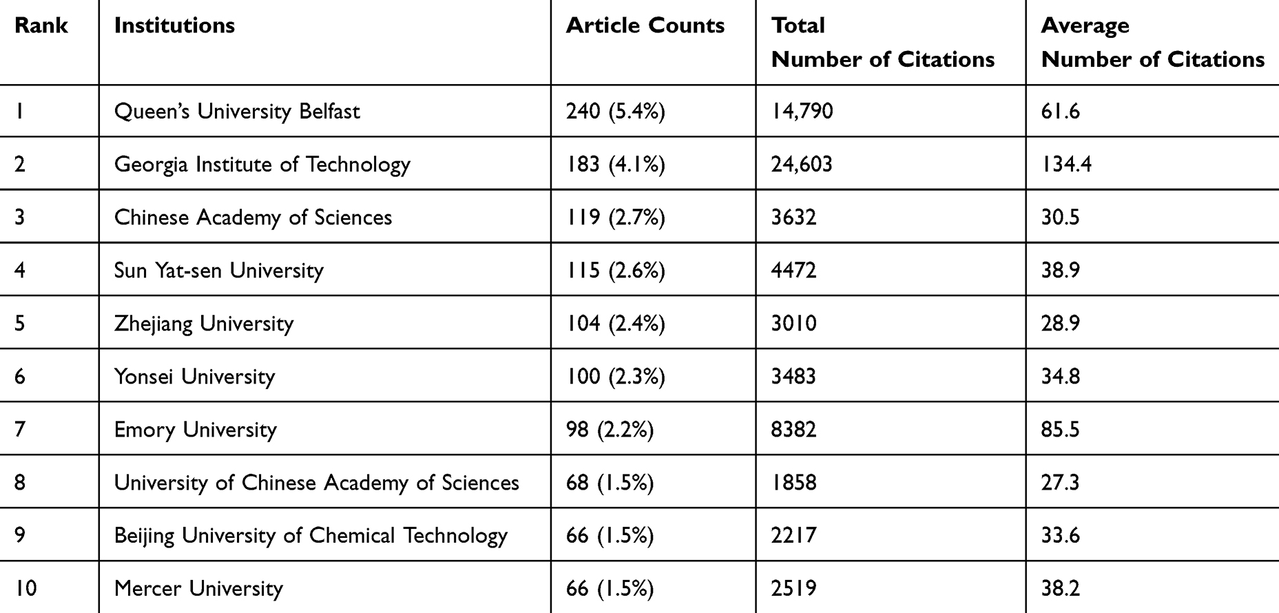 |
Table 1 Top 10 Institutions in the Field of Microneedles and Drug Delivery |
 |
Figure 3 Geographical distribution of retrieved articles in microneedles and drug delivery. |
 |
Figure 4 The visualization of countries on research of microneedles and drug delivery. |
 |
Figure 5 The growth trends of the top 10 nations/regions in microneedles and drug delivery. |
The publications included in this study come from 3290 institutions, with the top 10 institutions distributed across four countries, five of which are located in China. The institutions with the highest publication output are: Queens University Belfast (n=240, 5.4%), Georgia Institute of Technology (n=183, 4.1%), Chinese Academy of Sciences (n=119, 2.7%), and Sun Yat-sen University (n=115, 2.6%) (Table 1). Subsequently, based on a minimum publication threshold of ≥30 articles, 40 institutions were selected for visualization, and a collaboration network was constructed from a temporal perspective based on each institution’s publication volume and publication relationships (Figure 6). The larger the institution’s name, the greater its contribution to the field of microneedles and drug delivery. The size of the institution’s name is proportional to its contribution in the field of microneedles and drug delivery. The thickness of the inter-node connections indicates the intensity of collaboration.
 |
Figure 6 Cluster density visualization map of institutions on microneedles and drug delivery. |
We exported the information of the 40 institutions filtered by VOSviewer and calculated the collaboration strength index between each pair of institutions based on the Salton coefficient using the following formula:
Cij: Number of collaborations between institutions i and j
Ni, Nj: Total number of collaborations of institutions i and j (with all institutions)
Table 2 displays the top eight pairs of institutions with the closest collaborations. Among them, Queen’s University Belfast and Hasanuddin University exhibited the most intensive collaboration among all institutions, while the collaboration strength between Queen’s University Belfast and Mercer University also ranked on the list (sixth place). Meanwhile, JUVIC Biotech Co., Ltd. from South Korea maintained close collaborations with Seoul National University and Yonsei University, ranking second and third respectively in terms of collaboration strength among all institutions.
 |
Table 2 Top Eight Institutions with the Closest Collaboration |
Journals and Co-Cited Journals
A total of 752 journals published literature related to microneedles and drug delivery. Journal of Controlled Release published the most articles (n=240, 5.5%), followed by International Journal of Pharmaceutics (n=218, 5.0%), Pharmaceutics (n=162, 3.7%), and Journal of Drug Delivery Science and Technology (n=128, 2.9%).
Among the top 15 journals, the journal with the highest impact factor is Journal of Controlled Release (IF=10.5), followed by Advanced Healthcare Materials (IF=10). We then selected 42 journals based on a minimum of 20 related publications and constructed a journal network diagram (Figure 7). We found that International Journal of Pharmaceutics has an active citation relationship with Journal of Controlled Release, Pharmaceutics, Journal of Drug Delivery Science and Technology, and other journals.
 |
Figure 7 The visualization of journals on research of microneedles and drug delivery. |
Among the top 15 most-cited journals, two journals had citation frequencies exceeding 10,000. The journal with the highest citation frequency was Journal of Controlled Release, followed by International Journal of Pharmaceutics, Advanced Drug Delivery Reviews, and Pharmaceutical Research. Additionally, Advanced Materials had the highest impact factor (IF=27.4), followed by Advanced Functional Materials (IF=18.5) (Table 3). By selecting journals with a minimum co-citation frequency of ≥1000, we constructed a co-citation network diagram (Figure 8). Journal of Controlled Release has a positive co-citation relationship with International Journal of Pharmaceutics, Biomaterials, and other journals.
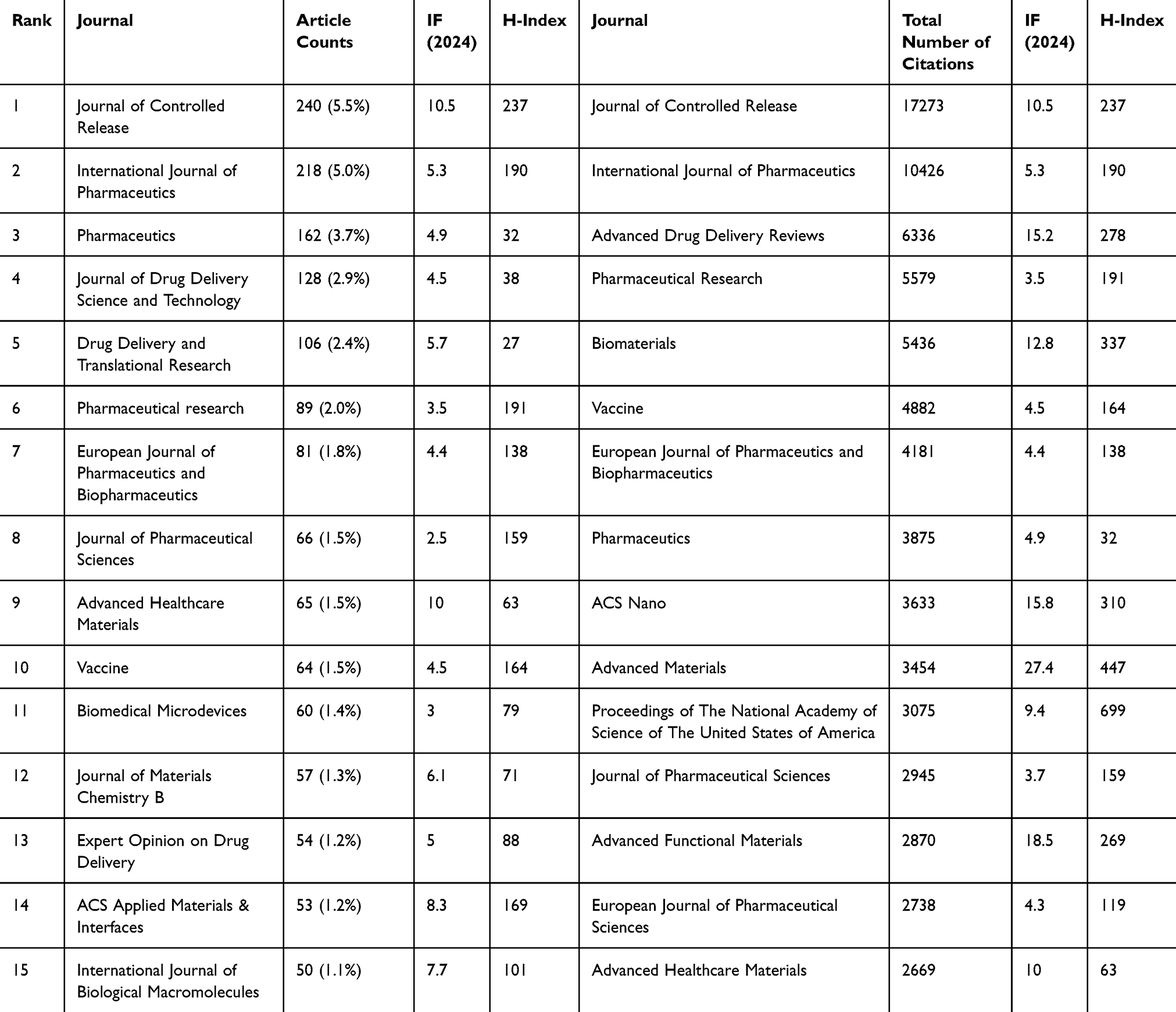 |
Table 3 Top 15 Journals and Co-Cited Journals in the Field of Microneedles and Drug Delivery |
 |
Figure 8 The visualization of co-cited journals on research of microneedles and drug delivery. |
Authors and Co-Cited Authors
A total of 16,697 authors have participated in research on microneedles and drug delivery. Among the top 15 authors, 9 have published more than 50 articles (Table 4). Notably, two authors have made outstanding contributions to the field of microneedles and drug delivery, each publishing over 100 articles: Ryan F. Donnelly from School of Pharmacy, Queens University Belfast, Medical Biology Centre, Antrim, Northern Ireland (n=226) and Mark R. Prausnitz from the Department of Biomolecular Engineering, Georgia Institute of Technology, Atlanta, Georgia, USA (n=155). Andi Dian Permana (n=62) from the Department of Pharmaceutical Science and Technology, Faculty of Pharmacy, Hasanuddin University, Makassar, Indonesia, and Helen O. McCarthy (n=62) from the same institution as Ryan F. Donnelly shared the third position in publication output. Eneko Larrañeta (n=58), ranked fifth in publication volume, is also affiliated with the School of Pharmacy, Queen’s University Belfast. We constructed a collaboration network based on authors with ≥20 publications (Figure 9). Ryan F. Donnelly and Mark R. Prausnitz have the largest nodes, as they have published the most relevant articles. The network as a whole formed a collaborative framework centered approximately around these two authors. Both have collaborated with numerous authors. Notably, Andi Dian Permana, Helen O. McCarthy, and Eneko Larrañeta maintained close collaborative relationships with Ryan F. Donnelly.
 |
Table 4 Top 10 Authors and Co-Cited Authors |
 |
Figure 9 The visualization of authors on research of microneedles and drug delivery. |
Among the 89,525 co-cited authors, 6 authors have been cited more than 1000 times. The author with the highest citation frequency is Donnelly, Ryan F. (n=2438), followed by Prausnitz, Mark R. (n=2283), and Kim, Yeu-Chun (n=1692) (Table 4). We selected authors with ≥300 citations and constructed a co-citation network diagram (Figure 10). Donnelly, Ryan F., and Larraneta, Eneko, as well as Prausnitz, Mark R. and Park, Jung-Hwan, among other co-cited authors, also show active collaboration.
 |
Figure 10 The visualization of co-cited Authors on research of microneedles and drug delivery. |
Co-Cited References
From 1997 to the present search, a total of 143,443 co-cited articles on microneedles and drug delivery have been identified. Among the top 10 most co-cited articles, 7 articles have a co-citation frequency of ≥500, and one article has a co-citation frequency of ≥800 (Table 5). We selected publications with ≥300 co-citations and constructed a co-citation network diagram (Figure 11). Notably, “Kim YC, 2012, Adv Drug Deliver Rev” shows a strong co-citation relationship with “Prausnitz MR, 2008, Nat Biotechnol” and “Park JH, 2005, J Control Release”, among others.
 |
Table 5 Top 10 Co-Cited References |
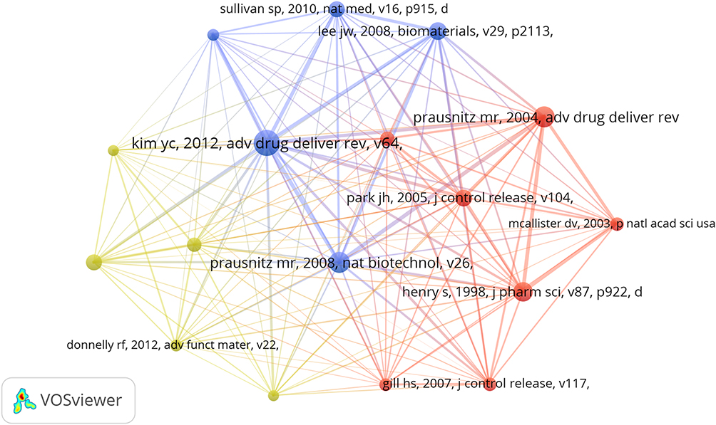 |
Figure 11 The visualization of co-cited references on the research of microneedles and drug delivery. |
Hotspots and Frontiers
Using VOSviewer, we filtered keywords with a frequency of ≥30 and excluded keywords that were identical to the search terms. Through keyword co-occurrence analysis, we captured the research hotspots related to microneedles and drug delivery (Figure 12). The thicker the line between the nodes, the stronger the relationship between the keywords. A total of four clusters were identified, representing three research directions. The red cluster includes keywords such as dissolving microneedles, release, nanopractices, insulin, diabetes, cancer, immunotherapy, hydrogel, and others. The blue cluster contains keywords such as skin, in-vitro, formulation, penetration, permeation, electroporation, and others. The green cluster includes keywords like immunization, vaccine, immune responses, antigen, intradermal delivery, and others. The yellow cluster features keywords such as fabrication, polymer microneedles, arrays, patches, 3D printing, and others. The purple cluster, which has fewer keywords, includes eye, injection, ocular drug delivery, suprachoroidal space, posterior segment, triamcinolone acetonide, molecular weight, and others. Table 6 shows the top 20 most frequent keywords in the research on microneedles and drug delivery.
 |
Table 6 Top 20 Keywords in the Field of Microneedles and Drug Delivery |
 |
Figure 12 Keyword cluster analysis on research of microneedles and drug delivery. |
We used CiteSpace software to analyze the keywords extracted from the literature in this search and examined the temporal trends of the top 25 burst terms (Figure 13). The figure shows the emergence time and burst strength of these terms, with the red bars indicating years when the occurrence rate of the keyword was particularly high.
 |
Figure 13 The top 25 burst words in research of microneedles and drug delivery. |
Discussion
General Information
Bibliometric analysis reveals that, overall, the literature on microneedle-mediated drug delivery has shown a continuous growth trend, with an increasing number of related research findings and growing interest from scholars in this field. Between 1998 and 2002, the annual publication volume was relatively low, averaging 3.7 papers per year, marking the initiation phase of research on microneedle-mediated drug delivery. From 2003 to 2019, research on microneedle-mediated drug delivery began to grow steadily. Between 2020 and 2024, the volume of literature on microneedle-mediated drug delivery experienced significant growth. Based on the statistical trends, we can predict that research on microneedle-mediated drug delivery will continue to increase, and this field will play an increasingly important role in biomedical research in the future.
China and the United States are the leading countries in microneedle-mediated drug delivery research, with China taking the lead. Among the top 10 research institutions, half are located in China (n=5), followed by the United States (n=3), the UK (n=1), and South Korea (n=1). Although there is some collaboration between countries, the breadth and strength of inter-institutional collaboration are not ideal. In the long run, this could hinder the development of the research field. Regarding research institutions, some have established strong collaborations. For example, Queens University Belfast in the UK and Georgia Institute of Technology in the US have become central hubs for numerous collaborative institutions, and these two institutions also maintain close cooperation. However, we also observed that some Chinese institutions, such as the Chinese Academy of Sciences, despite publishing the most papers, have limited collaboration with other institutions. Additionally, cross-national institutional collaboration remains relatively low. Therefore, we recommend that research institutions from different countries enhance their collaboration and communication to jointly promote the development of microneedle-mediated drug delivery research.
The journal that publishes the most literature on microneedle-mediated drug delivery is Journal of Controlled Release (n=238, IF= 10.5), which is also the most cited journal and has the highest impact factor among the top 15 journals publishing research on microneedle-mediated drug delivery. This indicates that the journal is currently the most popular in this research field. Among the top 15 journals publishing the most literature on microneedle-mediated drug delivery, two have an impact factor ≥10, and eight have an impact factor ≥5, indicating that they are high-quality international journals that support research in this area. Currently, research on microneedle-mediated drug delivery is primarily published in pharmaceutical and biological journals, with few studies appearing in clinical journals. This suggests that most research is still in the basic research stage and has not yet seen widespread clinical application.
From the perspective of the authors, two researchers have made outstanding contributions to the field of microneedle-mediated drug delivery, publishing more than 100 papers: Ryan F. Donnelly (n=224) and Mark R. Prausnitz (n=152). Ryan F. Donnelly and his team’s research primarily focuses on novel polymer microneedle arrays for transdermal drug delivery of “hard-to-deliver” drugs, as well as intradermal delivery of vaccines and photosensitizers. His research spans a wide range of areas, including cancer treatment,26 antimicrobial drug delivery,27 HIV prevention,28 ocular drug delivery,8 and 3D-printed microneedles.29 Similarly, Mark R. Prausnitz and his team have made significant contributions in areas such as insulin delivery for diabetes patients,30 microneedle-mediated vaccination,31 and drug delivery to ocular tissues.32 In terms of co-cited authors, these two researchers are also the most frequently co-cited, indicating their significant influence in the field of microneedle-mediated drug delivery.
Co-cited literature refers to documents that are cited by multiple publications and can be considered as foundational research in a given field.25 In this search, five of the ten most co-cited papers are review articles, which provide a comprehensive summary of the research foundations for microneedle-based drug and vaccine delivery. The review article “Microneedles for Drug and Vaccine Delivery” by Mark R. Prausnitz’s team is the most co-cited paper. This review discusses the use of microneedles for delivering various low molecular-weight drugs, biotherapeutics, and vaccines prior to the year of its publication. Among the most co-cited original studies, topics include microfabricated microneedles,3 dissolving microneedles,33 biodegradable polymer microneedles,34 dissolving polymer microneedles for influenza vaccination,35 and research on measuring and predicting insertion forces and needle breakage forces.36 These studies have laid the foundation for the development of microneedle-mediated drug delivery.
Hotspots and Frontiers
Bibliometrics is an important method for identifying research hotspots and predicting future development trends. This article, by analyzing the changes in emerging terms in recent years, examines the development trends of microneedle research. Cluster analysis identified four key research hotspots in microneedle studies.
The red cluster reveals the correlation between microneedle-mediated drug delivery and the treatment of various diseases. Our analysis shows that diseases such as diabetes, cancer, and melanoma are the main focus of microneedle-related drug delivery research. Type 1 diabetes is a very common chronic disease worldwide, and the only current treatment method is long-term subcutaneous insulin injections to maintain normal blood glucose levels.37 Due to its advantages of being pain-free and minimally invasive, microneedles have the potential to replace subcutaneous insulin injections in the treatment of Type 1 diabetes. The first report of using microneedles to deliver insulin appeared in 2003.38 Researchers used solid microneedles to penetrate the skin’s outer layer, increasing the permeability of insulin and thereby reducing blood glucose levels in diabetic rats. Since then, research on the combination of insulin and microneedles has expanded, including solid microneedles,39 coated microneedles,40 hollow microneedles,41 dissolving microneedles,42 and hydrogel microneedles.43 With continuous optimization of microneedle performance, microneedle-mediated insulin delivery is expected to play a significant role in the future diabetes treatment market, potentially replacing subcutaneous injections as the primary treatment method. In the prevention and treatment of cancer, microneedles have been used to deliver anticancer drugs (chemotherapy) to skin tumors, as well as for photodynamic therapy and photothermal therapy. Keyword emergence analysis reveals that since 2021, microneedle-mediated drug delivery has become a key development direction in the field of immunotherapy. In tumor immunotherapy, compared to traditional subcutaneous injections, using microneedles to implant tumor vaccines and adjuvants subcutaneously induces a stronger antigen-specific antibody response and enhances antibody memory.44 Additionally, combination chemotherapy-photothermal therapy can improve the therapeutic effect on tumors. Microneedle delivery systems can facilitate the localized delivery of drugs in combination therapies, avoiding the issues associated with the systemic delivery of anticancer drugs.45
The blue cluster highlights the correlation between microneedle-mediated drug delivery and the enhancement of transdermal/intradermal drug absorption. Many researchers have focused on improving the transdermal penetration efficiency of microneedle-mediated drug delivery, employing various strategies to enhance its effectiveness. Microneedles can break through the skin barrier, ensuring the precise delivery of both small molecules and large macromolecules, which promotes localized or systemic therapeutic effects.45 Moreover, when combined with nanoparticles, microneedles not only enhance the skin permeability of drugs but also leverage the inherent advantages of nanoparticles, such as protecting therapeutic payloads from chemical or enzymatic degradation, enabling sustained release, and promoting targeted drug delivery. This synergistic approach improves the overall efficiency and effectiveness of dermal/transdermal therapies.5 Hydrogel microneedles can regulate drug release by controlling the crosslinking density of the matrix material, allowing for sustained and controlled drug release.46 These systems have been applied in research on various diseases, including cancer,47 diabetes,48 and diabetes.49 Additionally, some researchers have investigated the impact of different microneedle shapes on the skin permeability of drugs.50
The green cluster highlights the correlation between microneedle-mediated drug delivery and vaccine delivery. Burst keyword analysis also indicates that vaccine delivery has been an important research focus in the microneedle field since 2013. Studies have shown that, compared to traditional injectable formulations, microneedles offer more effective immune responses, broader immunogenicity, and higher seroconversion rates.51 Additionally, vaccines can be stored at room temperature and transported in solid form, which reduces transport costs and the likelihood of contamination during storage.52 Soluble microneedles can incorporate vaccines into soluble polymer matrices, which dissolve in tissues without generating harmful medical waste, thus reducing the risk of blood-borne diseases.53 Current research on microneedle vaccines has focused on the development of vaccines for various diseases, including influenza,54 encephalitis,55 measles, rubella,56 gonorrhea,57 and malaria.58 During the global COVID-19 pandemic, numerous studies on COVID-19 microneedle vaccines significantly advanced the development of microneedle vaccine technology. For example, Kim et al found that soluble microneedles containing the SARS-CoV-2 S1 subunit vaccine elicited a strong antigen-specific antibody response two weeks after administration. Furthermore, the microneedle array vaccine maintained its immunogenicity even after gamma radiation sterilization, demonstrating the considerable advantages of microneedle vaccines.59 Although microneedle vaccines have not yet become a mainstream tool, their advantages are expected to expand as technology advances, and they hold great promise for the future.
The yellow cluster highlights the correlation between microneedle-mediated drug delivery and materials and manufacturing. The choice of materials, microneedle configuration, and manufacturing methods are all closely related to the intended application of microneedles. Silicon and silicon-based materials are widely used in microneedle drug delivery systems, primarily prepared through a combination of isotropic and anisotropic etching processes.60 To address their fragility and relatively poor biocompatibility, methods such as gold plating on silicon through metal sputtering61 and optimization of etching techniques62 have been used to improve their mechanical strength and biocompatibility. Metallic microneedles offer excellent mechanical and physical properties, resistance to fracture, low cost, and good biocompatibility. Metals such as stainless steel,12 titanium,63 titanium64 are commonly used for the manufacture of hollow, coated, and solid microneedles. Polymer materials, on the other hand, are easy to shape, have short processing times, low cost, and a wide variety of available materials, making them suitable for large-scale production. Additionally, polymers offer good needle flexibility. Furthermore, many polymer materials exhibit biocompatibility and biodegradability, making polymer-based microneedles an important research direction in the current field of medical microneedle fabrication. Currently, there are many types of polymer microneedles, including Polyvinylpyrrolidone (PVP),65 Polylactide (PLA),66 Acrylate,67 Hydrogels,68 Polyvinylalcohol (PVA),69 Polycaprolactone (PCL),69 Carboxymethylcellulose (CMC),70 Cellulose acetate phthalate (CAP),71 among others. Polymer microneedles are continuously evolving and innovating, representing a key development direction in microneedle-mediated drug delivery. The manufacturing methods for microneedles include selective etching photolithography, chemical etching processing, micro-milling, laser cutting, 3D printing, soft lithography, micro-injection molding, hot embossing, electrohydrodynamic spraying, laser processing, laser discharge processing, soft lithography, stretchable lithography, centrifugal lithography, and droplet transfer gas-blowing, among others.72 Researchers can adjust the drug release rate of microneedles by altering the design, materials, and drug carriers according to the specific needs of patients. This allows microneedle-mediated drug delivery research to progress toward more personalized and precise treatments.
Although “smart microneedles” did not appear among the high-frequency keywords in our analysis, existing prominent terms reveal widespread research trends focusing on demands such as controlled drug release, precise targeted delivery, multifunctional applications across multiple organs, long-term wearability, and user-friendly operation. Consequently, designing novel smart microneedles endowed with superior characteristics holds significant research value. Researchers have already developed various intelligent microneedle systems, including adhesive microneedles,73 rapidly separating dissolving microneedles,74 separable microneedles,75 and responsive microneedles.76 These innovative materials or fabrication techniques have further amplified the advantages of microneedle-mediated drug delivery, enabling their application across a broad spectrum of biomedical fields. Simultaneously, the emergence of novel smart microneedles has created opportunities for integrating microneedles with practical devices, opening new research directions in combination therapies and functional expansions.
This study has several unique advantages. We systematically analyzed the research on microneedles in drug delivery using bibliometric methods for the first time, providing comprehensive guidance for scholars interested in this field. Additionally, we employed multiple bibliometric tools to investigate and analyze the literature from various dimensions. The bibliometric analysis offers comprehensive statistical data and visualizations, providing a more objective and complete insight into the hotspots and cutting-edge developments in microneedle-mediated drug delivery, compared to traditional reviews.
However, there are some limitations to this study. The data used were exclusively sourced from the WoSCC database, and only English-language studies were included, which may result in the omission of some relevant research. Furthermore, recently published articles may not have been cited as frequently in a short period, potentially excluding high-quality articles with insufficient citation counts. Therefore, conducting bibliometric studies on the same research direction at different time intervals will help researchers continually identify emerging research hotspots.
Conclusion
In recent years, microneedle-mediated drug delivery has garnered increasing attention from researchers worldwide, with the number of related publications continuing to grow. China and the United States are leading the research in this field, and several teams are focusing on advancing this research, yielding exceptional results. However, collaboration and communication between countries and institutions still require improvement. Additionally, we recommend that researchers pay attention to the translation of research findings into clinical applications, further promoting the development of microneedles in drug delivery. Microneedle-mediated drug delivery has generated numerous research hotspots in disease treatment and diagnosis, vaccine delivery, and the discovery of new materials and technologies. As an emerging technology, microneedle-mediated drug delivery holds significant research value and promising application prospects. This paper aims to provide researchers in this field with effective references and suggestions for future development.
Data Sharing Statement
The original contributions presented in this study are included in the article. Further inquiries can be directed to the corresponding authors.
Acknowledgments
We would like to express our sincere gratitude to the Datawrapper online tool, VOSviewer software, CiteSpace software, Scimago Graphica software, and Pajek software for their significant contributions to the creation of figures in this study. Their user-friendly platforms enabled us to efficiently visualize and present complex geographical data, greatly enhancing the clarity and impact of our findings.
Author Contributions
All authors made a significant contribution to the work reported, whether that is in the conception, study design, execution, acquisition of data, analysis and interpretation, or in all these areas; took part in drafting, revising or critically reviewing the article; gave final approval of the version to be published; have agreed on the journal to which the article has been submitted; and agree to be accountable for all aspects of the work.
Disclosure
The authors report no conflicts of interest in this work.
References
1. Prausnitz MR, Mitragotri S, Langer R. Current status and future potential of transdermal drug delivery. Nat Rev Drug Discov. 2004;3(2):115–124. doi:10.1038/nrd1304
2. Prausnitz MR, Langer R. Transdermal drug delivery. Nat Biotechnol. 2008;26(11):1261–1268. doi:10.1038/nbt.1504
3. Henry S, McAllister DV, Allen MG, Prausnitz MR. Microfabricated microneedles: a novel approach to transdermal drug delivery. J Pharma Sci. 1998;87(8):922–925.
4. Miyano T, Tobinaga Y, Kanno T, et al. Sugar micro needles as transdermic drug delivery system. Biomed Microdevices. 2005;7(3):185–188. doi:10.1007/s10544-005-3024-7
5. Qu F, Geng R, Liu Y, Zhu J. Advanced nanocarrier- and microneedle-based transdermal drug delivery strategies for skin diseases treatment. Theranostics. 2022;12(7):3372–3406. doi:10.7150/thno.69999
6. Chen X, Wang L, Yu H, et al. Preparation, properties and challenges of the microneedles-based insulin delivery system. J Control Release. 2018;288:173–188. doi:10.1016/j.jconrel.2018.08.042
7. Ganeson K, Alias AH, Murugaiyah V, Amirul AA, Ramakrishna S, Vigneswari S. Microneedles for Efficient and Precise Drug Delivery in Cancer Therapy. Pharmaceutics. 2023;15(3):744. doi:10.3390/pharmaceutics15030744
8. Glover K, Mishra D, Gade S, et al. Microneedles for advanced ocular drug delivery. Adv Drug Delivery Rev. 2023;201:115082. doi:10.1016/j.addr.2023.115082
9. Balmert SC, Carey CD, Falo GD, et al. Dissolving undercut microneedle arrays for multicomponent cutaneous vaccination. J Control Release. 2020;317:336–346. doi:10.1016/j.jconrel.2019.11.023
10. Saeed Al-Japairai K A, Mahmood S, Hamed Almurisi S, et al. Current trends in polymer microneedle for transdermal drug delivery. Int J Pharm. 2020;587:119673.
11. Li Y, Zhang H, Yang R, et al. Fabrication of sharp silicon hollow microneedles by deep-reactive ion etching towards minimally invasive diagnostics. Microsys Nanoeng. 2019;5(1):41. doi:10.1038/s41378-019-0077-y
12. Cahill EM, Keaveney S, Stuettgen V, et al. Metallic microneedles with interconnected porosity: a scalable platform for biosensing and drug delivery. Acta Biomater. 2018;80:401–411. doi:10.1016/j.actbio.2018.09.007
13. Lee H, Choi TK, Lee YB, et al. A graphene-based electrochemical device with thermoresponsive microneedles for diabetes monitoring and therapy. Nature Nanotechnol. 2016;11(6):566–572. doi:10.1038/nnano.2016.38
14. Donnelly RF, Raj Singh TR, Woolfson AD. Microneedle-based drug delivery systems: microfabrication, drug delivery, and safety. Drug Delivery. 2010;17(4):187–207. doi:10.3109/10717541003667798
15. Luo X, Yang L, Cui Y. Microneedles: materials, fabrication, and biomedical applications. Biomed Microdevices. 2023;25(3):20. doi:10.1007/s10544-023-00658-y
16. Jamaledin R, Yiu CKY, Zare EN, et al. Advances in Antimicrobial Microneedle Patches for Combating Infections. Adv Mater. 2020;32(33):e2002129. doi:10.1002/adma.202002129
17. Waghule T, Singhvi G, Dubey SK, et al. Microneedles: a smart approach and increasing potential for transdermal drug delivery system. Biomed Pharmacother. 2019;109:1249–1258. doi:10.1016/j.biopha.2018.10.078
18. Ninkov A, Frank JR, Maggio LA. Bibliometrics: methods for studying academic publishing. Perspect Med Educ. 2022;11(3):173–176. doi:10.1007/S40037-021-00695-4
19. Ke L, Lu C, Shen R, Lu T, Ma B, Hua Y. Knowledge Mapping of Drug-Induced Liver Injury: a Scientometric Investigation (2010–2019). Front Pharmacol. 2020;11:842. doi:10.3389/fphar.2020.00842
20. Shi S, Gao Y, Liu M, et al. Top 100 most-cited articles on exosomes in the field of cancer: a bibliometric analysis and evidence mapping. Clin Exp Med. 2021;21(2):181–194. doi:10.1007/s10238-020-00624-5
21. Sun S, Mao Z, Wang H. Relationship between periodontitis and diabetes: a bibliometrics analysis. Ann Translat Med. 2022;10(7):401. doi:10.21037/atm-22-1067
22. Chen X, Xiao H, Zhao Q, Xu X, Cen Y, Xiao D. Research hotspot and trend of microneedles in biomedical field: a bibliometric analysis from 2011 to 2020. Burns. 2022;48(4):959–972. doi:10.1016/j.burns.2022.04.004
23. van Eck NJ, Waltman L. Software survey: vOSviewer, a computer program for bibliometric mapping. Scientometrics. 2010;84(2):523–538. doi:10.1007/s11192-009-0146-3
24. Arruda H, Silva ER, Lessa M, Proença Jr. D Jr, Bartholo R. VOSviewer and Bibliometrix. JMLA. 2022;110(3):392–395. doi:10.5195/jmla.2022.1434
25. Synnestvedt MB, Chen C, Holmes JH. CiteSpace II: visualization and knowledge discovery in bibliographic databases. AMIA Annual Symposium Proceedings AMIA Symposium. 2005;2005:724–728.
26. Abd-El-Azim H, Tekko IA, Ali A, et al. Hollow microneedle assisted intradermal delivery of hypericin lipid nanocapsules with light enabled photodynamic therapy against skin cancer. J Control Release. 2022;348:849–869. doi:10.1016/j.jconrel.2022.06.027
27. Abraham AM, Anjani QK, Adhami M, Hutton ARJ, Larrañeta E, Donnelly RF. Novel SmartReservoirs for hydrogel-forming microneedles to improve the transdermal delivery of rifampicin. J Mater Chem B. 2024;12(18):4375–4388. doi:10.1039/D4TB00110A
28. Volpe-Zanutto F, Vora LK, Tekko IA, et al. Hydrogel-forming microarray patches with cyclodextrin drug reservoirs for long-acting delivery of poorly soluble cabotegravir sodium for HIV Pre-Exposure Prophylaxis. J Control Release. 2022;348:771–785. doi:10.1016/j.jconrel.2022.06.028
29. Uddin MJ, Scoutaris N, Economidou SN, et al. 3D printed microneedles for anticancer therapy of skin tumours. Mater Sci Eng C. 2020;107:110248. doi:10.1016/j.msec.2019.110248
30. Chen Q, Xiao Z, Wang C, et al. Microneedle Patches Loaded with Nanovesicles for Glucose Transporter-Mediated Insulin Delivery. ACS nano. 2022;16(11):18223–18231. doi:10.1021/acsnano.2c05687
31. Kim MC, Lee JW, Choi HJ, et al. Microneedle patch delivery to the skin of virus-like particles containing heterologous M2e extracellular domains of influenza virus induces broad heterosubtypic cross-protection. J Control Release. 2015;210:208–216. doi:10.1016/j.jconrel.2015.05.278
32. Patel SR, Berezovsky DE, McCarey BE, Zarnitsyn V, Edelhauser HF, Prausnitz MR. Targeted administration into the suprachoroidal space using a microneedle for drug delivery to the posterior segment of the eye. Invest Ophthalmol Visual Sci. 2012;53(8):4433–4441. doi:10.1167/iovs.12-9872
33. Lee JW, Park JH, Prausnitz MR. Dissolving microneedles for transdermal drug delivery. Biomaterials. 2008;29(13):2113–2124. doi:10.1016/j.biomaterials.2007.12.048
34. Park JH, Allen MG, Prausnitz MR. Biodegradable polymer microneedles: fabrication, mechanics and transdermal drug delivery. J Control Release. 2005;104(1):51–66. doi:10.1016/j.jconrel.2005.02.002
35. Sullivan SP, Koutsonanos DG, Del Pilar Martin M, et al. Dissolving polymer microneedle patches for influenza vaccination. Nat Med. 2010;16(8):915–920. doi:10.1038/nm.2182
36. Davis SP, Landis BJ, Adams ZH, Allen MG, Prausnitz MR. Insertion of microneedles into skin: measurement and prediction of insertion force and needle fracture force. J Biomech. 2004;37(8):1155–1163. doi:10.1016/j.jbiomech.2003.12.010
37. Tuomi T, Santoro N, Caprio S, Cai M, Weng J, Groop L. The many faces of diabetes: a disease with increasing heterogeneity. Lancet. 2014;383(9922):1084–1094. doi:10.1016/S0140-6736(13)62219-9
38. McAllister DV, Wang PM, Davis SP, et al. Microfabricated needles for transdermal delivery of macromolecules and nanoparticles: fabrication methods and transport studies. Proc Natl Acad Sci USA. 2003;100(24):13755–13760. doi:10.1073/pnas.2331316100
39. Zhang P, Zhang Y, Liu CG. Polymeric nanoparticles based on carboxymethyl chitosan in combination with painless microneedle therapy systems for enhancing transdermal insulin delivery. RSC Adv. 2020;10(41):24319–24329. doi:10.1039/D0RA04460A
40. Zhao X, Coulman SA, Hanna SJ, Wong FS, Dayan CM, Birchall JC. Formulation of hydrophobic peptides for skin delivery via coated microneedles. J Control Release. 2017;265:2–13. doi:10.1016/j.jconrel.2017.03.015
41. Bolton CJW, Howells O, Blayney GJ, et al. Hollow silicon microneedle fabrication using advanced plasma etch technologies for applications in transdermal drug delivery. Lab Chip. 2020;20(15):2788–2795. doi:10.1039/D0LC00567C
42. Fonseca DFS, Costa PC, Almeida IF, et al. Pullulan microneedle patches for the efficient transdermal administration of insulin envisioning diabetes treatment. Carbohydr Polym. 2020;241:116314. doi:10.1016/j.carbpol.2020.116314
43. Chen X, Yu H, Wang L, Shen D, Li C, Zhou W. Cross-Linking-Density-Changeable Microneedle Patch Prepared from a Glucose-Responsive Hydrogel for Insulin Delivery. ACS Biomater Sci Eng. 2021;7(10):4870–4882. doi:10.1021/acsbiomaterials.1c01073
44. Duong HTT, Yin Y, Thambi T, Kim BS, Jeong JH, Lee DS. Highly potent intradermal vaccination by an array of dissolving microneedle polypeptide cocktails for cancer immunotherapy. J Mater Chem B. 2020;8(6):1171–1181. doi:10.1039/C9TB02175B
45. Moreira AF, Rodrigues CF, Jacinto TA, Miguel SP, Costa EC, Correia IJ. Poly (vinyl alcohol)/chitosan layer-by-layer microneedles for cancer chemo-photothermal therapy. Int J Pharm. 2020;576:118907. doi:10.1016/j.ijpharm.2019.118907
46. Dimatteo R, Darling NJ, Segura T. In situ forming injectable hydrogels for drug delivery and wound repair. Adv Drug Delivery Rev. 2018;127:167–184. doi:10.1016/j.addr.2018.03.007
47. Huang S, Liu H, Huang S, Fu T, Xue W, Guo R. Dextran methacrylate hydrogel microneedles loaded with doxorubicin and trametinib for continuous transdermal administration of melanoma. Carbohydr Polym. 2020;246:116650. doi:10.1016/j.carbpol.2020.116650
48. Chen S, Matsumoto H, Moro-Oka Y, et al. Smart Microneedle Fabricated with Silk Fibroin Combined Semi-interpenetrating Network Hydrogel for Glucose-Responsive Insulin Delivery. ACS Biomater Sci Eng. 2019;5(11):5781–5789. doi:10.1021/acsbiomaterials.9b00532
49. Cao J, Su J, An M, et al. Novel DEK-Targeting Aptamer Delivered by a Hydrogel Microneedle Attenuates Collagen-Induced Arthritis. Mol Pharmaceut. 2021;18(1):305–316. doi:10.1021/acs.molpharmaceut.0c00954
50. De Martino S, Battisti M, Napolitano F, et al. Effect of microneedles shape on skin penetration and transdermal drug administration. Biomater Adv. 2022;142:213169. doi:10.1016/j.bioadv.2022.213169
51. Arya J, Prausnitz MR. Microneedle patches for vaccination in developing countries. J Control Release. 2016;240:135–141. doi:10.1016/j.jconrel.2015.11.019
52. Ali AA, McCrudden CM, McCaffrey J, et al. DNA vaccination for cervical cancer; a novel technology platform of RALA mediated gene delivery via polymeric microneedles. Nanomedicine. 2017;13(3):921–932. doi:10.1016/j.nano.2016.11.019
53. Li J, Zeng M, Shan H, Tong C. Microneedle Patches as Drug and Vaccine Delivery Platform. Curr Med Chem. 2017;24(22):2413–2422. doi:10.2174/0929867324666170526124053
54. Iwata H, Kakita K, Imafuku K, et al. Safety and dose-sparing effect of Japanese encephalitis vaccine administered by microneedle patch in uninfected, healthy adults (MNA-J): a randomised, partly blinded, active-controlled, Phase 1 trial. Lancet Microbe. 2022;3(2):e96–e104. doi:10.1016/S2666-5247(21)00269-X
55. Kim YC, Quan FS, Compans RW, Kang SM, Prausnitz MR. Formulation and coating of microneedles with inactivated influenza virus to improve vaccine stability and immunogenicity. J Control Release. 2010;142(2):187–195. doi:10.1016/j.jconrel.2009.10.013
56. Adigweme I, Yisa M, Ooko M, et al. A measles and rubella vaccine microneedle patch in The Gambia: a phase 1/2, double-blind, double-dummy, randomised, active-controlled age de-escalation trial. Lancet. 2024;403(10439):1879–1892. doi:10.1016/S0140-6736(24)00532-4
57. Bagwe P, Bajaj L, Menon I, et al. Gonococcal microparticle vaccine in dissolving microneedles induced immunity and enhanced bacterial clearance in infected mice. Int J Pharm. 2023;642:123182. doi:10.1016/j.ijpharm.2023.123182
58. Yenkoidiok-Douti L, Barillas-Mury C, Jewell CM. Design of Dissolvable Microneedles for Delivery of a Pfs47-Based Malaria Transmission-Blocking Vaccine. ACS Biomater Sci Eng. 2021;7(5):1854–1862. doi:10.1021/acsbiomaterials.0c01363
59. Kim E, Erdos G, Huang S, et al. Microneedle array delivered recombinant coronavirus vaccines: immunogenicity and rapid translational development. EBioMed. 2020;55:102743. doi:10.1016/j.ebiom.2020.102743
60. Vinayakumar KB, Hegde GM, Nayak MM, Dinesh NS, Rajanna K. Fabrication and characterization of gold coated hollow silicon microneedle array for drug delivery. Microelectron Eng. 2014;128:12–18. doi:10.1016/j.mee.2014.05.039
61. Narayanan SP, Raghavan S. Fabrication and characterization of gold-coated solid silicon microneedles with improved biocompatibility. Int J Adv Manuf Technol. 2019;104(9–12):3327–3333. doi:10.1007/s00170-018-2596-3
62. Li WZ, Huo MR, Zhou JP, et al. Super-short solid silicon microneedles for transdermal drug delivery applications. Int J Pharm. 2010;389(1–2):122–129. doi:10.1016/j.ijpharm.2010.01.024
63. Ameri M, Kadkhodayan M, Nguyen J, et al. Human Growth Hormone Delivery with a Microneedle Transdermal System: preclinical Formulation, Stability, Delivery and PK of Therapeutically Relevant Doses. Pharmaceutics. 2014;6(2):220–234. doi:10.3390/pharmaceutics6020220
64. Chen J, Cheng P, Sun Y, et al. A Minimally Invasive Hollow Microneedle With a Cladding Structure: ultra-Thin but Strong, Batch Manufacturable. IEEE Transact Bio-Med Eng. 2019;66(12):3480–3485. doi:10.1109/TBME.2019.2906571
65. Liu D, Yu B, Jiang G, Yu W, Zhang Y, Xu B. Fabrication of composite microneedles integrated with insulin-loaded CaCO(3) microparticles and PVP for transdermal delivery in diabetic rats. Mater Sci Eng C. 2018;90:180–188. doi:10.1016/j.msec.2018.04.055
66. Kim MJ, Park SC, Rizal B, et al. Fabrication of Circular Obelisk-Type Multilayer Microneedles Using Micro-Milling and Spray Deposition. Front Bioeng Biotechnol. 2018;6:54. doi:10.3389/fbioe.2018.00054
67. Gittard SD, Chen B, Xu H, et al. The Effects of Geometry on Skin Penetration and Failure of Polymer Microneedles. J Adhes Sci Technol. 2013;27(3):227–243. doi:10.1080/01694243.2012.705101
68. Bok M, Zhao ZJ, Jeon S, Jeong JH, Lim E. Ultrasonically and Iontophoretically Enhanced Drug-Delivery System Based on Dissolving Microneedle Patches. Sci Rep. 2020;10(1):2027. doi:10.1038/s41598-020-58822-w
69. He R, Niu Y, Li Z, et al. A Hydrogel Microneedle Patch for Point-of-Care Testing Based on Skin Interstitial Fluid. Adv Healthc Mater. 2020;9(4):e1901201. doi:10.1002/adhm.201901201
70. Silva ACQ, Pereira B, Lameirinhas NS, et al. Dissolvable Carboxymethylcellulose Microneedles for Noninvasive and Rapid Administration of Diclofenac Sodium. Macromol Biosci. 2023;23(1):e2200323. doi:10.1002/mabi.202200323
71. Anderson A, Hegarty C, Casimero C, Davis J. Electrochemically Controlled Dissolution of Nanocarbon-Cellulose Acetate Phthalate Microneedle Arrays. ACS Appl Mater Interfaces. 2019;11(39):35540–35547. doi:10.1021/acsami.9b09674
72. Jung JH, Jin SG. Microneedle for transdermal drug delivery: current trends and fabrication. J Pharm Invest. 2021;51(5):503–517. doi:10.1007/s40005-021-00512-4
73. Zhang X, Chen G, Yu Y, Sun L, Zhao Y. Bioinspired Adhesive and Antibacterial Microneedles for Versatile Transdermal Drug Delivery. Research. 2020;2020(2020):3672120. doi:10.34133/2020/3672120
74. Yang Y, Li Z, Huang P, et al. Rapidly separating dissolving microneedles with sustained-release colchicine and stabilized uricase for simplified long-term gout management. Acta Pharma Sin B. 2023;13(8):3454–3470. doi:10.1016/j.apsb.2023.02.011
75. Zhao E, Xiao T, Tan Y, et al. Separable Microneedles with Photosynthesis-Driven Oxygen Manufactory for Diabetic Wound Healing. ACS Appl Mater Interfaces. 2023;15(6):7725–7734. doi:10.1021/acsami.2c18809
76. Chu H, Xue J, Yang Y, Zheng H, Luo D, Li Z. Advances of Smart Stimulus-Responsive Microneedles in Cancer Treatment. Small Methods. 2024;8(9):e2301455. doi:10.1002/smtd.202301455
 © 2025 The Author(s). This work is published and licensed by Dove Medical Press Limited. The
full terms of this license are available at https://www.dovepress.com/terms.php
and incorporate the Creative Commons Attribution
- Non Commercial (unported, 4.0) License.
By accessing the work you hereby accept the Terms. Non-commercial uses of the work are permitted
without any further permission from Dove Medical Press Limited, provided the work is properly
attributed. For permission for commercial use of this work, please see paragraphs 4.2 and 5 of our Terms.
© 2025 The Author(s). This work is published and licensed by Dove Medical Press Limited. The
full terms of this license are available at https://www.dovepress.com/terms.php
and incorporate the Creative Commons Attribution
- Non Commercial (unported, 4.0) License.
By accessing the work you hereby accept the Terms. Non-commercial uses of the work are permitted
without any further permission from Dove Medical Press Limited, provided the work is properly
attributed. For permission for commercial use of this work, please see paragraphs 4.2 and 5 of our Terms.