")
Back to Journals » International Journal of Nanomedicine » Volume 20
Advancements in Cell Membrane-Derived Biomimetic Nanotherapeutics for Breast Cancer
Authors Zeng M, Hu C, Chen T, Zhao T, Dai X
Received 13 November 2024
Accepted for publication 11 April 2025
Published 12 May 2025 Volume 2025:20 Pages 6059—6083
DOI https://doi.org/10.2147/IJN.S502144
Checked for plagiarism Yes
Review by Single anonymous peer review
Peer reviewer comments 3
Editor who approved publication: Professor Farooq A. Shiekh
Mingtang Zeng,1,2,* Chenji Hu,1,* Tao Chen,3 Tingrui Zhao,4 Xinhua Dai2
1Department of Pharmacy, West China Hospital, Sichuan University, Chengdu, 610041, People’s Republic of China; 2Department of Laboratory Medicine, West China Hospital, Sichuan University, Chengdu, 610041, People’s Republic of China; 3Pharmacy Department, Chongqing Emergency Medical Center, Chongqing University Central Hospital, Chongqing, 400014, People’s Republic of China; 4Department of Pharmacy, The Third Hospital of Mianyang, Sichuan Mental Health Center, Mianyang, 621000, People’s Republic of China
*These authors contributed equally to this work
Correspondence: Tingrui Zhao, Department of Pharmacy, The Third Hospital of Mianyang, Sichuan Mental Health Center, Mianyang, 621000, People’s Republic of China, Email [email protected] Xinhua Dai, Department of Laboratory Medicine, West China Hospital, Sichuan University, Chengdu, 610041, People’s Republic of China, Email [email protected]
Abstract: Breast cancer remains the leading cause of female mortality worldwide, necessitating innovative and multifaceted approaches to address its various subtypes. Nanotechnology has attracted considerable attention due to its nanoscale dimensions, diverse carrier types, suitability for hydrophobic drug delivery, and capacity for controlled and targeted administration. Nano-sized particles have become prevalent carriers for therapeutic agents targeting breast cancer, thanks to their reproducible synthesis and adjustable properties, including size, shape, and surface characteristics. In addition, certain nanoparticles can enhance therapeutic effects synergistically. However, the immune system often detects and removes these nanoparticles, limiting their efficacy. As a promising alternative, cell membrane-based delivery systems have gained attention due to their biocompatibility and targeting specificity. These membrane-coated drug delivery systems are derived from various cell sources, including blood cells, cancer cells, and stem cells. Leveraging the unique properties of these cell membranes enables precise targeting of breast cancer tumors and associated biomarkers. Inspired by natural structures, cell membranes disguise nanoparticles in the bloodstream, enhancing their retention time in vivo and improving tumor targeting. Consequently, cell membrane-derived nanoparticles (CMDNPs) have been investigated for their potential applications in breast cancer diagnostics, photothermal therapy (PTT), and vaccine development. This review comprehensively explores the potential and limitations of cell membrane-derived drug delivery systems in clinical applications against breast cancer.
Keywords: cell membrane-derived nanoparticles, breast cancer, active targeting, combination therapy, biomimetic delivery systems
Introduction
Breast cancer remains one of the most prevalent and deadly malignancies affecting women worldwide. According to recent statistics, over 2.3 million new cases of breast cancer were diagnosed globally in 2022, and it accounted for approximately 665,000 deaths.1 This makes breast cancer not only the most commonly diagnosed cancer in women but also a significant public health challenge. Despite advances in early detection and treatment, the mortality rate remains high due to the complex nature of the disease and the limitations of current therapeutic strategies.2 Traditional breast cancer treatments such as surgery, chemotherapy, and radiotherapy have been the cornerstone of clinical management for decades.3–5 However, these treatments are often associated with several drawbacks, including limited efficacy, high recurrence rates, significant adverse reactions, and narrow therapeutic windows.6–9 These limitations highlight the urgent need for the development of novel therapeutic approaches that can provide prolonged in vivo circulation, enhanced bioavailability, and more precise targeting of tumor cells to improve patient outcomes.10–12
In the past decade, nanoparticles (NPs) have emerged as promising drug carriers for targeted breast cancer therapy.13 Nanoparticles can be designed for both active and passive targeting. Active targeting involves the use of specific ligands, such as antibodies, to facilitate the uptake of nanoparticles by breast cancer cells, thereby improving the precision of drug delivery and minimizing damage to healthy tissues.14 Passive targeting, on the other hand, relies on the enhanced permeability and retention effect, which allows nanoparticles to accumulate preferentially in breast tumor tissues due to their leaky vasculature.15 While passive targeting nanoparticles are more commonly used in clinical settings, active targeting remains challenging due to the high interstitial fluid pressure within the breast tumor microenvironment, which hampers efficient nanoparticle accumulation.16 Moreover, nanoparticle-based therapies face issues related to toxicity and limited efficacy in breast cancer patients.17
In recent years, there has been a growing interest in biomimetic drug delivery systems, particularly those derived from cell membranes, to address the challenges of breast cancer treatment.18 These systems, known as cell membrane-derived biomimetic nanotherapeutics, exploit the natural properties of cell membranes to enhance drug delivery to breast tumors.19 By harnessing the homotypic and heterotypic adhesive properties of cells, these biomimetic carriers can be engineered through physical or chemical modifications to create drug delivery systems with active targeting capabilities specifically for breast cancer cells.20 These carriers exhibit high drug loading capacities, controlled release properties, excellent biocompatibility, low immunogenicity, and inherent targeting abilities.21 Additionally, by mimicking the flexible morphology of living cells, these nanotherapeutics can enhance drug accumulation and efficacy in breast tumor tissues.22
Common cell membrane sources for these biomimetic carriers include stem cells, red blood cells, cancer cells, bacteria, and exosomes, each offering unique therapeutic advantages and targeting properties for breast cancer therapy. This comprehensive literature review aims to summarize the drug-loading characteristics and therapeutic applications of various cell membrane-derived biomimetic nanotherapeutics in breast cancer therapy. The literature search for this review was conducted using databases including PubMed, ScienceDirect, Google Scholar, and Web of Science, ensuring a broad and exhaustive collection of pertinent articles. The following keywords were used in various combinations: “cell membrane-derived nanotherapeutics”, “biomimetic nanoparticles”, “breast cancer therapy”, “cell membrane coating”, “biomimetic drug delivery”, and “tumor targeting”. By exploring the latest advancements and current challenges, this review seeks to provide a holistic understanding of the potential of these innovative drug delivery systems in improving breast cancer treatment outcomes.
Stem Cell Membrane-Derived Biomimetic Nanotherapeutics
Stem cells, particularly mesenchymal stem cells (MSCs), have garnered significant attention in the field of drug delivery due to their unique biological properties, including self-renewal, tumor-homing capabilities, and low immunogenicity.23–25 These properties make MSCs highly attractive for targeted breast cancer therapies.26 Recent studies have demonstrated that MSC-derived membranes can encapsulate synthetic nanoparticles, forming biomimetic drug delivery systems with great potential for breast cancer treatment. For instance, Cao et al showed that MSCs could effectively deliver SiO2 nanoparticles containing photosensitizers (PS) to breast tumors for PTT, achieving efficient tumor destruction with minimal side effects.27 However, the challenge remains in enhancing the penetration depth of PTT, particularly for tumors that are deep-seated or resistant to heat-induced apoptosis. The same challenge applies to other approaches, such as the use of synthetic microcapsules loaded into MSCs, as explored by Litvinova et al, who highlighted MSCs’ ability to carry these microcapsules while maintaining their migratory and tumor-homing abilities.28 However, issues related to the mechanical stability of the microcapsules and the consistency of drug release still require optimization.29 Despite these challenges, MSCs’ tumor-targeting ability is crucial for efficient drug delivery.
Another promising approach involves MSC-derived membranes coated onto nanoparticles, such as doxorubicin (Dox)-loaded hollow gold nanoparticles, which have demonstrated significant tumor growth inhibition and improved CT imaging, along with a favorable safety profile.30 The addition of MUC1 aptamers further enhances targeted drug delivery, although variability in tumor responses, even within the same patient, poses a challenge due to differences in MUC1 expression levels. This suggests that targeting multiple tumor markers in combination with MSC-derived nanocarriers could improve therapeutic efficacy. Photodynamic therapy (PDT) has also emerged as a potential treatment due to its high selectivity and minimal off-target effects. However, the hydrophobic nature of PS makes their solubility and circulation in the bloodstream a challenge. Recent advancements have led to the development of biomimetic PDT platforms using MSC-coated nanoparticles, which show improved tumor-targeting efficiency and reduced macrophage uptake compared to uncoated PS.31 Still, the limited light penetration in solid tumors, particularly in advanced stages of breast cancer, remains a significant hurdle that needs to be addressed through the optimization of light delivery systems.
While MSC-based drug delivery systems show considerable promise, issues related to drug loading, release kinetics, and long-term safety need to be addressed. Studies by Kalimuthu et al have shown that Dox-loaded MSCs do not impair their tumor-homing ability, but challenges related to long-term toxicity and immune response need to be better understood.32 Therefore, future research should focus on improving the mechanical properties of MSC-derived carriers, fine-tuning drug release mechanisms, and exploring combination therapies to optimize therapeutic outcomes. Despite the challenges, MSC-derived nanocarriers offer a promising avenue for advancing breast cancer treatment, particularly through enhanced drug targeting, improved biocompatibility, and better overall efficacy.
Blood Cells as Targeted Drug Delivery Carriers
Based on blood cells, carriers possess numerous characteristics that render them ideal for targeted drug delivery in breast cancer.33 Such carriers are advantageous because they reduce immunogenicity and prolong the circulation time of drugs within the body. Generally, blood cells are biodegradable and do not produce toxic byproducts as a result of carrier biodegradation.34 Due to specific protein markers on their membranes, targeted delivery using blood cells is an innate function of the organism.35 Additionally, drugs are released in a controlled manner due to the constraints or affinities of the blood cell membranes, which significantly reduces fluctuations in the steady-state concentration. Consequently, blood cells constitute a vital source for drug-carrying cells, offering an effective strategy for enhancing the therapeutic efficacy and safety of drug delivery systems in breast cancer treatment (Table 1).
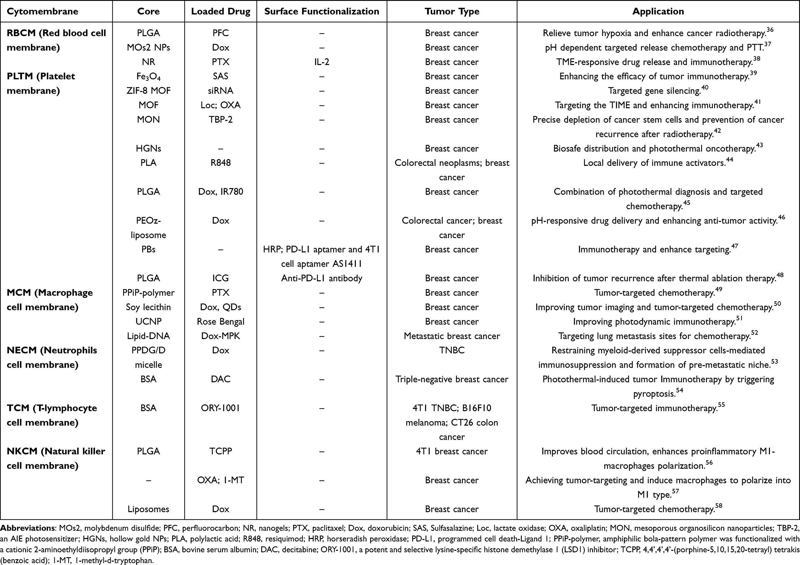 |
Table 1 The Application of Blood Cell Membrane-Coated Nanoparticles in Breast Cancer Therapy |
Red Blood Cell Membrane-Derived Biomimetic Nanotherapeutics
Red blood cells (RBCs) are highly promising drug delivery platforms due to their unique biological properties. Their biconcave disc shape provides an approximate membrane surface area of 135 um², facilitating the transport of various compounds.59 Coupled with the CD47-SIRPα mediated immune evasion mechanism, this theoretical advantage allows for a long circulation half-life of around 120 days.60 RBCs are the most prevalent type of blood cell, distinguished by their biocompatibility, substantial drug-loading capacity, and minimal immunogenicity.61 The interaction between the CD47 protein on the surface of RBCs and SIRPa regulates the phagocytic uptake of macrophages, enabling prolonged circulation within the body.62 However, their “natural stealth” property may be limited in terms of solid tumor penetration due to abnormal vascular structures within the tumor microenvironment. To harness the potential of RBCs for drug delivery, engineering strategies can be classified into genetic and non-genetic approaches.63 Genetic engineering involves using transfection technology to express specific membrane proteins, such as enzyme recognition sites, on the RBC surface to enable covalent anchoring of drugs.64 However, caution must be exercised as gene editing may alter the mechanical properties of RBC membranes, increasing membrane stiffness by 15%-20% post-transfection, which may affect their deformability within capillaries. Non-genetic engineering includes drug encapsulation, surface absorption, and membrane fusion techniques.65 For example, Pierige et al encapsulated 2-Fluoro-ara-AMP within RBCs and tested it on breast cancer cell lines (MCF-7, MDA-MB435 cells), demonstrating the drug’s ability to inhibit cancer cell proliferation and achieve slow and sustained delivery into the bloodstream.66 Despite encapsulation efficiencies reaching 60%-85%, in vitro experiments show that 30% of the drug may be released within the first six hours, necessitating the optimization of membrane stability through cross-linking agents.
Recent advancements have led to innovative applications in breast cancer targeted therapy, including metabolic activation carriers and multifunctional nano-composite systems. The 2-Fluoro-ara-AMP/RBC system utilizes intracellular phosphatases to convert the non-permeable prodrug into an active form, reducing IC50 by three times in MCF-7 and MDA-MB435 cells compared to the free drug.66 However, the lack of in vivo data raises doubts about its clinical translation value, and its responsiveness to the acidic conditions of the tumor microenvironment remains unverified. Multifunctional nano-composite systems, such as RBCM@MSNR/DOX/ICG, leverage the high drug-loading capacity of mesoporous silica nanorods (~1200 mg/g) and the long circulation characteristics of RBC membranes, enabling pH/NIR dual-responsive release.67 The biodegradability of MSNR is controversial, with its silicon-based skeleton showing removal periods exceeding 30 days in vivo. Further innovation is seen with ARISP nanovesicles, which utilize an LDLR receptor-mediated hypoxia targeting mechanism to significantly increase tumor accumulation.68 However, the downregulation of HIF-1α by Salidroside might interfere with the normalization process of tumor vasculature, presenting potential therapeutic contradictions. Jiang et al also developed a multifunctional biomimetic nanoplatform to enhance chemotherapy, chemodynamic therapy, and PTT, employing Cu-doped zeolitic imidazolate frameworks-8 (ZIF-8) coated with polydopamine (PDA) and RBCM. This platform shows effective synergistic therapies under 808 nm laser irradiation but faces challenges in scaling up production and ensuring therapeutic consistency.69
Upconversion nanoparticles (UCNPs) have garnered significant interest for their roles in tumor imaging through magnetic resonance imaging (MRI) and upconversion luminescence (UCL) imaging. The limited in vivo applications of UCNPs are attributed to their short blood clearance time and immunogenicity. Coating UCNPs with RBCs presents a promising strategy for pre-targeted multimodal imaging of triple-negative breast cancer, addressing these challenges.70 Further studies are required to validate these preclinical findings and ensure the safety and efficacy of these platforms in clinical settings. Despite significant progress, key issues such as carrier heterogeneity due to individual differences in RBC sources, metabolic evasion mechanisms through CD47-independent pathways, and scale-up production barriers with current membrane coating techniques must be resolved. Notably, components within the RBCM are directly related to breast cancer therapy. For instance, the presence of n-3 polyunsaturated fatty acids within the RBCM is negatively correlated with the risk of breast cancer among Chinese women.71 Omega-6 polyunsaturated fatty acids within the RBCM are associated with xerostomia and taste loss in breast cancer patients.72 Future research directions include developing intelligent responsive systems based on ROS/MMP-9 for “membrane shedding-drug release”, employing microfluidic technology for high-throughput preparation of RBCM biomimetic vesicles, and co-delivering PD-L1 inhibitors with RBC-carried antigens to enhance immune responses by utilizing lymph node homing characteristics. Comprehensive analyses of these challenges and potential solutions aim to facilitate the clinical translation and therapeutic success of RBC-derived biomimetic nanotherapeutics in the targeted treatment of breast cancer.
Platelet Membrane-Derived Biomimetic Nanotherapeutics
Platelets (PLT), like red blood cells, are anucleate cells with a lifespan of approximately 9–10 days, making them highly suitable as drug delivery carriers.73 Their primary physiological function is hemostasis, a critical process for responding to vascular injury resulting from surgery, inflammation, and infection.74 In addition to this, PLT are rich in bioactive molecules, including growth factors, chemokines, and cytokines, which contribute to vascular regeneration and cell proliferation. These inherent regenerative properties and biological compatibility suggest that PLT offer a highly favorable platform for drug delivery, with a notable safety profile.75 Furthermore, platelet-derived microparticles are the most abundant particles in the bloodstream, providing a natural, biocompatible alternative to synthetic and polymeric drug delivery systems, which are often associated with adverse immune responses and toxicity. As drug carriers, PLT can be engineered ex vivo to carry and release therapeutic agents into the bloodstream, or they can facilitate targeted drug delivery through the incorporation of prodrugs or nanoparticles in vivo. This versatility allows PLT to be tailored for precise therapeutic applications, including tumor-specific targeting.76
In the context of breast cancer, PLT have gained attention for their role in the tumor microenvironment, where they not only maintain vascular integrity but also actively promote tumor progression and metastasis. PLT facilitate chemokine secretion, which contributes to the development of a pro-metastatic environment, aiding the spread of breast cancer cells to distant organs. This dual role (both protective and metastatic) makes PLT a complex yet promising therapeutic target.77 Recent studies have explored the potential of PLT-coated nanoparticles in enhancing cancer treatment efficacy. For example, Gao et al developed platelet membrane (PLTM)-coated chlorin e6 liposomes, significantly improving tumor-specific targeting and anti-tumor responses in vivo.78 The PLTM coating enhanced the uptake of chlorin e6-loaded liposomes by cancer cells through immune evasion and targeting capabilities, resulting in significant inhibition of breast cancer growth. Similarly, the Van-ICG@PLT nano-sensing platform, developed using PLTM camouflage and a small-molecule drug self-assembly technique, showed promising results in localized delivery to surgical wound sites, exhibiting substantial cytotoxicity against 4T1 breast cancer cells when exposed to near-infrared laser irradiation.79 In a postoperative recurrence model using 4T1 tumor-bearing mice, Van-ICG@PLT demonstrated an impressive tumor inhibition rate of approximately 83% and also exhibited notable anti-infective properties in a mouse abscess model (Figure 1A and B). Additionally, recent research has focused on utilizing platelet membranes (PMs) to create a biochemotactic-targeting nanotherapeutic platform. This platform, based on dendritic large pore mesoporous silica nanoparticles (DLMSNs) co-loaded with chlorin e6 (Ce6) and lapatinib (LAP), combines PDT with EGFR inhibition for targeted breast cancer treatment. The PM@DLMSN/Ce6/Lap nanoparticles effectively target breast tumor cells, enhance drug delivery, and recruit more nanoparticles to the tumor site after PDT-induced vascular damage. This innovative approach not only inhibits tumor cell migration and metastasis but also offers a promising strategy for improving the targeted delivery of anti-tumor drugs, providing a new avenue for the clinical treatment of breast cancer.80 These findings highlight the potential of platelet-based drug delivery systems in addressing challenges related to tumor recurrence, metastasis, and post-surgical infection. However, further research is needed to better understand the dual role of PLT in tumor progression and to explore their broader clinical applications in oncology and regenerative medicine.
 |
Figure 1 Schematic illustrations of synthetic procedure for Van-ICG@PLT and the mechanism of synergistic therapy. (A) The assembly processes of Van-ICG@PLT. (B) After intravenous injection, phototherapy and PLT-mediated cascaded delivery of ICG and Van toward synergistically against post-surgical tumor recurrence and wound infection. Reproduced from Liu Y, Qi Y, Chen C, et al. Platelet-mimetic nano-sensor for combating postoperative recurrence and wound infection of triple-negative breast cancer. J Controlled Release. 2023;362:396–408. Copyright 2023, with permission from Elsevier.79 |
White Blood Cell Membrane-Derived Biomimetic Nanotherapeutics
White blood cells (WBC), as a type of blood cell, are essential to the immune response inside the circulatory system, demonstrating both free circulation and targeted action against inflammation to combat diseases like cancer.81 The prominent function of WBC lies in their immunological capabilities. Based on their morphological and staining properties, WBC used as immune cells in tumor-targeted drug delivery systems can be classified into three types: neutrophils in polymorphonuclear leukocytes, T cells in lymphocytes, and macrophages in monocytes.82 These immune cells exhibit inherent and precise targeting skills, minimal immunogenicity, and improved therapeutic effectiveness.83 Due to their role in the tumor microenvironment and the physical properties they share with tumor cells, WBC can be used as carriers to deliver nanotherapeutic drugs to tumor sites. Nanoparticles can be incorporated into WBC through a “hitchhiking strategy”, attached to their surface, or enveloped by membrane components derived from WBC.84
Neutrophil-Mediated Nanodrug Delivery
Neutrophils (PMNs) represent the largest population of WBCs in humans, comprising 50%-70% of circulating WBCs. These immune cells are the first responders to inflammatory stimuli and exhibit remarkable selectivity and mobility, rapidly migrating from the bloodstream to sites of inflammation.85 Due to these properties, PMNs serve as excellent cellular vehicles for drug and nanomedicine delivery, extending the circulation time of therapeutic agents while minimizing clearance by the immune system.86,87 Importantly, studies have shown that the internalization of nanoparticles does not impair the viability, apoptosis, or activation of PMNs, allowing these cells to effectively transport nanomedicine complexes and address issues related to drug toxicity.88 In addition to their role in inflammation, PMNs are also present in the tumor microenvironment, where they exhibit anti-tumor functions, intrinsic tumor-homing abilities, and the capacity to carry therapeutic agents.89 Nanoparticles can enhance therapeutic efficacy by exploiting these properties, either by “hijacking” PMNs in vivo or by promoting neutrophil infiltration through modulation of the tumor microenvironment during cancer treatment.90
For instance, Chu et al developed biomimetic supramolecular nanoconstructs consisting of a poly(vinyl pyrrolidone)-tannic acid (PVP-TA) core biofunctionalized with neutrophil cell membranes, resulting in the formation of PVT-NEU NPs.91 These biomimetic nanoparticles increased the interaction and targeting of breast cancer cells, thereby enhancing the therapeutic efficacy of the model drug, paclitaxel (PTX). This study highlights the potential of PMN-based nanoparticles as a targeted drug delivery platform for advanced breast cancer (Figure 2). Further research has demonstrated that neutrophil membrane-coated biomimetic platforms can actively target lung metastasis in triple-negative breast cancer (TNBC), significantly reducing metastasis to the lungs.92 Additionally, a multi-site attack nano-platform, camouflaged with neutrophil membranes and encapsulating the hypoxia-responsive dimeric prodrug hQ-MMAE2 (hQNM-PLGA), has been developed to enhance both cancer therapy and anti-metastasis treatment.93 Neutrophil membrane-coated immunomagnetic nanoparticles (IMNs) have also been employed for the efficient isolation and analysis of circulating tumor cells (CTCs) in breast cancer.94 Compared to bare IMNs, the neutrophil membrane-coated IMNs (Neu-IMNs) exhibited exceptional separation efficiency, ranging from 41.36% to 96.82%, and significantly increased purity from 40.25% to 90.68%. This approach holds promise for non-invasive early detection of breast cancer. While the use of neutrophil-based drug delivery systems offers substantial promise, several considerations must be addressed to fully realize their potential in clinical settings. One key challenge lies in the potential variability in neutrophil function depending on the tumor microenvironment. Neutrophils are highly dynamic cells whose behavior can be influenced by local factors such as cytokine levels, hypoxia, and the presence of other immune cells. These factors may alter the cells’ ability to efficiently target tumor sites or affect their therapeutic efficacy. Therefore, optimizing the conditions under which neutrophils are activated or infiltrate the tumor is critical for maximizing the effectiveness of PMN-based delivery systems.
 |
Figure 2 Schematic illustration of neutrophil-biomimic platform for eradicating metastatic breast cancer stem-like cells by redox microenvironment modulation and hypoxia-triggered differentiation therapy. Reproduced from Chu Y, Luo Y, Su B, et al. A neutrophil-biomimic platform for eradicating metastatic breast cancer stem-like cells by redox microenvironment modulation and hypoxia-triggered differentiation therapy. Acta pharmaceutica Sinica B. 2023;13(1):298–314. Copyright 2023, with permission from Elsevier.92 |
T Cell-Mediated Nanodrug Delivery
T cells are the second most abundant subset of WBCs in the human body.95 As integral components of the adaptive immune system, they are capable of recognizing antigens with remarkable specificity, with each T cell responding to only a single antigen. This specificity positions T cells as highly effective targeting cells for therapeutic purposes.96 Upon encountering their specific antigens, T cell receptors initiate a signaling cascade that alters the cell surface potential, leading to the secretion of proteins that induce apoptosis in antigen-presenting cells.97 These two key properties (specific antigen recognition and the ability to trigger apoptosis) highlight the considerable potential of T cells in drug delivery and cancer therapy.98 Chimeric antigen receptor (CAR) T cells have garnered considerable attention in cancer research, especially for their applications in breast cancer therapy. Stephan et al demonstrated that the combination of nanoparticles loaded with immune adjuvants and adoptive T cell therapy could substantially enhance tumor eradication, showcasing the potential of T cells as effective vehicles for targeted drug delivery.99 Additionally, Liu et al explored the efficacy of a cuproptosis-immunotherapy system using PD-1 overexpressing T cell membrane-coated nanosheets for the treatment of TNBC.100 This multifaceted approach simultaneously induced cuproptosis, PTT, and immunotherapy in a murine model, demonstrating a promising combination strategy for cancer treatment. In a similar context, a PD-1 receptor-presenting PTX prodrug nanoparticle system has shown significant promise in chemotherapeutic immunotherapy for TNBC. These biomimetic PTX nanoparticles not only exhibit superior cytotoxicity against TNBC cells but also disrupt the PD-L1/PD-1 axis, which is crucial in immune evasion by tumors. By selectively targeting PD-L1 ligands on breast cancer cells, these nanoparticles enhance the delivery of the chemotherapeutic agent directly to the tumor site. In vivo studies have revealed that PD-1@PTX2 nanoparticles achieve a remarkable 71.3% tumor growth inhibition in 4T1 xenograft models and significantly prolong survival in these models. Furthermore, treatment with these nanoparticles leads to a 3.2-fold increase in CD8 T cell infiltration within the tumor and a 73.7% depletion of regulatory T cells (Tregs), fostering a potent antitumor immune response.101 Together, these findings highlight the growing potential of immune checkpoint receptor-targeted nanoparticles in combination with chemotherapy, providing an innovative approach to enhance cancer treatment. The combination of increased cytotoxicity, immune modulation, and prolonged survival underscores the clinical promise of this strategy in overcoming current limitations in cancer therapy.
Macrophage-Mediated Nanodrug Delivery
Macrophages are the largest type of WBCs and serve as the primary phagocytic cells of the innate immune system. Depending on their location and the specific requirements of the host, macrophages can display distinct phenotypic and functional characteristics, which are modulated by the surrounding microenvironment, thus exhibiting remarkable functional plasticity.102 They can be polarized into different functional phenotypes under various stimuli, typically categorized as M1 and M2 types. M1 macrophages are capable of capturing, engulfing, and lysing tumor cells, while also secreting a range of immune-stimulating cytokines. In contrast, M2 macrophages act as immunosuppressors within the tumor microenvironment by expressing anti-inflammatory molecules.103 Due to their excellent biocompatibility, abundant surface receptors, chemotactic properties that enable efficient tumor localization, and their ability to extend drug circulation, enhance stability, and reduce immunogenicity in vivo, macrophages hold immense promise in the delivery of therapeutic drugs for breast cancer.104,105
Macrophage-targeted drug delivery systems using nano-carriers can be broadly classified into two types: passive targeting, which is primarily driven by the mononuclear phagocytic system rich in macrophages, and active targeting, which is primarily driven by ligand-receptor interactions.106 The tumor microenvironment is typically characterized by low pH and hypoxic conditions, which attract macrophages to accumulate in hypoxic regions of the tumor vasculature.107 Quijia et al utilized macrophage membrane-modified nanostructures to enhance immune evasion capabilities.108 They proposed an innovative approach with promising potential for breast cancer therapy, wherein metal-organic frameworks (MOFs) encapsulating piperine (PIP) were coated with macrophage membrane, demonstrating enhanced cytotoxicity compared to free PIP. Similarly, nano-sized dendritic alkaloids coated with macrophage membranes, as well as PTX-loaded nanoparticles with mannose-decorated/macrophage membrane coatings (UCNP@mSiO2-PFC/Ce6@RAW-Man/PTX), effectively suppressed breast cancer growth and metastatic burden without inducing systemic toxicity.109 Tumor-associated macrophages in the tumor microenvironment play a crucial role in enhancing or mediating the anti-tumor effects of cytotoxins and checkpoint inhibitors, contributing significantly to cancer progression, metastasis, and relapse. In a study focused on the postoperative recurrence of TNBC, Qiu et al used macrophages as carriers, incorporating PTX and resveratrol into R8-modified pegylated liposomes. This strategy facilitated macrophage uptake, resulting in cell-mediated carriers with high drug loading and a targeted response to inflammation and tumors, thereby improving therapy for postoperative tumor recurrence.110 Nevertheless, multimodal treatments that combine chemotherapy, PDT, and immunotherapy have demonstrated the most effective anti-tumor outcomes. A multifunctional biomimetic nanoplatform (mTSeIR) has been developed by researchers, incorporating selenium conjugates as an ultrasound sensitizer and tirapazamine (TPZ), encapsulated within M1 macrophage membranes. This platform utilizes hypoxia-driven chemotherapy to enhance the therapeutic efficacy of sonodynamic therapy, while simultaneously boosting adaptive immunotherapy through the activation of innate immunity and remodeling the immunosuppressive tumor microenvironment. Notably, mTSeIR facilitates the repolarization of M2 macrophages toward the M1 phenotype, further augmenting the anti-tumor immune response.111
Despite significant improvements in tumor-localized drug concentration achieved by existing technologies such as macrophage membrane-modified MOFs and R8 liposomes through passive/active targeting strategies (eg, a 52% increase in PIP cytotoxicity in the Quijia study), their therapeutic efficacy remains limited by the complexity of macrophage phenotype dynamics. Future studies should employ multi-omics approaches (such as single-cell sequencing and spatial transcriptomics) to dissect the receptor expression profiles of macrophage subpopulations within the tumor microenvironment. This will aid in the design of precise targeting ligands, such as specific antibodies targeting TREM2+ tumor-associated macrophages. Furthermore, the development of cross-scale efficacy assessment models is essential, integrating organ-on-a-chip platforms (to simulate vascular leakage effects) with artificial intelligence-based predictions (for drug release kinetics), in order to optimize the spatiotemporal controlled-release properties of delivery systems.
Natural Killer Cell-Mediated Nanodrug Delivery
Natural killer cells are key components of the body’s innate immune system and serve as primary defense cells against malignancies.112 These cells can directly recognize and destroy tumor cells through the perforin/granzyme and Fas/FasL pathways, without the need for antigen pre-sensitization, distinguishing them from T and B lymphocytes.113 Natural killer cell activity is regulated by a balance of activation and inhibitory receptors, enabling them to effectively target tumors while maintaining self-tolerance.114 As effector cells, natural killer cells offer immense potential for adoption in immunotherapy for cancer. However, treatment outcomes may be influenced by immune suppression within the tumor microenvironment. Research is ongoing to develop innovative nanomedicines capable of modulating the interactions between tumor cells and immune cells.115 Nanoparticles coated with natural killer cell membranes (NKCM-NPs) have demonstrated considerable promise in enhancing tumor-targeting capabilities within nanoparticle drug delivery systems (NDDS), benefiting from the inherent tumor-recognition efficiency of natural killer cells. For instance, Deng et al designed natural killer cell-mimicking nanoparticles to improve anti-tumor immunity and the efficacy of PDT.56 This biomimetic NDDS mirrors the antigen profile of natural killer cells and induces the polarization of pro-inflammatory M1 macrophages, effectively suppressing and eliminating breast cancer tumors in animal models. Similarly, Du et al developed self-assembled NKCM-NPs that treated breast cancer through chemotherapeutic immunotherapy.57 The natural killer cell membrane-mimicking modification of these NDDS effectively targets breast cancer cells and induces the polarization of tumor-inhibitory M1 macrophages, thereby initiating tumor-specific immune responses. Another promising approach utilizing natural killer cell membrane-coated NEM-NDDS demonstrated excellent biocompatibility and extended drug circulation times, further improving tumor-targeting efficiency.58
In recent years, natural killer cells have garnered significant attention as potential targets for immunotherapy, highlighting their growing importance in advancing cancer treatment strategies. Immunotherapy is rapidly gaining attention as a promising approach for the treatment of TNBC when combined with chemical agents and genetic engineering tools. Recent studies have demonstrated that lipid-based nanoparticles (LNPs) exhibit natural killer cell-like functionality, enabling targeted tumor-specific therapies and effectively inhibiting tumor growth. These nanoparticles facilitate the precise delivery of HIC1 plasmid DNA and modulate immune cell functions. The therapeutic genes delivered via LNPs can suppress metastasis in TNBC and induce apoptotic cell death, while also targeting macrophages to promote cytokine release. Moreover, LNPs can fuse with natural killer cell membrane proteins, allowing for the simultaneous delivery of both therapeutic chemicals and genes, thus offering significant potential for anti-cancer applications in treating a wide range of refractory cancers.116 The modification of nanoparticles with natural killer cell membranes enhances their ability to target and engage with tumors more effectively, as evidenced by the successful animal trials and the polarization of macrophages toward a pro-inflammatory phenotype. However, further research is needed to optimize these strategies for clinical applications, particularly in terms of scalability and the long-term stability of these nanoparticles.
Cancer Cell Membrane-Derived Biomimetic Nanotherapeutics
Currently, cell-mediated drug delivery primarily relies on living cell carriers that leverage the tumor microenvironment and the intrinsic properties and functions of cells for cancer therapy. However, there is still a need to improve drug efficacy and targeting specificity. Challenges include potential alterations in cell characteristics and surface protein dynamics upon drug loading, which may affect cell viability and increase immunogenicity.117 Furthermore, live cells used for targeted drug delivery and cancer therapy may lack homologous targeting compared to cancer cells loaded with therapeutic agents.118 As a result, therapeutic dead cells, such as cancer cells in hypothermic shock, have emerged as a novel approach for breast cancer treatment (Table 2). Zeng et al developed a cancer cell membrane (CCM) biomimetic nanodrug delivery method to enhance breast cancer chemophotothermal synergy.119 Cell membrane-coated nanoparticles inhibited macrophage internalization and enhanced 4T1 cell uptake through homologous targeting.
 |
Table 2 The Application of Cancer Cell Membrane-Coated Nanoparticles in Breast Cancer Therapy |
The ability of cancer cell-coated nanomedicines to selectively interact with cells of the same origin is known as homotypic targeting. This feature is valuable for targeting drug delivery to malignancies. Homotypic affinity among cancer cells is associated with interactions between galectin-3 and cancer embryonic antigens on cell surfaces.130 Fang et al explored the homotypic targeting of MDA-MB-435 CCM-coated PLGA nanoparticles as drug delivery vehicles. The coated PLGA nanoparticles accumulated 20 times more in MDA-MB-435 cells than uncoated ones, but showed minimal accumulation in human foreskin fibroblast cells.131 In contrast, PLGA nanoparticles coated with nonspecific RBCs exhibited lower binding to cancer cells, indicating that the CCM coating enhanced particle-cell adhesion.131 Additionally, lipid vesicles disguised with 4T1 cell membrane fragments encapsulating IR1048 (MLI) exhibited the highest uptake by 4T1 cells during tumor therapy. MLI enabled precise PTT through homologous tumor targeting and dual-modal imaging using NIR-II.132
Sun et al used a similar approach to create 4T1 cell membrane-coated PTX polymeric core biomimetic DDS (CPPNs) for the treatment of breast cancer and its lung metastasis.133 Unlike lung fibroblast WML2 cells or macrophage RAW264.7 cells, CPPNs coated with 4T1 cell membranes selectively targeted 4T1 tumor cells. These nanomedicines showed a 3.3-fold increase in accumulation at primary tumor sites and a 2.5-fold increase at lung metastatic sites compared to bare NPs. Furthermore, red blood cell-coated PPNs (RPPNs) and synthetic liposome vesicle-coated PPNs (LPPNs) showed lower uptake than CPPNs. This suggests that the improved internalization of CPPNs may be due to tumor cell membrane proteins specific to 4T1 tumor cells. During metastatic colonization, membrane proteins involved in adhesion, such as TF antigen and E-cadherin, also influence homotypic interactions among tumor cells.134,135 Surface markers like CD44 and CD326 on 4T1 cells are significant, playing a major role in the distant adhesion of metastatic cells.136,137
The specific interaction capabilities of cancer cell-coated drug delivery systems for breast cancer treatment have been the subject of numerous exciting results in recent years.123,138 Cell membrane-coated nanoparticles can also be used to create innovative bioengineered nanocarriers for vaccines, in addition to cancer-targeted drug delivery. Tumor cell membranes can stimulate the immune system to detect and eradicate malignant tumor cells by recognizing the expression of variant antigens, as many tumor antigens serve as surface markers.139 Strategies relying on single tumor-associated antigens may be inadequate when dealing with highly heterogeneous cancer cells and high mutation rates. In contrast, the therapeutic efficacy may be compromised when intracellular housekeeping proteins obscure relevant antigens when cell lysates are used to activate the immune system in a multi-antigen-based strategy.140 Cancer cell-coated nanoparticles offer a promising strategy by combining homotypic recognition of tumor cells with the active delivery of tumor-associated antigens to dendritic cells for immune processing. This mechanism enables the subsequent activation of T cells specific to tumor antigens. Ni et al created a multifunctional CCM-coated calcium carbonate (CC) nanoparticle (MC) capable of producing tumor-associated antigens (TAAs) for DC vaccination.141 In order to cause immunogenic cell death (ICD), low-dose Dox can be encapsulated in the CC core of MC. In the interim, Ce6 is a frequently employed photosensitizer that generates reactive oxygen species (ROS) to facilitate the development of a vaccine (MC/Dox/Ce6). However, while homotypic targeting has shown great promise, the complexity of tumor heterogeneity and the presence of multiple cell subtypes within a tumor may complicate the universal applicability of this approach. Additionally, the challenge of achieving efficient immune activation via tumor cell-coated nanoparticles needs to be carefully managed, especially considering the potential for immune evasion by tumors. The combination of CCM-based targeting with immunotherapeutic strategies, such as dendritic cell vaccination, offers a promising avenue for improving both localized treatment effects and overall patient outcomes.
Hybrid Cell Membrane-Derived Biomimetic Nanotherapeutics
Nanocarriers derived from engineered cancer cells present a promising approach for the targeted delivery of chemotherapeutic agents to breast cancer or metastatic sites, helping to minimize undesirable side effects. Moreover, the incorporation of novel antigens, other active targeting moieties, or chemotherapeutic agents is facilitated by surface modifications of these carriers, achieved through metabolic or genetic engineering. Hybrid cell membrane-derived carriers, which are created by fusing membranes from different cell types, enable the combination of diverse biological properties. For example, the integration of erythrocyte membrane fragments into cancer cell membranes (CCMs) extends the circulation time of these cells, while incorporating platelet cell membrane components allows for the targeting of specific CTCs.142 The fusion of membrane fragments from multiple cell types into CCMs provides hybrid membranes with multi-targeting capabilities, effectively addressing multi-organ metastases in breast cancer treatment.143 Zhang et al developed a hybrid biomimetic coating called TRM, which combines membranes from murine-derived 4T1 breast cancer cells with RAW264.7 (RAW) cells.144 This hybrid membrane-coated platform was then used to create Fe3O4 nanoparticles loaded with imiquimod (R837) and indocyanine green for combined breast cancer therapy. In vitro studies demonstrated that the RIFe@TRM nanoparticles exhibited superior cell-specific recognition of 4T1 cells compared to bare Fe3O4 nanoparticles. This resulted in an extended circulation time and improved in vivo targeting. The biomimetic RIFe@TRM nanoplatform induced tumor necrosis through the Fenton reaction and photothermal effects, while R837 promoted the uptake of tumor-associated antigens, activating CD8 cytotoxic T cells and enhancing antitumor immunotherapy.
In addition to cancer cell hybrid membrane biomimetic drug delivery systems, hybrid cell membrane nanoparticles (CM-NPs) designed for breast cancer treatment encompass RBC-PLT and PLT-WBC hybrid CM-coated NPs (Table 3). In 2017, the initial hybrid membrane encapsulation for nanodrug delivery systems in breast cancer therapy was established, utilizing a combination of RBC and PLT hybrid cell membranes.145 Since that time, the application of hybrid CM for coating nanoparticles has been progressively documented in numerous studies. Rao et al demonstrated the fusion of platelet and white blood cell membranes, followed by the attachment of the PLT-WBC hybrid membrane to magnetic beads, which were further enhanced with specific antibodies.146 The resulting PLT-WBC hybrid membrane-coated immunomagnetic beads (HM-IMB) showed improved capacity for binding cancer cells derived from platelets, reduced interactions with homologous white blood cells, and facilitated the precise isolation of CTCs. Additionally, HM-IMB successfully detected high-purity CTCs in 19 out of 20 clinical blood samples from breast cancer patients. Researchers have also integrated engineered membranes with natural cell membranes to enhance the functionality of biomedicine nanocarrier drug delivery systems. This approach imparts beneficial biological properties to nanoparticles by combining two types of natural cell membranes.147 Studies using mouse models have shown the effectiveness of a HER2-targeted DNA-aptamer-modified DNA tetrahedron camouflaged with hybrid membranes, in conjunction with maytansine (DM1), for the treatment of HER2-positive breast cancer. Hybrid membrane vesicles demonstrated enhanced anti-tumor efficacy, further improving the specificity and effectiveness of the treatment while minimizing systemic side effects. However, one potential challenge is the complexity of manufacturing these hybrid systems at a large scale, which could limit their clinical application. Although these carriers show excellent targeting properties in preclinical models, further clinical validation is necessary to fully assess their effectiveness across diverse patient populations and tumor types. Additionally, the integration of natural cell membranes could be optimized to reduce immune responses or off-target effects that might arise from fusing multiple cell types.
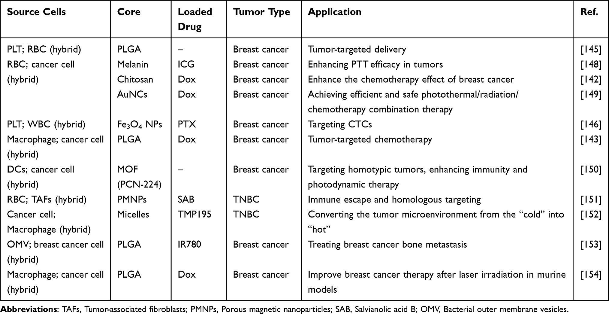 |
Table 3 The Application of Nanoparticles Encapsulated Within Hybrid Cells for the Treatment of Breast Cancer |
Bacterial Outer Membrane Vesicle-Derived Biomimetic Nanotherapeutics
Pathogenic cells, along with mammalian cells, can serve as sources for nanoparticle coatings. Bacteria have been explored in various studies as an innovative delivery system for biomedical applications.155 Unlike traditional NP-based vaccine strategies, which incorporate immune-stimulating ligands, bacterial membranes are inherently immunogenic. They contain potent ligands for pattern recognition receptors (PRRs), which are crucial for stimulating the innate immune system and enhancing adaptive immune responses.156 Encapsulating nanoparticles with these molecular patterns imparts intrinsic adjuvant properties.157 As a result, these nanoparticles, when co-administered with antigens, are processed by antigen-presenting cells (APCs) in a manner similar to the source pathogenic cells. Cui’s group demonstrated anti-breast cancer efficacy by encapsulating over-expressed pre-miRNA, using Escherichia coli as a model pathogen.158 They utilized bacterial outer membrane vesicles (OMVs), also known as extracellular vesicles. OMVs are nanoscale lipid bilayer vesicles naturally produced by all Gram-negative bacteria, and they contain various immune-stimulatory components.159 Research has shown that bacterial membrane vesicles (B-MVs) secreted by the Gram-positive bacterium Bifidobacterium can induce apoptosis in TNBC cells, thereby inhibiting tumor growth.160 In a similar vein, Cui et al developed a highly effective miRNA nano-delivery system for breast cancer treatment by coating zeolitic imidazolate framework-8 with OMVs.161 This system demonstrated precise tumor targeting and high miRNA delivery efficacy in a murine breast cancer model. Furthermore, the therapeutic effect was enhanced by the synergistic impact of the miR-34a payload within the carrier, which promoted OMV-PD1-induced immune activation and checkpoint inhibition. As a result, OMVs have emerged as promising nanoscale carriers with inherent adjuvant functions.
Combining OMVs with other therapeutic modalities holds great potential for enhancing anti-tumor outcomes. Tumors are known to recruit platelets, and leveraging this phenomenon, He et al have innovatively utilized platelets as “messengers” to improve the tumor-targeted delivery efficiency of OMVs and photothermal agents. Specifically, they designed nanoparticles (IR780-SLN@O-P) based on OMVs, incorporating platelet-binding capabilities. These “delivery” platelets use P-selectin to transport the IR780-SLN@O-P NP “cargo” to the tumor site, ensuring precise targeting.162 Upon laser irradiation, the photothermal agent induces a significant photothermal effect, which, combined with the immune-stimulatory properties of OMVs, elicits a robust anti-tumor immune response. Moreover, transferring and functionalizing OMVs onto other nanoparticle types that encapsulate pharmaceuticals or antigens could lead to the creation of highly effective multifunctional carriers. Despite the growing body of research focused on developing these bionic systems, the molecular interactions at the biological interface (particularly those between these membrane coatings and cells) require further investigation. Additionally, understanding the mechanisms of intracellular delivery is critical, as the cargo often includes therapeutics that must be effectively transported to intracellular targets.
Cell-Derived Extracellular Vesicles as Nanocarriers for Targeted Drug Delivery
Exosomes are endogenous extracellular vesicles derived from cells and are the most common type of vesicle found in cell-derived biological products, making them extensively utilized for drug delivery (Figure 3A and B). With a diameter ranging from 40 to 100 nm, exosomes carry proteins, lipids, and other biological molecules (Figure 3C).163 The lipid bilayer of exosomes plays a crucial role in safeguarding the proper transport of drugs, ensuring their circulation in body fluids with enhanced circulatory stability and long-term safety properties.164 Due to the diverse signaling molecules carried by exosomes secreted by different cells, as well as their inherently small size, exosomes serve as natural nanocarriers capable of targeted delivery.165 The primary sources of exosomes include stem cells, immune cells (such as T cells, natural killer cells, and macrophages), and exosomes derived from tumor cells.166 Additionally, exosomes exhibit a “homing” ability, enabling them to target the cells from which they originated. In breast cancer treatment, exosomes can selectively deliver anti-cancer drugs to tumor cells through interactions between their surface membrane proteins and cell receptors. This drug delivery method can help overcome multidrug resistance associated with proteins like P-glycoprotein.167
 |
Figure 3 (A) Exosomes can be secreted by nearly all tissues and organs, making them detectable in a variety of bodily fluids, including blood, saliva, and sweat. (B) The process of exosome production involves the maturation of early endosomes, which invaginate and eventually transform into late endosomes or multivesicular bodies (MVBs). (C) Exosomes contain a diverse array of molecular constituents, including DNA, RNA, lipids, and proteins. (D) The methods by which exosomes load drugs. |
Exosomes have been widely studied as carriers for small molecule drug delivery and are capable of transporting a variety of drugs, both hydrophilic and hydrophobic, such as Dox, dopamine, curcumin, and PTX.168,169 The methods for loading drugs into exosomes are generally categorized into several main types: direct loading into donor cells via transfection or co-incubation, and post-exosomal formation methods such as electroporation and direct mixing (Figure 3D). Research has demonstrated the effectiveness of exosome-mediated drug delivery in various models, showing promising anti-breast cancer effects. Haney et al demonstrated the high anti-cancer effects of macrophage-derived extracellular vesicles loaded with PTX and Dox in a mouse model of lung metastasis. They also observed anti-tumor activity targeting TNBC in immune-active BALB/C mice and in situ T11 tumors in athymic nu/nu mice.170 Similarly, Sun et al loaded curcumin into extracellular vesicles derived from donor human adenocarcinoma cells and mouse breast tumor cells. Experimental data showed that curcumin delivered through extracellular vesicles was more stable in the blood at higher concentrations, enhancing its target specificity.171 However, it is essential to package extracellular vesicles with therapeutic nucleic acids, proteins, or drugs for effective breast cancer treatment due to the content similarity between donor and recipient cells (Table 4). Additionally, since unmodified extracellular vesicles from most cell sources have limited targeting capabilities, there is a growing focus on surface modifications using strategies such as genetic engineering and chemical modification. These modifications aim to enhance targeting and improve the efficiency of delivering therapeutic small molecules for breast cancer treatment.172
 |
Table 4 Extracellular Vesicles Loading Different Contents and Effects in the Treatment of Breast Cancer |
While exosomes have been extensively investigated in preclinical studies for drug delivery in breast cancer, most clinical trials are currently in the early stages and potentially limited by low exosome yields and purification methods. Standard techniques like ultracentrifugation, ultrafiltration, precipitation, and immunoaffinity have been shown to compromise exosome structural integrity and functional properties.172 Furthermore, exosome structure and composition are complex, influenced by precursor cell type, physiological conditions, and production methods.172 Therefore, there is an urgent need to develop stable, effective and more scalable exosome-mimicking nanoparticles for advanced applications in drug delivery and breast cancer therapy.
Application of Other Membrane-Derived Biomimetic Nanotherapeutics
For the purpose of drug delivery, numerous specialized cell types have been developed. Tumor-associated fibroblasts (TAFs) are a critical element of the tumor microenvironment, which is characterized by their ability to secrete a variety of inflammatory and growth factors, as well as their ability to remodel the extracellular matrix. Li et al created semiconductor polymer NPs that absorb near-infrared light and are camouflaged with activated fibroblasts (AFs) for the purpose of combining photodynamic and PTT.188 By facilitating the accumulation of NPs within the tumor, the cellular membrane coating enhances the efficacy of photodiagnostics and phototherapy by providing homologous targeting to cancer-associated fibroblasts. Additionally, Zhang et al employed HEK293T cell membranes that express PD-1 to encapsulate a sinophoryrin sodium-binding human serum albumin-perfluorotributylamine nanoemulsion, with the objective of achieving synergistic photodynamic immunotherapy targeting hypoxic 4T1 breast tumors and their distant metastases.189 Recently, there has been a growing interest in the concept of replicating natural viral intracellular infection mechanisms within the context of anti-tumor immunotherapy. Zhao et al created HSV-NP analogs aimed at cancer-targeted therapy, employing herpes simplex virus (HSV) to initiate innate immune responses as a mechanism of action.190 This system involves the engineering of DNA enzyme-loaded Mn-ZIF-90 nanoparticles (ZM@TD) to mimic virus capsids containing genomes, while RBCMs are modified with RGD and HA2 functional peptides to imitate virus envelopes. This biomimetic platform effectively circumvents rapid clearance in the bloodstream while simultaneously mimicking several processes associated with HSV infection. Activation of the cGAS-STING pathway led to a significant stimulation of the innate immune system, achieving a 68% regression in primary tumors and extending median survival by 32 days in rodents bearing 4T1 tumors.
The Future Prospective
Personalized therapy is a primary goal for the future of breast cancer treatment. Cell-derived nanocarriers serve as versatile biomimetic nano-platforms for advanced personalized therapies, combining their intrinsic properties with established methods like phototherapy, immunotherapy, gene therapy, and chemotherapy (Table 5).191 The tumor microenvironment is composed of various immune-related cells, including natural killer cells, macrophages, and dendritic cells. Recent studies have also highlighted the role of bacteria within the tumor microbiome.192 By selecting appropriate cells (eg, immune cells, cancer cells, microbes), it is possible to replicate their natural characteristics and functions, thus facilitating breast cancer therapy. Most studies have focused on the in-vivo applications of cell-derived nanocarriers in murine models, which have demonstrated excellent biocompatibility and safety. However, understanding their cellular behavior is crucial for developing successful clinical applications. Fundamental research on cell-derived nanocarriers and their interactions within tissues and cells is still incomplete and requires more comprehensive and systematic analysis. On one hand, developing efficient nanocarriers for specific tissues necessitates identifying key motifs responsible for binding to surface markers and cell receptors. On the other hand, a deeper understanding of cellular internalization mechanisms opens new avenues for intracellular applications, particularly those requiring endosomal escape.
 |
Table 5 Advantages and Anti-Breast Cancer Applications of Different Drug-Loaded Cells |
There is a need for more advanced and reliable 3D cell culture models, such as spheroids and organoids grown under flow conditions, to better mimic natural environments for investigating and predicting biomolecular mechanisms. These models enhance our understanding of the complex cellular behavior exhibited by cell-derived nanocarriers in vivo. As discussed in this review, cell membrane-derived nanocarriers show significant promise for improving drug delivery. However, several critical challenges must be addressed before advancing to clinical trials. Drug loading within cells can alter cell properties, affecting cell surface proteins and membrane fluidity, which may impact cell viability and increase immunogenicity.193 For example, RBCs may affect biocompatibility when used as vectors, leading to potential drug exposure risks.117 Certain cell types, such as macrophages, must maintain circulation in body fluids to enable migration and homeostasis when used as vectors and to release drugs in large quantities at tumor sites.194 However, intracellular protective mechanisms may cause the excretion of free drugs, neutralizing their endocytotic effects and leading to premature drug release before reaching the tumor site. Furthermore, when cells are used for targeted drug delivery and breast cancer therapy, they may not possess the same homologous targeting properties as cancer cell-loaded drugs, which can reduce the efficacy of drug release at tumor sites. Currently, there is limited research on the delivery of therapeutic agents to dead cells, which introduces a degree of uncertainty. Further investigation is needed into the metabolism and safety of cancer cells in vivo, including the potential for tumor formation.
The large-scale production and standardization of manufacturing and characterization methods for these biomaterials present significant challenges. Various cell types require distinct culture conditions, and sample quantity and quality can be affected by factors such as cell type, cell cycle, lifespan, and channel number. This is critical not only for reducing inter-batch variability but also for achieving consistent biomimetic nanocarriers across different populations. Due to cell surface heterogeneity, nanocarriers with specific proteins or varied epitope sequences and densities can be generated within a single batch. Therefore, precise characterization tools are essential to analyze surface composition and biological performance. Ensuring the integrity of the cell surface, as well as the proper orientation, presence, and stability of key membrane proteins, is crucial following the assembly of cell membrane fragments. Targeting effectiveness may decrease due to protein loss or malfunction during membrane extraction, purification, or storage.
Data Sharing Statement
All data generated or analyzed during this study are included in this manuscript.
Consent for Publication
All authors have reviewed and approved the final version of the manuscript and have given their consent for the publication of this work.
Funding
This research was supported by National Key Clinical Specialties Construction Program, the Chongqing Kewei Joint Medical Science Foundation (2025QNXM019).
Disclosure
The authors declare that they have no competing interests.
References
1. Bray F, Laversanne M, Sung H, et al. Global cancer statistics 2022: GLOBOCAN estimates of incidence and mortality worldwide for 36 cancers in 185 countries. CA. 2024;74(3):229–263. doi:10.3322/caac.21834
2. Giaquinto AN, Sung H, Newman LA, et al. Breast cancer statistics 2024. CA. 2024;74(6):477–495. doi:10.3322/caac.21863
3. Al-Hilli Z, Wilkerson A. Breast surgery: management of postoperative complications following operations for breast cancer. The Surgical Clinics of North America. 2021;101(5):845–863. doi:10.1016/j.suc.2021.06.014
4. Provenzano E. Neoadjuvant chemotherapy for breast cancer: moving beyond pathological complete response in the molecular age. Acta Medica Academica. 2021;50(1):88–109. doi:10.5644/ama2006-124.328
5. Upadhyay R, Bazan JG. Advances in radiotherapy for breast cancer. Surgical Oncol Clin of North America. 2023;32(3):515–536. doi:10.1016/j.soc.2023.03.002
6. Schirrmacher V. From chemotherapy to biological therapy: a review of novel concepts to reduce the side effects of systemic cancer treatment (Review). Int J Oncol. 2019;54(2):407–419. doi:10.3892/ijo.2018.4661
7. Köksal M, Streppel R, Hauser S, et al. Impact of patient nationality on the severity of early side effects after radiotherapy. J Cancer Res Clin Oncol. 2023;149(9):5573–5582. doi:10.1007/s00432-022-04505-0
8. Esfahani K, Roudaia L, Buhlaiga N, Del Rincon SV, Papneja N, Miller WH. A review of cancer immunotherapy: from the past, to the present, to the future. Curr Oncol. 2020;27(Suppl 2):S87–s97. doi:10.3747/co.27.5223
9. Zhao M, Chen C, Zhang C, et al. Cardiotoxicity with human epidermal growth factor receptor-2 inhibitors in breast cancer: disproportionality analysis of the FDA adverse event reporting system. Int J Cardiol. 2023;375:87–93. doi:10.1016/j.ijcard.2022.12.043
10. Li Z, Xiao C, Yong T, Li Z, Gan L, Yang X. Influence of nanomedicine mechanical properties on tumor targeting delivery. Chemical Society Rev. 2020;49(8):2273–2290. doi:10.1039/C9CS00575G
11. Kutova OM, Guryev EL, Sokolova EA, Alzeibak R, Balalaeva IV. Targeted delivery to tumors: multidirectional strategies to improve treatment efficiency. Cancers. 2019;11(1). doi:10.3390/cancers11010068
12. Li C, Wang J, Wang Y, et al. Recent progress in drug delivery. Acta Pharmaceutica Sinica B. 2019;9(6):1145–1162. doi:10.1016/j.apsb.2019.08.003
13. Chowdhury P, Ghosh U, Samanta K, Jaggi M, Chauhan SC, Yallapu MM. Bioactive nanotherapeutic trends to combat triple negative breast cancer. Bioactive Materials. 2021;6(10):3269–3287. doi:10.1016/j.bioactmat.2021.02.037
14. Zhu Y, Wang A, Zhang S, et al. Paclitaxel-loaded ginsenoside Rg3 liposomes for drug-resistant cancer therapy by dual targeting of the tumor microenvironment and cancer cells. Journal of Advanced Research. 2023;49:159–173. doi:10.1016/j.jare.2022.09.007
15. Dos Reis LR, Luiz MT, Sábio RM, et al. Design of rapamycin and resveratrol coloaded liposomal formulation for breast cancer therapy. Nanomedicine. 2023;18(10):789–801. doi:10.2217/nnm-2022-0227
16. Bhavya K, Agarwal K, Negi D, et al. Oxygen nanobubbles halt tumor aggression and metastasis by inhibiting hypoxia-induced epithelial-to-mesenchymal transition in lung and mammary adenocarcinoma. ACS Applied Nano Materials. 2024;7(21):25198–25211. doi:10.1021/acsanm.4c05295
17. Yang RQ, Wang PY, Lou KL, et al. Biodegradable nanoprobe for NIR-II fluorescence image-guided surgery and enhanced breast cancer radiotherapy efficacy. Adv Sci. 2022;9(12):e2104728. doi:10.1002/advs.202104728
18. Lu Y, Gu F, Ma Y, et al. Tumor cell membrane-camouflaged vortex magnetic nanoannulars programmed by low-frequency magnetic field: a novel anti-cancer delivery system in triple-negative breast cancer. Adv Functional Mat. 2024;34(42):2401940.
19. Yan J, Ji S, Chang T, et al. Construction of bionic nanoparticles camouflaged with macrophage membranes for drug delivery in breast cancer. Journal of Drug Delivery Science and Technology. 2023;84:104433. doi:10.1016/j.jddst.2023.104433
20. Yu H, Ben-Akiva E, Meyer RA, Green JJ. Biomimetic anisotropic-functionalized platelet-membrane-coated polymeric particles for targeted drug delivery to human breast cancer cells. ACS Applied Materials & Interfaces. 2025;17(1):351–362. doi:10.1021/acsami.4c15471
21. Wang R, Yan H, Yu A, Ye L, Zhai G. Cancer targeted biomimetic drug delivery system. J Drug Delivery Sci Technol. 2021;63:102530. doi:10.1016/j.jddst.2021.102530
22. Liu M, Sun Y, Wei Q, et al. 4T1 cell membrane biomimetic nanovehicle for enhanced breast cancer treatment. ACS Medicinal Chemistry Letters. 2025;16(1):51–58. doi:10.1021/acsmedchemlett.4c00425
23. Wang M, Xin Y, Cao H, et al. Recent advances in mesenchymal stem cell membrane-coated nanoparticles for enhanced drug delivery. Biomaterials Science. 2021;9(4):1088–1103. doi:10.1039/D0BM01164A
24. Takayama Y, Kusamori K, Nishikawa M. Mesenchymal stem/stromal cells as next-generation drug delivery vehicles for cancer therapeutics. Expert Opinion on Drug Delivery. 2021;18(11):1627–1642. doi:10.1080/17425247.2021.1960309
25. Ullah I, Subbarao RB, Rho GJ. Human mesenchymal stem cells - current trends and future prospective. Bioscience Reports. 2015;35(2). doi:10.1042/BSR20150025
26. Zhang H, Feng Y, Xie X, et al. Engineered mesenchymal stem cells as a biotherapy platform for targeted photodynamic immunotherapy of breast cancer. Adv Healthcare Mat. 2022;11(6):e2101375. doi:10.1002/adhm.202101375
27. Cao B, Yang M, Zhu Y, Qu X, Mao C. Stem cells loaded with nanoparticles as a drug carrier for in vivo breast cancer therapy. Advanced Materials (Deerfield Beach, Fla). 2014;26(27):4627–4631. doi:10.1002/adma.201401550
28. Litvinova LS, Shupletsova VV, Khaziakhmatova OG, et al. Human mesenchymal stem cells as a carrier for a cell-mediated drug delivery. Frontiers in Bioengineering Biotechnol. 2022;10:796111. doi:10.3389/fbioe.2022.796111
29. Yu Q, Tian Z, Li G, et al. Multifunctional composite capsules in drug delivery systems: bridging pharmaceutical and biomedical applications. Advanced Composites and Hybrid Materials. 2025;8(1):118. doi:10.1007/s42114-024-01203-y
30. Taghavi S, Tabasi H, Zahiri M, et al. Surface engineering of hollow gold nanoparticle with mesenchymal stem cell membrane and MUC-1 aptamer for targeted theranostic application against metastatic breast cancer. European J Pharmaceutics and Biopharmaceutics. 2023;187:76–86. doi:10.1016/j.ejpb.2023.04.014
31. Avancini G, Menilli L, Visentin A, Milani C, Mastrotto F, Moret F. Mesenchymal stem cell membrane-coated Tpcs(2a)-loaded nanoparticles for breast cancer photodynamic therapy. Pharmaceutics. 2023;15(6):1654. doi:10.3390/pharmaceutics15061654
32. Kalimuthu S, Zhu L, Oh JM, et al. Migration of mesenchymal stem cells to tumor xenograft models and in vitro drug delivery by doxorubicin. Int J Med Sci. 2018;15(10):1051–1061. doi:10.7150/ijms.25760
33. Al-Quteimat OM, Amer AM. The Impact of the COVID-19 Pandemic on Cancer Patients. American J Clin Oncol. 2020;43(6):452–455. doi:10.1097/COC.0000000000000712
34. Serafini S, Rossi L, Antonelli A, et al. Drug delivery through phagocytosis of red blood cells. Transfusion Med Hemother. 2004;31(2):92–101. doi:10.1159/000078042
35. Kolesnikova TA, Skirtach AG, Möhwald H. Möhwald H: red blood cells and polyelectrolyte multilayer capsules: natural carriers versus polymer-based drug delivery vehicles. Expert Opinion on Drug Delivery. 2013;10(1):47–58. doi:10.1517/17425247.2013.730516
36. Gao M, Liang C, Song X, et al. Erythrocyte-membrane-enveloped perfluorocarbon as nanoscale artificial red blood cells to relieve tumor hypoxia and enhance cancer radiotherapy. Adv Mat. 2017;29(35). doi:10.1002/adma.201701429
37. Li JQ, Zhao RX, Yang FM, Qi XT, Ye PK, Xie M. An erythrocyte membrane-camouflaged biomimetic nanoplatform for enhanced chemo-photothermal therapy of breast cancer. J Mat Chemistry B. 2022;10(12):2047–2056. doi:10.1039/D1TB02522H
38. Song Q, Yin Y, Shang L, et al. Tumor microenvironment responsive nanogel for the combinatorial antitumor effect of chemotherapy and immunotherapy. Nano Letters. 2017;17(10):6366–6375. doi:10.1021/acs.nanolett.7b03186
39. Jiang Q, Wang K, Zhang X, et al. Platelet membrane-camouflaged magnetic nanoparticles for ferroptosis-enhanced cancer immunotherapy. Small. 2020;16(22):e2001704. doi:10.1002/smll.202001704
40. Zhuang J, Gong H, Zhou J, et al. Targeted gene silencing in vivo by platelet membrane-coated metal-organic framework nanoparticles. Science Advances. 2020;6(13):eaaz6108. doi:10.1126/sciadv.aaz6108
41. Wang H, Wu C, Tong X, Chen S. A biomimetic metal-organic framework nanosystem modulates immunosuppressive tumor microenvironment metabolism to amplify immunotherapy. J Controlled Release. 2023;353:727–737. doi:10.1016/j.jconrel.2022.11.054
42. Ning S, Lyu M, Zhu D, et al. Type-I AIE photosensitizer loaded biomimetic system boosting cuproptosis to inhibit breast cancer metastasis and rechallenge. ACS nano. 2023;17(11):10206–10217. doi:10.1021/acsnano.3c00326
43. Zou J, He J, Wang X, et al. Glycoprotein Ib-regulated micro platelet ghost for biosafe distribution and photothermal oncotherapy. J Controlled Release. 2022;351:341–360. doi:10.1016/j.jconrel.2022.09.036
44. Bahmani B, Gong H, Luk BT, et al. Intratumoral immunotherapy using platelet-cloaked nanoparticles enhances antitumor immunity in solid tumors. Nature Communications. 2021;12(1):1999. doi:10.1038/s41467-021-22311-z
45. Pei W, Huang B, Chen S, Wang L, Xu Y, Niu C. Platelet-mimicking drug delivery nanoparticles for enhanced chemo-photothermal therapy of breast cancer. Int J Nanomedicine. 2020;15:10151–10167. doi:10.2147/IJN.S285952
46. Liu G, Zhao X, Zhang Y, et al. Engineering biomimetic platesomes for pH-responsive drug delivery and enhanced antitumor activity. Adv Mat. 2019;31(32):e1900795. doi:10.1002/adma.201900795
47. Li W, Li F, Li T, et al. Self-actuated biomimetic nanocomposites for photothermal therapy and PD-L1 immunosuppression. Frontiers in Chemistry. 2023;11:1167586. doi:10.3389/fchem.2023.1167586
48. Han X, Chen J, Chu J, et al. Platelets as platforms for inhibition of tumor recurrence post-physical therapy by delivery of anti-PD-L1 checkpoint antibody. J Controlled Release. 2019;304:233–241. doi:10.1016/j.jconrel.2019.05.008
49. Zhang Y, Cai K, Li C, et al. Macrophage-membrane-coated nanoparticles for tumor-targeted chemotherapy. Nano Letters. 2018;18(3):1908–1915. doi:10.1021/acs.nanolett.7b05263
50. Liang B, Deng T, Li J, Ouyang X, Na W, Deng D. Biomimetic theranostic strategy for anti-metastasis therapy of breast cancer via the macrophage membrane camouflaged superparticles. Materials Science & Engineering C, Materials for Biological applications. 2020;115:111097. doi:10.1016/j.msec.2020.111097
51. Chen C, Song M, Du Y, et al. Tumor-associated-macrophage-membrane-coated nanoparticles for improved photodynamic immunotherapy. Nano Letters. 2021;21(13):5522–5531. doi:10.1021/acs.nanolett.1c00818
52. Li Y, Yan T, Chang W, Cao C, Deng D. Fabricating an intelligent cell-like nano-prodrug via hierarchical self-assembly based on the DNA skeleton for suppressing lung metastasis of breast cancer. Biomaterials Science. 2019;7(9):3652–3661. doi:10.1039/C9BM00630C
53. Xia C, Bai W, Deng T, et al. Sponge-like nano-system suppresses tumor recurrence and metastasis by restraining myeloid-derived suppressor cells-mediated immunosuppression and formation of pre-metastatic niche. Acta biomaterialia. 2023;158:708–724. doi:10.1016/j.actbio.2023.01.009
54. Yu X, Xing G, Sheng S, et al. Neutrophil camouflaged stealth nanovehicle for photothermal-induced tumor immunotherapy by triggering pyroptosis. Adv Sci. 2023;10(15):e2207456. doi:10.1002/advs.202207456
55. Zhai Y, Wang J, Lang T, et al. T lymphocyte membrane-decorated epigenetic nanoinducer of interferons for cancer immunotherapy. Nature Nanotechnol. 2021;16(11):1271–1280. doi:10.1038/s41565-021-00972-7
56. Deng G, Sun Z, Li S, et al. Cell-membrane immunotherapy based on natural killer cell membrane coated nanoparticles for the effective inhibition of primary and abscopal tumor growth. ACS nano. 2018;12(12):12096–12108. doi:10.1021/acsnano.8b05292
57. Du W, Chen C, Sun P, et al. Eliciting an immune hot tumor niche with biomimetic drug-based multi-functional nanohybrids augments immune checkpoint blockade-based breast cancer therapy. Nanoscale. 2020;12(5):3317–3329. doi:10.1039/C9NR09835F
58. Pitchaimani A, Nguyen TDT, Aryal S. Natural killer cell membrane infused biomimetic liposomes for targeted tumor therapy. Biomaterials. 2018;160:124–137. doi:10.1016/j.biomaterials.2018.01.018
59. Villa CH, Pan DC, Zaitsev S, Cines DB, Siegel DL, Muzykantov VR. Delivery of drugs bound to erythrocytes: new avenues for an old intravascular carrier. Therapeutic Delivery. 2015;6(7):795–826. doi:10.4155/tde.15.34
60. Oldenborg PA, Gresham HD, Lindberg FP. CD47-signal regulatory protein alpha (SIRPalpha) regulates Fcgamma and complement receptor-mediated phagocytosis. J Experimental Med. 2001;193(7):855–862. doi:10.1084/jem.193.7.855
61. Zhang W, Zhao M, Gao Y, et al. Biomimetic erythrocytes engineered drug delivery for cancer therapy. Chemical Engineering Journal. 2022;433:133498. doi:10.1016/j.cej.2021.133498
62. Oldenborg PA, Zheleznyak A, Fang YF, Lagenaur CF, Gresham HD, Lindberg FP. Role of CD47 as a marker of self on red blood cells. Science. 2000;288(5473):2051–2054. doi:10.1126/science.288.5473.2051
63. Han X, Wang C, Liu Z. Red Blood Cells as Smart Delivery Systems. Bioconjugate Chemistry. 2018;29(4):852–860. doi:10.1021/acs.bioconjchem.7b00758
64. Shi J, Kundrat L, Pishesha N, et al. Engineered red blood cells as carriers for systemic delivery of a wide array of functional probes. Proceedings of the National Academy of Sciences of the United States of America. 2014;111(28):10131–10136. doi:10.1073/pnas.1409861111
65. Wang Q, Cheng H, Peng H, Zhou H, Li PY, Langer R. Non-genetic engineering of cells for drug delivery and cell-based therapy. Adv Drug Delivery Rev. 2015;91:125–140. doi:10.1016/j.addr.2014.12.003
66. Pierigè F, De Marco C, Orlotti N, et al. Cytotoxic activity of 2-Fluoro-ara-AMP and 2-Fluoro-ara-AMP-loaded erythrocytes against human breast carcinoma cell lines. Int J Oncol. 2010;37(1):133–142. doi:10.3892/ijo_00000661
67. Zong S, Cao C, Chen K, Cui Y, Li J, Wang Z. Red Blood cell membrane camouflaged mesoporous silica nanorods as nanocarriers for synergistic chemo-photothermal therapy. IEEE Transactions on Nanobioscience. 2023;22(3):655–663. doi:10.1109/TNB.2022.3233378
68. Pan Y, He Y, Zhao X, et al. Engineered red blood cell membrane-coating salidroside/indocyanine green nanovesicles for high-efficiency hypoxic targeting phototherapy of triple-negative breast cancer. Adv Healthcare Mat. 2022;11(17):e2200962. doi:10.1002/adhm.202200962
69. Ren L, Sun Y, Zhang J, et al. Red blood cell membrane-coated functionalized Cu-doped metal organic framework nanoformulations as a biomimetic platform for improved chemo-/chemodynamic/photothermal synergistic therapy. Int J Pharmaceutics. 2024;652:123811. doi:10.1016/j.ijpharm.2024.123811
70. Li M, Fang H, Liu Q, et al. Red blood cell membrane-coated upconversion nanoparticles for pretargeted multimodality imaging of triple-negative breast cancer. Biomaterials Science. 2020;8(7):1802–1814. doi:10.1039/D0BM00029A
71. Zhang ZL, Ho SC, Shi DD, et al. Erythrocyte membrane n-3 PUFA are inversely associated with breast cancer risk among Chinese women. British J Nutrition. 2024;131(1):103–112. doi:10.1017/S0007114523001447
72. Amézaga J, Ugartemendia G, Larraioz A, et al. Omega 6 polyunsaturated fatty acids in red blood cell membrane are associated with xerostomia and taste loss in patients with breast cancer. Prostaglandins, Leukotrienes, and Essential Fatty Acids. 2021;173:102336. doi:10.1016/j.plefa.2021.102336
73. Li QR, Xu HZ, Xiao RC, et al. Platelets are highly efficient and efficacious carriers for tumor-targeted nano-drug delivery. Drug Delivery. 2022;29(1):937–949. doi:10.1080/10717544.2022.2053762
74. Kapur R, Semple JW. The nonhemostatic immune functions of platelets. Seminars in Hematology. 2016;53 Suppl 1:S2–6. doi:10.1053/j.seminhematol.2016.04.002
75. Lu Y, Hu Q, Jiang C, Gu Z. Platelet for drug delivery. Curr Opinion Biotechnol. 2019;58:81–91. doi:10.1016/j.copbio.2018.11.010
76. Dovizio M, Ballerini P, Fullone R, Tacconelli S, Contursi A, Patrignani P. Multifaceted functions of platelets in cancer: from tumorigenesis to liquid biopsy tool and drug delivery system. International J Mol Sci. 2020;21(24):9585. doi:10.3390/ijms21249585
77. Hinterleitner C, Zhou Y, Tandler C, et al. Platelet-Expressed TNFRSF13B (TACI) predicts breast cancer progression. Front Oncol. 2021;11:642170. doi:10.3389/fonc.2021.642170
78. Song Y, Tan X, Gao Y. Platelet-biomimetic nanoparticles for in vivo targeted photodynamic therapy of breast cancer. J Biomaterials Science Polymer Edition. 2022;33(11):1383–1397. doi:10.1080/09205063.2022.2056942
79. Liu Y, Qi Y, Chen C, et al. Platelet-mimetic nano-sensor for combating postoperative recurrence and wound infection of triple-negative breast cancer. J Controlled Release. 2023;362:396–408. doi:10.1016/j.jconrel.2023.08.057
80. Wan G, Chen X, Gou R, et al. Platelet membrane-based biochemotactic-targeting nanoplatform combining PDT with EGFR inhibition therapy for the treatment of breast cancer. Biomaterials Science. 2024;12(3):691–709. doi:10.1039/D3BM01627G
81. Molinaro R, Martinez JO, Zinger A, et al. Leukocyte-mimicking nanovesicles for effective doxorubicin delivery to treat breast cancer and melanoma. Biomaterials Science. 2020;8(1):333–341. doi:10.1039/C9BM01766F
82. Dong X, Chu D, Wang Z. Leukocyte-mediated delivery of nanotherapeutics in inflammatory and tumor sites. Theranostics. 2017;7(3):751–763. doi:10.7150/thno.18069
83. Chen Y, Qin D, Zou J, et al. Living leukocyte-based drug delivery systems. Adv Mat. 2023;35(17):e2207787. doi:10.1002/adma.202207787
84. Huang Y, Gao X, Chen J. Leukocyte-derived biomimetic nanoparticulate drug delivery systems for cancer therapy. Acta pharmaceutica Sinica B. 2018;8(1):4–13. doi:10.1016/j.apsb.2017.12.001
85. Chu D, Dong X, Shi X, Zhang C, Wang Z. Neutrophil-based drug delivery systems. Adv Mat. 2018;30(22):e1706245.
86. Han Y, Zhao R, Xu F. Neutrophil-based delivery systems for nanotherapeutics. Small. 2018;14(42):e1801674. doi:10.1002/smll.201801674
87. Wang H, Zang J, Zhao Z, Zhang Q, Chen S. The advances of neutrophil-derived effective drug delivery systems: a key review of managing tumors and inflammation. Int J Nanomed. 2021;16:7663–7681. doi:10.2147/IJN.S328705
88. Zhang CY, Dong X, Gao J, Lin W, Liu Z, Wang Z. Nanoparticle-induced neutrophil apoptosis increases survival in sepsis and alleviates neurological damage in stroke. Sci Adv. 2019;5(11):eaax7964. doi:10.1126/sciadv.aax7964
89. Hosseinalizadeh H, Mahmoodpour M, Razaghi Bahabadi Z, Hamblin MR, Mirzaei H. Neutrophil mediated drug delivery for targeted glioblastoma therapy: a comprehensive review. Biomedicine & Pharmacotherapy. 2022;156:113841. doi:10.1016/j.biopha.2022.113841
90. Chu D, Dong X, Zhao Q, Gu J, Wang Z. Photosensitization priming of tumor microenvironments improves delivery of nanotherapeutics via neutrophil infiltration. Adv Mat. 2017;29(27). doi:10.1002/adma.201701021
91. Chowdhury P, Bhusetty Nagesh PK, Hollingsworth TJ, Jaggi M, Chauhan SC, Yallapu MM. Coating a self-assembly nanoconstruct with a neutrophil cell membrane enables high specificity for triple negative breast cancer treatment. ACS Applied Bio Materials. 2022;5(9):4554–4566. doi:10.1021/acsabm.2c00614
92. Chu Y, Luo Y, Su B, et al. A neutrophil-biomimic platform for eradicating metastatic breast cancer stem-like cells by redox microenvironment modulation and hypoxia-triggered differentiation therapy. Acta pharmaceutica Sinica B. 2023;13(1):298–314. doi:10.1016/j.apsb.2022.05.027
93. Huang R, Fan D, Cheng H, et al. Multi-site attack, neutrophil membrane-camouflaged nanomedicine with high drug loading for enhanced cancer therapy and metastasis inhibition. Int J Nanomedicine. 2023;18:3359–3375. doi:10.2147/IJN.S415139
94. Wu X, Lin Z, Zhao C, et al. Neutrophil membrane-coated immunomagnetic nanoparticles for efficient isolation and analysis of circulating tumor cells. Biosensors & Bioelectronics. 2022;213:114425. doi:10.1016/j.bios.2022.114425
95. Masopust D, Soerens AG. Tissue-resident T cells and other resident leukocytes. Annual Rev Immunol. 2019;37(1):521–546. doi:10.1146/annurev-immunol-042617-053214
96. Jones RB, Mueller S, Kumari S, et al. Antigen recognition-triggered drug delivery mediated by nanocapsule-functionalized cytotoxic T-cells. Biomaterials. 2017;117:44–53. doi:10.1016/j.biomaterials.2016.11.048
97. Tang L, Zheng Y, Melo MB, et al. Enhancing T cell therapy through TCR-signaling-responsive nanoparticle drug delivery. Nature Biotechnology. 2018;36(8):707–716. doi:10.1038/nbt.4181
98. Li Y, Zheng Y, Liu T, Liao C, Shen G, He Z. The potential and promise for clinical application of adoptive T cell therapy in cancer. J Transl Med. 2024;22(1):413. doi:10.1186/s12967-024-05206-7
99. Stephan MT, Moon JJ, Um SH, Bershteyn A, Irvine DJ. Therapeutic cell engineering with surface-conjugated synthetic nanoparticles. Nature Medicine. 2010;16(9):1035–1041. doi:10.1038/nm.2198
100. Liu T, Zhou Z, Zhang M, et al. Cuproptosis-immunotherapy using PD-1 overexpressing T cell membrane-coated nanosheets efficiently treats tumor. J Controlled Release. 2023;362:502–512. doi:10.1016/j.jconrel.2023.08.055
101. Hu N, Xue H, Zhang T, et al. Harnessing PD-1 cell membrane-coated paclitaxel dimer nanoparticles for potentiated chemoimmunotherapy. Biomed Pharmacother. 2024;174:116482. doi:10.1016/j.biopha.2024.116482
102. Wang HF, Liu Y, Yang G, Zhao CX. Macrophage-mediated cancer drug delivery. Materials Today Sustainability. 2021;11-12:100055. doi:10.1016/j.mtsust.2020.100055
103. Liu J, Geng X, Hou J, Wu G. New insights into M1/M2 macrophages: key modulators in cancer progression. Cancer Cell International. 2021;21(1):389. doi:10.1186/s12935-021-02089-2
104. Zhang W, Wang M, Tang W, et al. Nanoparticle-laden macrophages for tumor-tropic drug delivery. Advanced Materials. 2022;34(6):e2109925. doi:10.1002/adma.202109925
105. Liang T, Zhang R, Liu X, et al. Recent advances in macrophage-mediated drug delivery systems. Int J Nanomedicine. 2021;16:2703–2714. doi:10.2147/IJN.S298159
106. Li H, Tatematsu K, Somiya M, Iijima M, Kuroda S. Development of a macrophage-targeting and phagocytosis-inducing bio-nanocapsule-based nanocarrier for drug delivery. Acta biomaterialia. 2018;73:412–423. doi:10.1016/j.actbio.2018.04.023
107. Anderson NM, Simon MC. The tumor microenvironment. Current Biology. 2020;30(16):R921–R925. doi:10.1016/j.cub.2020.06.081
108. Quijia CR, Navegante G, Sábio RM, et al. Macrophage cell membrane coating on piperine-loaded mil-100(fe) nanoparticles for breast cancer treatment. J Functional Biomaterials. 2023;14(6):319. doi:10.3390/jfb14060319
109. Ying K, Zhu Y, Wan J, et al. Macrophage membrane-biomimetic adhesive polycaprolactone nanocamptothecin for improving cancer-targeting efficiency and impairing metastasis. Bioactive Materials. 2023;20:449–462. doi:10.1016/j.bioactmat.2022.06.013
110. Yoon J, Le XT, Kim J, et al. Macrophage-reprogramming upconverting nanoparticles for enhanced TAM-mediated antitumor therapy of hypoxic breast cancer. J Controlled Release. 2023;360:482–495. doi:10.1016/j.jconrel.2023.07.009
111. Xue Y, Wang Q, Chen Y, et al. Biomimetic diselenide-sonosensitizer nanoplatform for enhanced sonodynamic therapy and in situ remodeling immunosuppressive microenvironment via activating innate and adaptive immunotherapy. Adv Healthcare Mat. 2025;14(6):e2403998. doi:10.1002/adhm.202403998
112. Hazeldine J, Lord JM. The impact of ageing on natural killer cell function and potential consequences for health in older adults. Ageing Research Reviews. 2013;12(4):1069–1078. doi:10.1016/j.arr.2013.04.003
113. Baier C, Fino A, Sanchez C, et al. Natural killer cells modulation in hematological malignancies. Frontiers in Immunology. 2013;4:459. doi:10.3389/fimmu.2013.00459
114. Kim S, Poursine-Laurent J, Truscott SM, et al. Licensing of natural killer cells by host major histocompatibility complex class I molecules. Nature. 2005;436(7051):709–713. doi:10.1038/nature03847
115. Pan S, Guan J, Xianyu B, Tan Y, Li T, Xu H. A nanotherapeutic strategy to reverse NK cell exhaustion. Advanced Materials (Deerfield Beach, Fla). 2023;35(23):e2211370. doi:10.1002/adma.202211370
116. Won EJ, Lee M, Lee EK, Baek SH, Yoon TJ. Lipid-based nanoparticles fused with natural killer cell plasma membrane proteins for triple-negative breast cancer therapy. Pharmaceutics. 2024;16(9):1142. doi:10.3390/pharmaceutics16091142
117. Malhotra S, Dumoga S, Singh N. Red blood cells membrane-derived nanoparticles: applications and key challenges in their clinical translation. Wiley Interdisciplinary Reviews Nanomedicine and Nanobiotechnology. 2022;14(3):e1776. doi:10.1002/wnan.1776
118. Xu L, Wu S, Wang J. Cancer cell membrane–coated nanocarriers for homologous target inhibiting the growth of hepatocellular carcinoma. J Bioactive and Compatible Polymers. 2018;34(1):58–71. doi:10.1177/0883911518819107
119. Zeng H, Yan G, Zheng R, Wang X. Cancer cell membrane-biomimetic nanoparticles based on gelatin and mitoxantrone for synergetic chemo-photothermal therapy of metastatic breast cancer. ACS Biomaterials Science & Engineering. 2024;10(2):875–889. doi:10.1021/acsbiomaterials.3c01325
120. Chen Z, Zhao P, Luo Z, et al. Cancer cell membrane-biomimetic nanoparticles for homologous-targeting dual-modal imaging and photothermal therapy. ACS nano. 2016;10(11):10049–10057. doi:10.1021/acsnano.6b04695
121. Zheng D, Zhou J, Qian L, et al. Biomimetic nanoparticles drive the mechanism understanding of shear-wave elasticity stiffness in triple negative breast cancers to predict clinical treatment. Bioactive Materials. 2023;22:567–587. doi:10.1016/j.bioactmat.2022.10.025
122. Shen J, Karges J, Xiong K, Chen Y, Ji L, Chao H. Cancer cell membrane camouflaged iridium complexes functionalized black-titanium nanoparticles for hierarchical-targeted synergistic NIR-II photothermal and sonodynamic therapy. Biomaterials. 2021;275:120979. doi:10.1016/j.biomaterials.2021.120979
123. Li Z, Cai H, Li Z, et al. A tumor cell membrane-coated self-amplified nanosystem as a nanovaccine to boost the therapeutic effect of anti-PD-L1 antibody. Bioactive Materials. 2023;21:299–312. doi:10.1016/j.bioactmat.2022.08.028
124. Jin F, Qi J, Liu D, et al. Cancer-cell-biomimetic Upconversion nanoparticles combining chemo-photodynamic therapy and CD73 blockade for metastatic triple-negative breast cancer. J Controlled Release. 2021;337:90–104. doi:10.1016/j.jconrel.2021.07.021
125. Li SY, Cheng H, Xie BR, et al. Cancer cell membrane camouflaged cascade bioreactor for cancer targeted starvation and photodynamic therapy. ACS Nano. 2017;11(7):7006–7018. doi:10.1021/acsnano.7b02533
126. Meng X, Wang J, Zhou J, et al. Tumor cell membrane-based peptide delivery system targeting the tumor microenvironment for cancer immunotherapy and diagnosis. Acta biomaterialia. 2021;127:266–275. doi:10.1016/j.actbio.2021.03.056
127. Liu Y, Meng J, Cao W, et al. Homotypic membrane-camouflaged camptothecin nanorods combining photothermal and chemotherapy for synergistic antitumor therapy. Int J Pharmaceutics. 2025;671:125239. doi:10.1016/j.ijpharm.2025.125239
128. Wang D, Lin S, Li T, et al. Cancer cell membrane-coated siRNA-Decorated Au/MnO(2) nanosensitizers for synergistically enhanced radio-immunotherapy of breast cancer. Materials Today Bio. 2024;29:101275. doi:10.1016/j.mtbio.2024.101275
129. Graván P, Rojas S, Picchi DF, Galisteo-González F, Horcajada P, Marchal JA. Towards a more efficient breast cancer therapy using active human cell membrane-coated metal-organic frameworks. Nanomaterials. 2024;14(9). doi:10.3390/nano14090784
130. Siegel RL, Miller KD, Fedewa SA, et al. Colorectal cancer statistics, 2017. CA: a Cancer Journal for Clinicians. 2017;67(3):177–193. doi:10.3322/caac.21395
131. Fang RH, Hu CM, Luk BT, et al. Cancer cell membrane-coated nanoparticles for anticancer vaccination and drug delivery. Nano Letters. 2014;14(4):2181–2188. doi:10.1021/nl500618u
132. Xu M, Yang Y, Yuan Z. Breast cancer cell membrane camouflaged lipid nanoparticles for tumor-targeted NIR-II phototheranostics. Pharmaceutics. 2022;14(7):1367. doi:10.3390/pharmaceutics14071367
133. Sun H, Su J, Meng Q, et al. Cancer-cell-biomimetic nanoparticles for targeted therapy of homotypic tumors. Advanced Materials. 2016;28(43):9581–9588. doi:10.1002/adma.201602173
134. Heimburg J, Yan J, Morey S, et al. Rittenhouse-Olson K: inhibition of spontaneous breast cancer metastasis by anti-Thomsen-Friedenreich antigen monoclonal antibody JAA-F11. Neoplasia (New York, NY). 2006;8(11):939–948. doi:10.1593/neo.06493
135. Zhao Q, Barclay M, Hilkens J, et al. Interaction between circulating galectin-3 and cancer-associated MUC1 enhances tumour cell homotypic aggregation and prevents anoikis. Molecular Cancer. 2010;9(1):154. doi:10.1186/1476-4598-9-154
136. Naor D, Sionov RV, Ish-Shalom D. CD44: structure, function, and association with the malignant process. Advances in Cancer Research. 1997;71:241–319. doi:10.1016/s0065-230x(08)60101-3
137. Ito K, Ralph SJ. Inhibiting galectin-1 reduces murine lung metastasis with increased CD4(+) and CD8 (+) T cells and reduced cancer cell adherence. Clinical & Experimental Metastasis. 2012;29(6):561–572. doi:10.1007/s10585-012-9471-7
138. Liu Z, Wang F, Liu X, et al. Cell membrane-camouflaged liposomes for tumor cell-selective glycans engineering and imaging in vivo. Proceedings of the National Academy of Sciences of the United States of America. 2021;118(30).
139. Zhou Z, Zhang S, Xue N. Research progress of cancer cell membrane coated nanoparticles for the diagnosis and therapy of breast cancer. Front Oncol. 2023;13:1270407. doi:10.3389/fonc.2023.1270407
140. Han L, Xu Y, Guo X, Yuan C, Mu D, Xiao Y. Cancer cell membrane-coated biomimetic platform for targeted therapy of breast cancer in an orthotopic mouse model. J Biomaterials Science Polymer Edition. 2020;31(12):1538–1551. doi:10.1080/09205063.2020.1764163
141. Ni J, Song J, Wang B, et al. Dendritic cell vaccine for the effective immunotherapy of breast cancer. Biomedicine & Pharmacotherapy = Biomedecine & Pharmacotherapie. 2020;126:110046. doi:10.1016/j.biopha.2020.110046
142. Rezaei S, de Araújo Júnior RF, da Silva ILG, Schomann T, Eich C, Cruz LJ. Erythrocyte-cancer hybrid membrane-coated reduction-sensitive nanoparticles for enhancing chemotherapy efficacy in breast cancer. Biomaterials Advances. 2023;151:213456. doi:10.1016/j.bioadv.2023.213456
143. Gong C, Yu X, You B, et al. Macrophage-cancer hybrid membrane-coated nanoparticles for targeting lung metastasis in breast cancer therapy. J Nanobiotechnology. 2020;18(1):92. doi:10.1186/s12951-020-00649-8
144. Zhang S, Chen W, Zhou Y, et al. Intelligent nanoplatform integrating macrophage and cancer cell membrane for synergistic chemodynamic/immunotherapy/photothermal therapy of breast cancer. ACS Applied Materials & Interfaces. 2023;15(51):59117–59133. doi:10.1021/acsami.3c12560
145. Dehaini D, Wei X, Fang RH, et al. Erythrocyte-platelet hybrid membrane coating for enhanced nanoparticle functionalization. Advanced Materials. 2017;29(16). doi:10.1002/adma.201606209
146. Rao L, Meng Q-F, Huang Q, et al. Platelet–leukocyte hybrid membrane-coated immunomagnetic beads for highly efficient and highly specific isolation of circulating tumor cells. Advanced Functional Materials. 2018;28(34):1803531. doi:10.1002/adfm.201803531
147. Ma W, Yang Y, Zhu J, et al. Biomimetic nanoerythrosome-coated aptamer-DNA tetrahedron/maytansine conjugates: pH-responsive and targeted cytotoxicity for HER2-positive breast cancer. Advanced Mat. 2022;34(46):e2109609. doi:10.1002/adma.202109609
148. Jiang Q, Liu Y, Guo R, et al. Erythrocyte-cancer hybrid membrane-camouflaged melanin nanoparticles for enhancing photothermal therapy efficacy in tumors. Biomaterials. 2019;192:292–308. doi:10.1016/j.biomaterials.2018.11.021
149. Sun M, Duan Y, Ma Y, Zhang Q. Cancer cell-erythrocyte hybrid membrane coated gold nanocages for near infrared light-activated photothermal/radio/chemotherapy of breast cancer. Int J Nanomedicine. 2020;15:6749–6760. doi:10.2147/IJN.S266405
150. Liu WL, Zou MZ, Liu T, et al. Expandable immunotherapeutic nanoplatforms engineered from cytomembranes of hybrid cells derived from cancer and dendritic cells. Adv Mat. 2019;31(18):e1900499. doi:10.1002/adma.201900499
151. Cheng N, Zhou Q, Jia Z, et al. Functionalized biomimetic nanoparticles loaded with salvianolic acid B for synergistic targeted triple-negative breast cancer treatment. Materials Today Bio. 2025;30:101441. doi:10.1016/j.mtbio.2024.101441
152. Gong J, Cheng D, Liu C, et al. Hybrid cell membrane-coated nanoparticles for synergizing sonodynamic therapy and immunotherapy against triple-negative breast cancer. Advanced Healthcare Materials. 2025;14(3):e2404184. doi:10.1002/adhm.202404184
153. Wang J, Liang S, Chen S, et al. Bacterial outer membrane vesicle-cancer cell hybrid membrane-coated nanoparticles for sonodynamic therapy in the treatment of breast cancer bone metastasis. J Nanobiotechnol. 2024;22(1):328. doi:10.1186/s12951-024-02619-w
154. Wang C, Wu S. Hybrid cell membranes camouflage liposomes containing payloads to improve breast cancer chemo and photodynamic therapy. Biomaterials Science. 2024;12(19):4980–4992. doi:10.1039/D4BM00772G
155. Li Z, Wang Y, Liu J, et al. Chemically and biologically engineered bacteria-based delivery systems for emerging diagnosis and advanced therapy. Advanced Materials. 2021;33(38):e2102580. doi:10.1002/adma.202102580
156. Lee EY, Bang JY, Park GW, et al. Global proteomic profiling of native outer membrane vesicles derived from Escherichia coli. Proteomics. 2007;7(17):3143–3153. doi:10.1002/pmic.200700196
157. Poetsch A, Wolters D. Bacterial membrane proteomics. Proteomics. 2008;8(19):4100–4122. doi:10.1002/pmic.200800273
158. Cui C, Guo T, Zhang S, et al. Bacteria-derived outer membrane vesicles engineered with over-expressed pre-miRNA as delivery nanocarriers for cancer therapy. Nanomedicine: Nanotechnology, Biology, and Medicine. 2022;45:102585. doi:10.1016/j.nano.2022.102585
159. Acevedo R, Fernández S, Zayas C, et al. Bacterial outer membrane vesicles and vaccine applications. Frontiers in Immunology. 2014;5:121. doi:10.3389/fimmu.2014.00121
160. Jiang Y, Wang L, Yang B, et al. Bifidobacterium-derived membrane vesicles inhibit triple-negative breast cancer growth by inducing tumor cell apoptosis. Molecular Biology Reports. 2023;50(9):7547–7556. doi:10.1007/s11033-023-08702-z
161. Cui C, He Q, Wang J, et al. Targeted miR-34a delivery with PD1 displayed bacterial outer membrane vesicles-coated zeolitic imidazolate framework nanoparticles for enhanced tumor therapy. International Journal of Biological Macromolecules. 2023;247:125692. doi:10.1016/j.ijbiomac.2023.125692
162. He J, Li J, Li M, et al. Platelet backpacking nanoparticles based on bacterial outer membrane vesicles enhanced photothermal-immune anti-tumor therapy. Nanoscale. 2025;17(3):1510–1523. doi:10.1039/D4NR02757D
163. Butreddy A, Kommineni N, Dudhipala N. Exosomes as naturally occurring vehicles for delivery of biopharmaceuticals: insights from drug delivery to clinical perspectives. Nanomaterials. 2021;11(6). doi:10.3390/nano11061481
164. Wang J, Chen D, Ho EA. Challenges in the development and establishment of exosome-based drug delivery systems. J Controlled Release. 2021;329:894–906. doi:10.1016/j.jconrel.2020.10.020
165. Li T, Li X, Han G, et al. The therapeutic potential and clinical significance of exosomes as carriers of drug delivery system. Pharmaceutics. 2022;15(1):21. doi:10.3390/pharmaceutics15010021
166. Kim H, Kim EH, Kwak G, Chi SG, Kim SH, Yang Y. Exosomes: cell-derived nanoplatforms for the delivery of cancer therapeutics. Int J Mol Sci. 2020;22(1):14. doi:10.3390/ijms22010014
167. Chen L, Wang L, Zhu L, et al. Exosomes as drug carriers in anti-cancer therapy. Frontiers in Cell and Developmental Biology. 2022;10:728616. doi:10.3389/fcell.2022.728616
168. Tran PHL, Wang T, Yin W, et al. Development of a nanoamorphous exosomal delivery system as an effective biological platform for improved encapsulation of hydrophobic drugs. International Journal of Pharmaceutics. 2019;566:697–707. doi:10.1016/j.ijpharm.2019.06.028
169. Chen Q, Wu D, Wang Y, Chen Z. Exosomes as novel delivery systems for application in traditional Chinese medicine. Molecules. 2022;27(22).
170. Haney MJ, Zhao Y, Jin YS, et al. Macrophage-derived extracellular vesicles as drug delivery systems for triple negative breast cancer (TNBC) therapy. J Neuroimmune Pharmacol. 2020;15(3):487–500. doi:10.1007/s11481-019-09884-9
171. Sun D, Zhuang X, Xiang X, et al. A novel nanoparticle drug delivery system: the anti-inflammatory activity of curcumin is enhanced when encapsulated in exosomes. Molecular Therapy. 2010;18(9):1606–1614. doi:10.1038/mt.2010.105
172. Zheng Y, Li M, Weng B, Mao H, Zhao J. Exosome-based delivery nanoplatforms: next-generation theranostic platforms for breast cancer. Biomaterials Science. 2022;10(7):1607–1625. doi:10.1039/D2BM00062H
173. Limoni SK, Moghadam MF, Moazzeni SM, Gomari H, Salimi F. Engineered exosomes for targeted transfer of siRNA to HER2 positive breast cancer cells. Applied Biochemistry and Biotechnology. 2019;187(1):352–364. doi:10.1007/s12010-018-2813-4
174. O’Brien K, Lowry MC, Corcoran C, et al. O’Driscoll L: miR-134 in extracellular vesicles reduces triple-negative breast cancer aggression and increases drug sensitivity. Oncotarget. 2015;6(32):32774–32789. doi:10.18632/oncotarget.5192
175. Shojaei S, Hashemi SM, Ghanbarian H, Sharifi K, Salehi M, Mohammadi-Yeganeh S. Delivery of miR-381-3p mimic by mesenchymal stem cell-derived exosomes inhibits triple negative breast cancer aggressiveness; an in vitro study. Stem Cell Reviews and Reports. 2021;17(3):1027–1038. doi:10.1007/s12015-020-10089-4
176. Zhou Y, Yamamoto Y, Takeshita F, Yamamoto T, Xiao Z, Ochiya T. Delivery of miR-424-5p via extracellular vesicles promotes the apoptosis of MDA-MB-231 TNBC cells in the tumor microenvironment. International Journal of Molecular Sciences. 2021;22(2).
177. Naseri Z, Oskuee RK, Forouzandeh-Moghadam M, Jaafari MR. Delivery of LNA-antimiR-142-3p by mesenchymal stem cells-derived exosomes to breast cancer stem cells reduces tumorigenicity. Stem Cell Reviews and Reports. 2020;16(3):541–556. doi:10.1007/s12015-019-09944-w
178. Pelisenco IA, Zizioli D, Guerra F, et al. Marchina E et al: miR-23b-3p, miR-126-3p and GAS5 delivered by extracellular vesicles inhibit breast cancer xenografts in zebrafish. Cell Communication and Signaling: CCS. 2024;22(1):552. doi:10.1186/s12964-024-01936-9
179. Ahmed SF, Das N, Sarkar M, Chatterjee U, Chatterjee S, Ghosh MK. Exosome-mediated delivery of the intrinsic C-terminus domain of PTEN protects it from proteasomal degradation and ablates tumorigenesis. Molecular Therapy. 2015;23(2):255–269. doi:10.1038/mt.2014.202
180. Yu M, Gai C, Li Z, et al. Targeted exosome-encapsulated erastin induced ferroptosis in triple negative breast cancer cells. Cancer Science. 2019;110(10):3173–3182. doi:10.1111/cas.14181
181. Melzer C, Rehn V, Yang Y, Bähre H, von der Ohe J, Hass R. Taxol-loaded MSC-derived exosomes provide a therapeutic vehicle to target metastatic breast cancer and other carcinoma cells. Cancers. 2019;11(6):798. doi:10.3390/cancers11060798
182. Wang P, Wang H, Huang Q, et al. Exosomes from M1-polarized macrophages enhance paclitaxel antitumor activity by activating macrophages-mediated inflammation. Theranostics. 2019;9(6):1714–1727. doi:10.7150/thno.30716
183. Meng W, Wang L, Du X, et al. Engineered mesenchymal stem cell-derived extracellular vesicles constitute a versatile platform for targeted drug delivery. Journal of Controlled Release. 2023;363:235–252. doi:10.1016/j.jconrel.2023.09.037
184. Hass R, von der Ohe J, Luo T. Human mesenchymal stroma/stem-like cell-derived taxol-loaded EVs/exosomes transfer anti-tumor microRNA signatures and express enhanced SDF-1-mediated tumor tropism. Cell Communication and Signaling: CCS. 2024;22(1):506. doi:10.1186/s12964-024-01886-2
185. Kazeminava F, Javanbakht S, Latifi Z, et al. Ultrasound-assisted encapsulating folic acid-based carbon quantum dots within breast cancer cell-derived exosomes as a co-receptors-mediated anticancer nanocarrier for enhanced breast cancer therapy. Scientific Reports. 2024;14(1):16941. doi:10.1038/s41598-024-67934-6
186. Gong C, Tian J, Wang Z, et al. Functional exosome-mediated co-delivery of doxorubicin and hydrophobically modified microRNA 159 for triple-negative breast cancer therapy. Journal of Nanobiotechnology. 2019;17(1):93. doi:10.1186/s12951-019-0526-7
187. Basak M, Kulkarni M, Narisepalli S, Chitkara D, Mittal A. Exosomal fragment enclosed polyamine-salt nano-complex for co-delivery of docetaxel and mir-34a exhibits higher cytotoxicity and apoptosis in breast cancer cells. Scientific Reports. 2024;14(1):21669. doi:10.1038/s41598-024-72226-0
188. Li J, Zhen X, Lyu Y, Jiang Y, Huang J, Pu K. Cell membrane coated semiconducting polymer nanoparticles for enhanced multimodal cancer phototheranostics. ACS Nano. 2018;12(8):8520–8530. doi:10.1021/acsnano.8b04066
189. Zhang Y, Liao Y, Tang Q, Lin J, Huang P. Biomimetic nanoemulsion for synergistic photodynamic-immunotherapy against hypoxic breast tumor. Angewandte Chemie. 2021;60(19):10647–10653. doi:10.1002/anie.202015590
190. Zhao X, Wang Y, Jiang W, et al. Herpesvirus-mimicking DNAzyme-loaded nanoparticles as a mitochondrial DNA stress inducer to activate innate immunity for tumor therapy. Advanced Materials (Deerfield Beach, Fla). 2022;34(37):e2204585. doi:10.1002/adma.202204585
191. Li Z, Wang Y, Ding Y, Repp L, Kwon GS, Hu Q. Cell-based delivery systems: emerging carriers for immunotherapy. Advanced Functional Materials. 2021;31(23):2100088. doi:10.1002/adfm.202100088
192. Krishnan N, Kubiatowicz LJ, Holay M, Zhou J, Fang RH, Zhang L. Bacterial membrane vesicles for vaccine applications. Adv Drug Delivery Rev. 2022;185:114294. doi:10.1016/j.addr.2022.114294
193. Yang L, Yang Y, Chen Y, Xu Y, Peng J. Cell-based drug delivery systems and their in vivo fate. Adv Drug Delivery Rev. 2022;187:114394. doi:10.1016/j.addr.2022.114394
194. Qiu Y, Ren K, Zhao W, et al. A “dual-guide” bioinspired drug delivery strategy of a macrophage-based carrier against postoperative triple-negative breast cancer recurrence. J Controlled Release. 2021;329:191–204. doi:10.1016/j.jconrel.2020.11.039
 © 2025 The Author(s). This work is published and licensed by Dove Medical Press Limited. The
full terms of this license are available at https://www.dovepress.com/terms.php
and incorporate the Creative Commons Attribution
- Non Commercial (unported, 4.0) License.
By accessing the work you hereby accept the Terms. Non-commercial uses of the work are permitted
without any further permission from Dove Medical Press Limited, provided the work is properly
attributed. For permission for commercial use of this work, please see paragraphs 4.2 and 5 of our Terms.
© 2025 The Author(s). This work is published and licensed by Dove Medical Press Limited. The
full terms of this license are available at https://www.dovepress.com/terms.php
and incorporate the Creative Commons Attribution
- Non Commercial (unported, 4.0) License.
By accessing the work you hereby accept the Terms. Non-commercial uses of the work are permitted
without any further permission from Dove Medical Press Limited, provided the work is properly
attributed. For permission for commercial use of this work, please see paragraphs 4.2 and 5 of our Terms.
Recommended articles
Lipid-Coated Ag@MnO2 Core-Shell Nanoparticles for Co-Delivery of Survivin siRNA in Breast Tumor Therapy
Zhang J, Zha M, Xiao S, Filipczak N, Yalamarty SSK, Wu X, Gong C, Li X
International Journal of Nanomedicine 2025, 20:6515-6531
Published Date: 22 May 2025