Back to Journals » Diabetes, Metabolic Syndrome and Obesity » Volume 17
Association Between Different Metabolic Obesity Phenotypes and Erosive Esophagitis: A Retrospective Study
Authors He T, Sun XY, Tong MH, Zhang MJ, Duan ZJ
Received 30 March 2024
Accepted for publication 10 August 2024
Published 16 August 2024 Volume 2024:17 Pages 3029—3041
DOI https://doi.org/10.2147/DMSO.S471499
Checked for plagiarism Yes
Review by Single anonymous peer review
Peer reviewer comments 2
Editor who approved publication: Prof. Dr. Juei-Tang Cheng
Tao He,1,2,* Xiao-Yu Sun,1,2,* Meng-Han Tong,1,2 Ming-Jie Zhang,1,2 Zhi-Jun Duan1,2
1Department of Gastroenterology, the First Affiliated Hospital of Dalian Medical University, Dalian, People’s Republic of China; 2Dalian Central Laboratory of Integrative Neuro-Gastrointestinal Dynamics and Metabolism Related Diseases Prevention and Treatment, Dalian, People’s Republic of China
*These authors contributed equally to this work
Correspondence: Zhi-Jun Duan, Department of Gastroenterology, the First Affiliated Hospital of Dalian Medical University, No. 222 Zhongshan Road, Dalian, 161000, People’s Republic of China, Tel +86 83635963, Email [email protected]
Background and Aim: Obesity is association with elevated risks of erosive esophagitis (EE), and metabolic abnormalities play crucial roles in its development. The aim of the study was to assess the association between metabolic obesity phenotypes and the risk of EE.
Methods: This retrospective study enrolled 11,599 subjects who had undergone upper gastrointestinal endoscopy at the First Affiliated Hospital of Dalian Medical University from January 1, 2008, to December 31, 2023. The enrolled individuals were grouped into four cohorts based on their metabolic health and obesity profiles, namely, metabolically healthy non-obesity (MHNO; n=2134, 18.4%), metabolically healthy obesity (MHO; n=1736, 15.0%), metabolically unhealthy non-obesity (MUNO; n=4290, 37.0%), and metabolically unhealthy obesity (MUO; n=3439, 29.6%). The relationships of the different phenotypes of metabolic obesity with the risks of developing EE in the different sexes and age groups were investigated by multivariate logistic regression analysis.
Results: The MUNO, MHO, and MUO cohorts exhibited elevated risks of developing EE than the MHNO cohort. The confounding factors were adjusted for, and the findings revealed that the MUO cohort exhibited the greatest risk of EE, with odds ratios (ORs) of 5.473 (95% CI: 4.181– 7.165) and 7.566 (95% CI: 5.718– 10.010) for males and females, respectively. The frequency of occurrence of EE increased following an increase in proportion of metabolic risk factors. Subgroup analyses showed that the individuals under and over 60 years of age in the MHO, MUNO, and MUO cohorts exhibited elevated risks of developing EE. Further analysis suggested that obesity has a stronger influence on the risks of developing EE compared to metabolic disorders.
Conclusion: Metabolic disorders and obesity are both related with an elevated risk of EE, in which obesity has a potentially stronger influence. Clinical interventions should target both obesity and metabolic disorders to reduce EE risk.
Keywords: metabolic obesity phenotype, erosive esophagitis, metabolic disorders
Graphical Abstract:

Introduction
Erosive esophagitis (EE) is primarily characterized by persistent and distressing heartburn and regurgitation, which result from the esophageal reflux of gastric contents.1 In recent years, the incidence of is rising yearly owing to an improvement in the standard of living, lifestyle alterations, and changing dietary habits.2 EE diminishes the quality of life of the affected individuals, and the rising occurrence of EE and the requirement for prolonged therapeutic management utilize significant healthcare resources and impose substantial expenses to society.3 It is therefore crucial to identify the components that contribute to the onset of EE and implement preventative strategies to mitigate the risk of developing this condition.
Currently, EE is recognized as a multifactorial disorder, with lifestyle factors playing a pivotal role in its etiology. Studies have identified several risk factors for EE, including smoking, psychosocial stress, dietary patterns such as coffee, fried and spicy foods, obesity, inactivity, and alcohol consumption.4,5 Obesity, the paramount menace to public health worldwide, poses as a key determinant of EE. Meta-analysis of 22 reports revealed that individuals with obesity exhibit elevated risks of suffering from EE.6 Obesity is inherently linked to multiple disorders of metabolism, namely, dyslipidemia, hypertension, and hyperglycemia.7 Retrospective case-control research conducted in China found that metabolic syndrome (Mets) was correlated with EE.8 However, the metabolic characteristics of obese individuals are different, and the effect on EE is not yet fully understood. Obesity without any metabolic disorders is categorized as metabolically healthy obesity (MHO).9 MHO individuals exhibit unique disease outcomes in comparison to metabolically unhealthy or metabolically healthy non-obesity (MHNO) phenotypes.10 Emerging research over the last ten years has suggested that people characterized as MHO might be predisposed to a greater likelihood of encountering cardiovascular ailments, non-alcoholic fatty liver disease (NAFLD), and specific malignancies, compared to individuals categorized as MHNO.11–13
However, few studies have investigated the combined effects of obesity and different metabolic phenotypes on the risk of developing EE to date. Furthermore, as EE and metabolic profiles frequently present variably across the different sexes and ages, it is crucial to examine whether sex and age influence this association. This study differentiated the effects of obesity and metabolic profiles on the development of EE by analyzing the components of abnormal metabolism and obesity. The relation of different obese phenotypes with the onset of EE was subsequently investigated, with a focus on gender-specific relationships as well as the potential impact of age as a modifying factor. Our ultimate objective was to provide valuable insights for preventing EE in the clinics and identifying intervention approaches.
Materials and Methods
Study Participants
Individuals who had undergone comprehensive medical examination at the First Affiliated Hospital of Dalian Medical University were enrolled in this retrospective analysis. A total of 24,368 subjects who received thorough medical evaluations, including physicals, blood tests, and upper gastrointestinal endoscopy (GIF-H260, -HQ260; Olympus; Tokyo, Japan) were recruited between January 1, 2008, and December 31, 2023. The exclusion criteria comprised cases with no data pertaining to height, weight, and information on metabolic syndrome (n = 7664), underweight subjects (body mass index (BMI)) < 18.5 kg/m2; n = 7415), patients currently receiving medications including H2-receptor antagonists or proton pump inhibitors (PPIs) (n = 1418), individuals with a history of gastric surgery (n = 389), and subjects with esophageal diseases, including malignancy, eosinophilic esophagitis, or peptic ulcers (n = 1783). Finally, 11,599 participants were enrolled for the analysis. They were categorized into two groups: 3125 individuals with EE and 8474 individuals without EE. Subsequently, individuals were further divided into four specific groups: MHNO with 2134 individuals, MHO with 1736 individuals, metabolically unhealthy non-obesity (MUNO) with 4290 individuals, and metabolically unhealthy obesity (MUO) with 3439 individuals (Figure 1).
 |
Figure 1 Flowchart of the study participants. |
Data Collection
Anthropometrics
Data pertaining to the demographic attributes, personal medical history, body weights, heights, and medications used were recorded by qualified nurses using standardized methods. All subjects underwent anthropometric assessments while donning lightweight undergarments and in a state of fasting following voiding. Blood pressure was measured using a digital sphygmomanometer (HEM-770A Fuzzy) at the conclusion of the physical examination when the participant was seated, and was allowed to rest for at least 10 min prior to the measurements. The demographic data collected herein comprised age, sex, smoking habit, and alcohol consumption. The personal medical history of the individuals, including the occurrence of diabetes mellitus, hypertension, surgeries, or malignancies, was additionally recorded. The present use of antihypertensives, hypoglycemic drugs, lipid-lowering medications, PPIs, and H2-receptor antagonists was recorded in the treatment history.
Laboratory Indicators
Samples of blood were collected after the individuals had fasted overnight for least 8 h. The study measured the serum biochemical parameters, including albumin (ALB), fasting blood glucose (FBG), total cholesterol (TC), low-density lipoprotein-cholesterol (LDL-C), triglyceride (TG), high-density lipoprotein-cholesterol (HDL-C), alanine aminotransferase (ALT), aspartate aminotransferase (AST), gamma-glutamyl transpeptidase (GGT), and uric acid (UA) using a Roche Cobas c701 automatic analyzer (Roche Diagnostics, Germany). The blood samples had been subjected to analyses within 24 h of collection at the Medical Laboratory Center of the First Affiliated Hospital of Dalian Medical University.
Evaluation of Helicobacter Pylori Infections
H. pylori infections were verified by positive outcomes in the 13C-urea breath test (UBT) or rapid urease tests. The UBTs were performed using 100 mg UBIT tablets, and a cut-off of 2.5% was selected for detecting H. pylori. The samples were collected by endoscopic biopsy, fixed in formalin, and verified by Giemsa staining. A positive result in the rapid urease tests was indicated by changes in the color of the gel to pink or red after 24 h at room temperature.
Gastrointestinal Endoscopy
The definition of EE was based on the results of the upper gastrointestinal endoscopy (GIF-H260, -HQ260; Olympus; Tokyo, Japan). In the endoscopic findings, EE presents with injuries in the mucosa or minor alterations, including erythema and/or whitish discoloration.14 The recruited individuals were categorized into two cohorts, namely, those with and those without EE. The endoscopic findings of all the individuals were verified in this study by two independent specialists.
Definitions
The BMIs of all the enrolled individuals were determined by dividing the weights of the individuals by the square of their heights (kg/m2). Obesity was defined based on the criteria published by the World Health Organization Criteria for East Asians (BMI ≥ 25 kg/m2).15,16 The metabolic profiles of the individuals were analyzed according to the criteria of Adult Treatment Panel III.17 The individuals with fewer than two of the following criteria were regarded as metabolically healthy: (1) systolic or diastolic blood pressure (DBP) ≥ 130 and ≥ 85 mm Hg, respectively; (2) FBG ≥ 5.6 mmol/L; (3) HDL-C < 1.03 and < 1.29 mmol/L for males and females, respectively; and (4) TG ≥ 1.7 mmol/L. The enrolled subjects were phenotypically categorized into four cohorts based on the BMI: (1) MHNO: BMI < 25 kg/m2 and fewer than two metabolic syndrome components; (2) MHO: BMI ≥ 25 kg/m2 and fewer than two metabolic syndrome components; (3) MUNO: BMI < 25 kg/m2 and a minimum of two metabolic syndrome components; (4) MUO: BMI ≥ 25 kg/m2 and a minimum of two metabolic syndrome components.
Statistical Analyses
The SPSS software, version 26.0 (SPSS Institute, Chicago, IL) was employed for statistical analyses in this study. The Shapiro–Wilk test was employed for determining whether the continuous variables were normally distributed, and the data denote the mean ± standard deviation (SD) or median (interquartile range). The categorical variables were denoted as frequencies or percentages. The gender-specific variations in the fundamental traits were compared with t- or Mann–Whitney U-tests for the continuous variables, and by chi-square analysis for categorical variables. The data from more than two cohorts were initially compared by one-way analysis of variance (ANOVA) and subsequently analyzed by Tukey’s test for between-group comparisons. The associations between the different phenotypes of metabolic obesity and the frequency of occurrence of EE were investigated by logistic regression analyses. The odds ratios (ORs) and 95% confidence intervals (CIs) of the MHO, MUNO, and MUO groups were determined with MHNO as reference. Furthermore, the frequency of occurrence of EE in individuals with different phenotypes of metabolic obesity were separately analyzed in terms of their gender and age. Statistical significance was regarded at p < 0.05 (two-tailed p-value).
Results
Baseline Characteristics of the Study Population
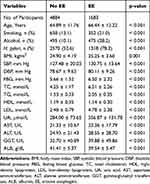
A total of 11,599 individuals, comprising 5032 males and 6567 females, were enrolled in the study. The frequency of occurrence of EE was 43.4% and 56.6% in the male and female subjects, respectively. The initial traits of the enrolled individuals are separately provided for males and females according to the prevalence of EE (Tables 1 and 2). Regardless of gender, individuals diagnosed with EE exhibited several notable differences compared to those without the diagnosis. Older individuals having higher levels of systolic blood pressure (SBP), DBP, FBG, TC, TG, LDL, UA, AST, ALT, and GGT, as well as elevated incidences of smoking, alcohol consumption, and H. pylori infections exhibited greater likelihoods of developing EE. Additionally, they showed reduced levels of ALB and HDL (p < 0.05).
 |
Table 1 Comparison of Baseline Characteristics of Females with and without EE |
 |
Table 2 Comparison of Baseline Characteristics of Males with and without EE |
Attributes of Enrolled Subjects in Different Metabolic Obesity Cohorts
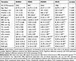
Table 3 summarizes the traits of the enrolled females (n = 6567) at baseline in terms of the different phenotypes of metabolic obesity. The MHNO, MHO, MUNO, and MUO cohorts comprised 1062 (16.2%), 893 (13.6%), 2644 (40.3%), and 1968 (29.9%) females, respectively, and the frequency of occurrence of EE in these cohorts was 9.8% 27.7%, 25.5%, and 33.4%, respectively (p < 0.05; Figure 2A). The frequency of occurrence of EE increased significantly in the female participants following an increase in the proportion of metabolic risk factors (p < 0.05; Figure 2B). The female individuals enrolled in the study had an average age of 65.23 ± 11.90 years. The SBP, DBP, FBG, TG, UA, AST, ALT, GGT, and frequency of smoking were significantly higher for the MUNO and MUO cohorts, while the levels of HDL and ALB were lower than those of the MHNO and MHO cohorts (p < 0.001). Additionally, the BMI was elevated for the MHO and MUO cohorts, compared to those of the MHNO and MUNO cohorts (p < 0.001). Furthermore, the frequency of occurrence of H. pylori infections was elevated in the MHO, MUNO, and MUO cohorts compared to that of the MHNO cohort (p < 0.001). The consumption of alcohol, LDL levels, and ages exhibited significant variations among the four cohorts of metabolic obesity (p < 0.001).
 |
Table 3 Traits of Enrolled Female Subjects at Baseline in the Different Cohorts of Metabolic Obesity |
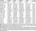
Table 4 summarizes the traits of the male subjects enrolled in the study (n = 5032) at baseline, in terms of the different phenotypes of metabolic obesity. The MHNO, MHO, MUNO, and MUO groups comprised 1072 (21.3%), 843 (16.8%), 1646 (32.7%), and 1471 (29.2%) subjects, respectively. The frequency of occurrence of EE in the MHNO, MHO, MUNO, and MUO cohorts was 13.3%, 28.6%, 26.1%, and 42.8%, respectively (p < 0.05; Figure 2A). The frequency of occurrence of EE was significantly elevated among the male participants following an increase in the proportion of metabolic risk factors (p < 0.05; Figure 2B). The male subjects enrolled in the study were aged 65.35 ± 13.06 years on average. The SBP, DBP, FBG, TG, UA, AST, ALT, GGT, and the frequency of smoking and alcohol consumption were significantly elevated in the MUNO and MUO cohorts than those of the other phenotypes, while the BMIs of the MHO and MUO cohorts were found to be higher (p < 0.001). Additionally, the subjects in the MHNO, MHO, and MUNO cohorts were younger than those in the MUO cohort (p < 0.001). The findings further revealed that the frequency of occurrence of H. pylori infections and the levels of TC, LDL, and ALB varied significantly among the four cohorts (p < 0.001).
 |
Table 4 Traits of Enrolled Male Subjects in the Different Cohorts of Metabolic Obesity |
Association Among the Different Phenotypes of Metabolic Obesity and Prevalence of EE Based on the Sex of the Individual
The findings of logistic regression analyses of the prevalence of EE in the different obesity phenotypes based on the sex of the individuals have been depicted in Figure 3. The findings demonstrated that, irrespective of sex, the subjects in the MHO, MUNO, and MUO cohorts exhibited elevated risks of developing EE than those in the MHNO cohort (p < 0.001). The age, BMI, smoking, alcohol intake, and H. pylori infections were adjusted for, and the adjusted ORs (95% CI) for the frequency of occurrence of EE in the MHO, MUNO, and MUO cohorts were determined to be 5.008 (4.057–6.181), 2.333 (2.000–2.721), and 6.385 (5.269–7.737), respectively, compared to that of the MHNO phenotype. The age, BMI, smoking, alcohol intake, and H. pylori infections of the male subjects were adjusted for, and it was observed that the male participants having the MHO phenotype (OR: 3.608; 95% CI: 2.697–4.827) possessed significantly greater risks of developing EE than those in the MHNO and MUNO cohorts (combined OR: 1.950; 95% CI: 1.572–2.420). The individuals with the MUO phenotype had the greatest OR of 5.473 (95% CI: 4.181–7.165) of all the cohorts. The age, BMI, smoking, alcohol intake, and H. pylori infections of the female subjects were adjusted for, and the findings similarly revealed that the female participants with the MHO phenotype (OR: 6.920; 95% CI: 5.084–9.420) exhibited significantly elevated risks of suffering from EE in comparison to those in the MHNO and MUNO cohorts (combined OR: 2.769; 95% CI: 2.204–3.479). The individuals with the MUO exhibited the greatest OR of 7.566 (95% CI: 5.718–10.010) of all the cohorts.
Relationships of Different Metabolic Obesity Phenotypes with the Prevalence of EE According to Age
The results of analyses of the relationships of the different metabolic obesity phenotypes with the prevalence of EE in terms of the age of the individuals are depicted in Figure 4. Regardless of the age, the occurrence of MHO, MUNO, or MUO was a risk factor for developing EE, unlike the MHNO phenotype (p < 0.001). The age, BMI, smoking, alcohol intake, and H. pylori infections were adjusted for, and the adjusted ORs (95% CI) for the prevalence of EE in the MHO, MUNO, and MUO cohorts were determined to be 5.008 (4.057–6.181), 2.333 (2.000–2.721), and 6.385 (5.269–7.737), respectively, compared to that of the MHNO phenotype. The sex, BMI, smoking, alcohol consumption, and H. pylori infections were adjusted for, and it was observed that participants aged less than 60 years and having the MHO phenotype (OR: 6.158; 95% CI: 4.047–9.371) possessed significantly elevated risks of developing EE than those in the MHNO and MUNO cohorts (combined OR: 2.822; 95% CI: 2.040–3.903). The individuals in the MUO cohort had the greatest OR of 9.639 (95% CI: 6.504–14.286) of all the cohorts. The age, BMI, smoking habits, alcohol consumption, and H. pylori infections of the participants aged above 60 years and having the MHO phenotype were adjusted for, and it was similarly observed that these individuals possessed significantly elevated risks of developing EE (OR: 4.712; 95% CI: 3.686–6.023) than those in the MHNO and MUNO cohorts (combined OR: 2.197; 95% CI: 1.843–2.621). The individuals in the MUO cohort had the greatest OR of 5.607 (95% CI: 4.493–6.997) of all the cohorts.
Discussion
The present retrospective study analyzed the relationship of the different phenotypes of metabolic obesity with the frequency of occurrence of EE. The findings demonstrated that unlike the MHNO phenotype, there is a significant association between the occurrence of MHO, MUNO, and MUO and elevated risks of developing EE. Altogether, our investigation substantiated that the incidence of EE is more pronounced among individuals characterized by obesity, irrespective of their metabolic health status. The result underscores the importance of weight management as a preventive measure against EE. Further investigations revealed that the occurrence of MHO, MUNO, or MUO are correlated with the elevated prevalence of EE, regardless of gender and age.
Previous research has shown a significant association between obesity and EE. Many processes have been suggested to explain this relationship, although the precise mechanisms that relate fat and EE are not yet known. Obese individuals experience an elevated pressure in the lower esophageal sphincter (LES), which impairs the anti-reflux barrier that subsequently triggers the gastroesophageal reflux (GER). This phenomenon may be associated with compensatory mechanisms triggered by heightened intra-abdominal pressure.18,19 Saliva secretion, gravity, and esophageal motility collectively determine the esophageal clearance rate. Obesity often results in reduced saliva secretion and impaired esophageal motility, compromising the function of esophageal clearance.20–22 Vicente Ortiz et al23 found that obese individuals demonstrate reduced esophageal sensitivity to acid perfusion, potentially affecting esophageal clearance function. A recent study suggested that the pathogenesis of EE is possibly mediated via cytokine-induced esophageal inflammation.24 Visceral adipose tissue functions as a significant depot of adipocyte-derived factors and release cytokines, including interleukin-1 (IL-1), IL-6, tumor necrosis factor-alpha (TNF-α), leptin, adiponectin, and other molecules. These mediators can induce systemic effects, influencing and amplifying systemic inflammatory responses.25–27 These findings demonstrate that obesity alone may serve as a significant risk factor for EE. The findings obtained in this study support the traditional perspective that obesity elevates the risk of developing EE. This association is likely attributable to factors such as heightened intra-abdominal pressure, increased episodes of transient LES relaxation, and heightened esophageal acid exposure, which are commonly associated with obesity.
Consistent with our findings, multiple studies have found metabolic disorders to be significantly associated with reflux esophagitis, though the mechanism underlying this association is uncertain.28–31 This may be associated with the administration of antihypertensive agents. Calcium channel blockers have been shown to have the power to suppress muscular contractions in the esophagus, which eases the pressure on the esophageal sphincter.30 Our study found that high SBP and DBP were associated with EE, regardless of sex. Earlier reports have investigated the relationship between the prevalence of EE and hyperglycemia.32,33 High glucose levels can lead to increased stomach acid production, which contributes to the development of gastroesophageal reflux symptoms.34 This finding correlates with the results observed herein, which revealed that the serum levels of glucose was elevated in the EE cohort compared to that of the non-EE category. An earlier investigation reported that there is significant association between hyperlipidemia and the onset of EE, and that high-fat diets can reduce the risk of depression in individuals with hyperlipidemia.35 Additionally, an earlier study reported that esophageal clearance is impaired by elevated lipid levels, which weakens the LES and ultimately contributes to the development of EE.36 Our research found that the risk factors for EE included higher TG, TC, and LDL, and lower HDL levels. The findings also revealed that metabolic disorders were more predominant in males than in the female individuals, especially in patients with EE. The present study demonstrated that dyslipidemia, hypertension, and hyperglycemia can elevate the risks of developing EE, which correlated with the observations of earlier reports. This investigation emphasizes the significance of modifying metabolic abnormalities irrespective of the obesity phenotype.
The effects of obesity and metabolic abnormalities on the risks of developing EE have not been compared in aforementioned reports. The present investigation therefore expanded upon the existing concept of obesity by concurrently assessing the metabolic status. This allowed us to propose a risk of EE assessment strategy according to the metabolic obesity phenotype. The study revealed that the prevalence of EE varied between the MUNO and MHO cohorts. The prevalence of EE was greater in the MHO cohort than in the MUNO cohort. However, the frequency of occurrence of EE in the MHO and MUNO cohorts was significantly elevated compared to that of the MHNO cohort, which suggested that obesity was the most significant risk factor for the onset of EE, independent of the occurrence of metabolically unhealthy phenotypes. The occurrence of obesity and presence of metabolic abnormalities jointly contributed to the risk of developing EE, and the prevalence of EE was most elevated for the MUO cohort. The present report therefore emphasizes the significance of altering the obesity profile of affected individuals, irrespective of their metabolic profiles. A retrospective analysis revealed that the occurrence of MHO is related to an elevated risk of EE, however, the presence of metabolic abnormalities alone was not a risk factor for EE.37 Moreover, another study using a large patient cohort speculated that there is an association between the occurrence of MHO and an elevation in the prevalence of EE.38 These results suggested that obesity and not the metabolic profile is a more significant risk factor for the development of EE. This phenomenon was possibly caused by the storage of visceral fat in the MHO phenotype.39 A prospective study revealed that patients with the MHO phenotype frequently underwent a deterioration in their metabolic health status over an extended period of follow-up, ultimately transitioning into the MUO phenotype. This investigation indicates that the MHO phenotype cannot be considered a consistently stable metabolic obesity phenotype.40 Therefore, we should keep a normal weight regardless of metabolic health status. Although the individuals in the MHO cohort exhibited elevated risks of suffering from EE than the subjects in the MUNO and MHNO cohorts, the individuals in the MUO cohort were at the greatest risk of suffering from EE, for both sexes. The age, SBP, DBP, FBG, and levels of TG, TC, LDL, UA, AST, ALT, GGT, and ALB significantly differed among the four groups, being more abnormal in MUO than MHNO, MHO, and MUNO in our study. Therefore, the individuals at a higher risk of developing EE can be identified more accurately by considering both the obesity and metabolic profiles during analysis of the metabolic obesity phenotypes. Physicians could make early interventions for abnormal obesity phenotypes by using the metabolic obesity phenotypes, which can reduce the financial burden of the therapeutic management of EE and its complications, typically EAC and BE.
Few reports have assessed the relationship of different obesity phenotypes with the development of EE in the different sexes, and the present report observed that after adjusting for the influencing factors, the females in the MHO, MUNO, and MUO cohorts exhibited increased risks of suffering from EE than the males in the corresponding cohorts. The present report further demonstrated that the frequency of occurrence of EE was significantly elevated in male subjects than in female individuals, in all the obesity phenotypes. The reason for this sex difference is unclear, although several possible explanations exist. First, men have a higher tendency to accumulate visceral adipose tissue compared to women, which highlights the increased risk of obesity-related health hazards in men.41 Secondly, the biological activity of visceral adipose tissue is higher than that of adipose deposits in other areas.42 A surplus accumulation of visceral adipose tissue contributes to chronic low-grade inflammation, leading to the onset of EE.36,43 Finally, estrogen enhances nitric oxide production, a vasodilator that promotes smooth muscle relaxation. This can relax the LES and subsequently increase reflux phenomena. Earlier reports have determined that age is one of major risk factors for EE.44 The relationship of the different metabolic obesity phenotypes with the onset of EE was additionally investigated herein across the different age groups. Interestingly, the findings revealed that individuals with MHO, MUNO, and MUO and aged less than 60 years exhibited higher risks of developing EE than the subjects older than 60 years; however, the causative factors underlying the age-dependent variations in the onset of EE remain unknown. We hypothesize that the variations could be associated with the fact that as aging is related to leptin resistance and the receptors for leptin decreases with age, the detrimental consequences of leptin on EE may in some way be alleviated in aged subjects.45 Additionally, aging is associated with alterations in body composition and muscular atrophy. Further studies using body composition data can enhance our understanding of the underlying mechanisms.
The present study has multiple constraints, which are described hereafter. Firstly, a cross-sectional approach was used in the investigation, as a result of which it was not possible to identify the causalities from the results. Secondly, the research focused on Asian population, which limits the general applicability of the findings obtained herein to different ethnicities. Thirdly, obesity was diagnosed herein solely based on the BMI as the routine data did not include waist circumference. Conducting additional research that includes waist circumference and other body composition measurements could offer thorough insights into the relationship of the different obesity phenotypes with the onset of EE. Fourthly, our study did not incorporate stricter triglyceride cutoff values. The European Atherosclerosis Society suggests that a fasting triglyceride level of < 1.1 mmol/L may offer a more precise evaluation of metabolic health and related risks in obese patients.46 Lastly, although the potential confounding parameters were adjusted for during the multivariable analysis, some unmeasured residual confounding factors, including dietary patterns, psychosocial stress, and socioeconomic status, which may have influenced our risk estimates, were not considered in this study.
Conclusions
The findings obtained herein indicate that unlike MHNO, the occurrence of MHO, MUNO, and MUO is related with an elevated risk of developing EE. Additionally, MHO is not a health status and is at higher risk of EE compared to MUNO, which implied that obesity significantly contributes to the occurrence of EE. The findings revealed the metabolic obesity phenotype is significantly correlated with the occurrence of EE, regardless of sex and age. The frequency of occurrence of EE was elevated following an increase in the number of metabolic risk factors. The results emphasize the significance of considering the metabolic health profiles of obese individuals for assessing the risk of EE. However, while focusing on patients with metabolic abnormalities, we must also recognize the importance of addressing MHO individuals. Individuals with MHO should retain a healthy weight and adopt a healthy lifestyle to mitigate the risks of developing EE.
Institutional Review Board Statement
This study was executed in conformity with the ethical principles stipulated in the Declaration of Helsinki and received approval from the Institutional Review Board of the First Affiliated Hospital of Dalian Medical University for all protocols involving human subjects (PJ-KS-KY-2020-04). Informed consent was obtained in writing from all participants before they participated in the study.
Data Sharing Statement
The data in support of the conclusions described herein are available from the corresponding author upon a legitimate request.
Funding
There is no funding to report.
Disclosure
The authors declare that they have no competing interests in this work.
References
1. Maret-Ouda J, Markar SR, Lagergren J. Gastroesophageal reflux disease: a review. JAMA. 2020;324(24):2536–2547. doi:10.1001/jama.2020.21360
2. Diab N, Patel M, O’Byrne P, Satia I. Narrative review of the mechanisms and treatment of cough in asthma, cough variant asthma, and non-asthmatic eosinophilic bronchitis. Lung. 2022;200(6):707–716. doi:10.1007/s00408-022-00575-6
3. Becher A, El-Serag H. Systematic review: the association between symptomatic response to proton pump inhibitors and health-related quality of life in patients with gastro-oesophageal reflux disease. Aliment. Pharmacol. Ther. 2011;34(6):618–627. doi:10.1111/j.1365-2036.2011.04774.x
4. Baklola M, Terra M, Badr A, et al. Prevalence of gastro-oesophageal reflux disease, and its associated risk factors among medical students: a nation-based cross-sectional study. BMC Gastroenterol. 2023;23(1):269. doi:10.1186/s12876-023-02899-w
5. Taraszewska A. Risk factors for gastroesophageal reflux disease symptoms related to lifestyle and diet. Roczniki Panstwowego Zakladu Higieny. 2021;72(1):21–28. doi:10.32394/rpzh.2021.0145
6. Eusebi LH, Ratnakumaran R, Yuan Y, Solaymani-Dodaran M, Bazzoli F, Ford AC. Global prevalence of, and risk factors for, gastro-oesophageal reflux symptoms: a meta-analysis. Gut. 2018;67(3):430–440. doi:10.1136/gutjnl-2016-313589
7. Lee YB, Kim DH, Kim SM, et al. Hospitalization for heart failure incidence according to the transition in metabolic health and obesity status: a nationwide population-based study. Cardiovasc Diabet. 2020;19(1):77. doi:10.1186/s12933-020-01051-2
8. Wu P, Ma L, Dai GX, et al. The association of metabolic syndrome with reflux esophagitis: a case-control study. Neurogastroenterol Motil: Official J European Gastrointestinal Motility Soc. 2011;23(11):989–994. doi:10.1111/j.1365-2982.2011.01786.x
9. Stefan N, Häring HU, Hu FB, Schulze MB. Metabolically healthy obesity: epidemiology, mechanisms, and clinical implications. Lancet Diabetes Endocrinol. 2013;1(2):152–162. doi:10.1016/S2213-8587(13)70062-7
10. Kramer CK, Zinman B, Retnakaran R. Are metabolically healthy overweight and obesity benign conditions?: a systematic review and meta-analysis. Ann Internal Med. 2013;159(11):758–769. doi:10.7326/0003-4819-159-11-201312030-00008
11. Man S, Lv J, Yu C, et al. Association between metabolically healthy obesity and non-alcoholic fatty liver disease. Hepatol Internat. 2022;16(6):1412–1423. doi:10.1007/s12072-022-10395-8
12. Yang H, Xia Q, Shen Y, Chen TL, Wang J, Lu YY. Gender-specific impact of metabolic obesity phenotypes on the risk of hashimoto’s thyroiditis: a retrospective data analysis using a health check-up database. J Inflamm Res. 2022;15:827–837. doi:10.2147/JIR.S353384
13. Cao Z, Zheng X, Yang H, et al. Association of obesity status and metabolic syndrome with site-specific cancers: a population-based cohort study. Br. J. Cancer. 2020;123(8):1336–1344. doi:10.1038/s41416-020-1012-6
14. Lundell LR, Dent J, Bennett JR, et al. Endoscopic assessment of oesophagitis: clinical and functional correlates and further validation of the Los Angeles classification. Gut. 1999;45(2):172–180. doi:10.1136/gut.45.2.172
15. Hsu WC, Araneta MR, Kanaya AM, Chiang JL, Fujimoto W. BMI cut points to identify at-risk Asian Americans for type 2 diabetes screening. Diabetes Care. 2015;38(1):150–158. doi:10.2337/dc14-2391
16. Organization WH. Appropriate body-mass index for Asian populations and its implications for policy and intervention strategies. Lancet. 2004;363:157–163.
17. Executive Summary of The Third Report of The National Cholesterol Education Program (NCEP). Expert panel on detection, evaluation, and treatment of high blood cholesterol in adults (adult treatment panel III). JAMA. 2001;285(19):2486–2497. doi:10.1001/jama.285.19.2486
18. Macías Valadez LC Z, Pescarus R, Hsieh T, et al. Laparoscopic limited Heller myotomy without anti-reflux procedure does not induce significant long-term gastroesophageal reflux. Surg Endosc. 2015;29(6):1462–1468. doi:10.1007/s00464-014-3824-z
19. Suter M, Dorta G, Giusti V, Calmes JM. Gastro-esophageal reflux and esophageal motility disorders in morbidly obese patients. Obes Surg. 2004;14(7):959–966. doi:10.1381/0960892041719581
20. Valezi AC, Herbella FAM, Schlottmann F, Patti MG. Gastroesophageal reflux disease in obese patients. J Laparoendoscopic Adv Surg Tech Part A. 2018;28(8):949–952. doi:10.1089/lap.2018.0395
21. Côté-Daigneault J, Leclerc P, Joubert J, Bouin M. High prevalence of esophageal dysmotility in asymptomatic obese patients. Can J Gastroenterol Hepatol. 2014;28(6):311–314. doi:10.1155/2014/960520
22. Koppman JS, Poggi L, Szomstein S, Ukleja A, Botoman A, Rosenthal R. Esophageal motility disorders in the morbidly obese population. Surg Endosc. 2007;21(5):761–764. doi:10.1007/s00464-006-9102-y
23. Ortiz V, Alvarez-Sotomayor D, Sáez-González E, et al. Decreased esophageal sensitivity to acid in morbidly obese patients: a cause for concern? Gut Liver. 2017;11(3):358–362. doi:10.5009/gnl16081
24. Dunbar KB, Agoston AT, Odze RD, et al. Association of acute gastroesophageal reflux disease with esophageal histologic changes. JAMA. 2016;315(19):2104–2112. doi:10.1001/jama.2016.5657
25. Gregor MF, Hotamisligil GS. Inflammatory mechanisms in obesity. Ann Rev Immunol. 2011;29(1):415–445. doi:10.1146/annurev-immunol-031210-101322
26. Nam SY, Choi IJ, Ryu KH, Park BJ, Kim HB, Nam BH. Abdominal visceral adipose tissue volume is associated with increased risk of erosive esophagitis in men and women. Gastroenterology. 2010;139(6):1902–1911.e1902. doi:10.1053/j.gastro.2010.08.019
27. Kord HV, Tinsley GM, Santos HO, et al. The influence of fasting and energy-restricted diets on leptin and adiponectin levels in humans: a systematic review and meta-analysis. Clinical Nutr. 2021;40(4):1811–1821. doi:10.1016/j.clnu.2020.10.034
28. Song HJ, Shim KN, Yoon SJ, et al. The prevalence and clinical characteristics of reflux esophagitis in koreans and its possible relation to metabolic syndrome. J Korean Med Sci. 2009;24(2):197–202. doi:10.3346/jkms.2009.24.2.197
29. Fu S, Xu M, Zhou H, Wang Y, Tan Y, Liu D. Metabolic syndrome is associated with higher rate of gastroesophageal reflux disease: a meta-analysis. Neurogastroenterol Motil: Official J European Gastrointestinal Motility Soc. 2022;34(5):e14234. doi:10.1111/nmo.14234
30. Mohammadi M, Ramezani Jolfaie N, Alipour R, Zarrati M. Is metabolic syndrome considered to be a risk factor for gastroesophageal reflux disease (non-erosive or erosive esophagitis)?: a systematic review of the evidence. Iran Red Crescent Med J. 2016;18(11):e30363. doi:10.5812/ircmj.30363
31. Fukunaga S, Nakano D, Tsutsumi T, et al. Lean/normal-weight metabolic dysfunction-associated fatty liver disease is a risk factor for reflux esophagitis. Hepatol Res: Official J Japan Society of Hepatol. 2022;52(8):699–711. doi:10.1111/hepr.13795
32. Bou Daher H, Sharara AI. Gastroesophageal reflux disease, obesity and laparoscopic sleeve gastrectomy: the burning questions. World J Gastroenterol. 2019;25(33):4805–4813. doi:10.3748/wjg.v25.i33.4805
33. Loke SS, Yang KD, Chen KD, Chen JF. Erosive esophagitis associated with metabolic syndrome, impaired liver function, and dyslipidemia. World J Gastroenterol. 2013;19(35):5883–5888. doi:10.3748/wjg.v19.i35.5883
34. Hirata A, Kishida K, Nakatsuji H, et al. High prevalence of gastroesophageal reflux symptoms in type 2 diabetics with hypoadiponectinemia and metabolic syndrome. Nutr Metab. 2012;9(1):4. doi:10.1186/1743-7075-9-4
35. Nam SY, Park BJ, Cho YA, et al. Different effects of dietary factors on reflux esophagitis and non-erosive reflux disease in 11,690 Korean subjects. J Gastroenterol. 2017;52(7):818–829. doi:10.1007/s00535-016-1282-1
36. Rieder F, Biancani P, Harnett K, Yerian L, Falk GW. Inflammatory mediators in gastroesophageal reflux disease: impact on esophageal motility, fibrosis, and carcinogenesis. Am J Physiol Gastrointest Liver Physiol. 2010;298(5):G571–581. doi:10.1152/ajpgi.00454.2009
37. Baeg MK, Ko SH, Ko SY, Jung HS, Choi MG. Obesity increases the risk of erosive esophagitis but metabolic unhealthiness alone does not: a large-scale cross-sectional study. BMC Gastroenterol. 2018;18(1):82. doi:10.1186/s12876-018-0814-y
38. Kim TJ, Lee H, Baek SY, et al. Metabolically healthy obesity and the risk of erosive esophagitis: a cohort study. Clin Transl Gastroenterol. 2019;10(9):e00077. doi:10.14309/ctg.0000000000000077
39. Elías-López D, Vargas-Vázquez A, Mehta R, et al. Natural course of metabolically healthy phenotype and risk of developing Cardiometabolic diseases: a three years follow-up study. BMC Endocr Disord. 2021;21(1):85. doi:10.1186/s12902-021-00754-1
40. Eshtiaghi R, Keihani S, Hosseinpanah F, Barzin M, Azizi F. Natural course of metabolically healthy abdominal obese adults after 10 years of follow-up: the Tehran lipid and glucose study. Int J Obesity (2005). 2015;39(3):514–519. doi:10.1038/ijo.2014.176
41. Tchernof A, Després JP. Pathophysiology of human visceral obesity: an update. Physiol Rev. 2013;93(1):359–404. doi:10.1152/physrev.00033.2011
42. Francisco V, Ruiz-Fernández C, Pino J, et al. Adipokines: linking metabolic syndrome, the immune system, and arthritic diseases. Biochem. Pharmacol. 2019;165:196–206. doi:10.1016/j.bcp.2019.03.030
43. Macdougall CE, Wood EG, Loschko J, et al. Visceral adipose tissue immune homeostasis is regulated by the crosstalk between adipocytes and dendritic cell subsets. Cell Metab. 2018;27(3):588–601.e584. doi:10.1016/j.cmet.2018.02.007
44. Richter JE, Rubenstein JH. Presentation and epidemiology of gastroesophageal reflux disease. Gastroenterology. 2018;154(2):267–276. doi:10.1053/j.gastro.2017.07.045
45. Gulcelik NE, Halil M, Ariogul S, Usman A. Adipocytokines and aging: adiponectin and leptin. Minerva Endocrinol. 2013;38(2):203–210.
46. Ginsberg HN, Packard CJ, Chapman MJ, et al. Triglyceride-rich lipoproteins and their remnants: metabolic insights, role in atherosclerotic cardiovascular disease, and emerging therapeutic strategies-A consensus statement from the European Atherosclerosis Society. Eur Heart J. 2021;42(47):4791–4806. doi:10.1093/eurheartj/ehab551
 © 2024 The Author(s). This work is published and licensed by Dove Medical Press Limited. The
full terms of this license are available at https://www.dovepress.com/terms.php
and incorporate the Creative Commons Attribution
- Non Commercial (unported, 3.0) License.
By accessing the work you hereby accept the Terms. Non-commercial uses of the work are permitted
without any further permission from Dove Medical Press Limited, provided the work is properly
attributed. For permission for commercial use of this work, please see paragraphs 4.2 and 5 of our Terms.


