")
Back to Journals » Cancer Management and Research » Volume 16
Bioinformatics Analysis Reveals CDK1 and DLGAP5 as Key Modulators of Tumor Immune Cell Infiltration in Hepatocellular Carcinoma
Authors Li J, Liu Q, Zhang T, Du Q
Received 18 June 2024
Accepted for publication 1 November 2024
Published 14 November 2024 Volume 2024:16 Pages 1597—1608
DOI https://doi.org/10.2147/CMAR.S478426
Checked for plagiarism Yes
Review by Single anonymous peer review
Peer reviewer comments 3
Editor who approved publication: Professor Yong Teng
Jiajing Li,1 Qi Liu,2 Ting Zhang,2 Qian Du3
1The Diagnostics Laboratory, Affiliated Hospital to Zunyi Medical University, Zunyi, Guizhou, 563000, People’s Republic of China; 2Affiliated Hospital to Zunyi Medical University, Zunyi, Guizhou, 563000, People’s Republic of China; 3Department of Endoscopy and Digestive System, Guizhou Provincial People’s Hospital, Guiyang, Guizhou, 550002, People’s Republic of China
Correspondence: Qian Du, Email [email protected]
Introduction: Hepatocellular carcinoma (HCC), a prevalent and aggressive form of cancer, poses significant challenges due to its limited therapeutic options. This study aims to leverage multi-omics data from liver cancer to identify potential therapeutic targets for HCC.
Methods: We employed an integrative approach by analyzing various omics datasets related to liver cancer. Through comprehensive data mining and analysis, we identified key genes that are significantly associated with HCC. To gain insights into their biological roles and underlying mechanisms, we constructed gene ontology (GO) and Kyoto Encyclopedia of Genes and Genomes (KEGG) pathway networks. Specifically, we focused on genes that exhibited high expression levels in HCC and were correlated with poor patient prognosis. Among these, CDK1 and DLGAP5 emerged as promising candidates and were further investigated for their potential involvement in tumor immune cell infiltration and HCC progression.
Results: Our analysis revealed that CDK1 and DLGAP5 are highly expressed in HCC tissues compared to normal liver tissues, and their elevated expression is associated with unfavorable clinical outcomes. Furthermore, through GO and KEGG pathway analyses, we found that these genes are implicated in critical biological processes and signaling pathways relevant to HCC pathogenesis. Notably, CDK1 and DLGAP5 were shown to be associated with tumor immune cell infiltration, suggesting their potential role in modulating the tumor microenvironment and promoting HCC progression.
Discussion: These findings provide valuable insights into the development of novel therapeutic approaches for HCC.
Keywords: Hepatocellular carcinoma, bioinformatics analysis, DLGAP5, CDK1, immune cell infiltration
Introduction
Hepatocellular carcinoma (HCC) is major global health concern, with rising incidence rates and limited treatment options. Despite advances in surgical resection, liver transplantation, and targeted therapies, the prognosis for HCC patients remains unsatisfactory.1,2 Therefore, there is an urgent need to identify novel therapeutic targets and develop innovative treatment strategies for this aggressive malignancy.3 In recent years, bioinformatics analysis has emerged as powerful tools in cancer research,4–6 enabling the identification of potential therapeutic targets and the discovery of novel treatment approaches.
HCC is a complex disease driven by genetic and epigenetic alterations, dysregulated signaling pathways, and intricate interactions between tumor cells and the tumor microenvironment.7–9 A previous study indicated regorafenib’s potential role in immune checkpoint blockade10 and Tyrosine kinase inhibitors (TKIs) in immunotherapy for hepatocellular carcinoma.11 The traditional “one gene, one drug” paradigm in drug discovery and development has proven to be insufficient in effectively targeting the heterogeneity and complexity of HCC.12,13 Therefore, there is a growing interest in adopting a systems biology approach that integrates multiomics data and network pharmacological analysis to identify key molecular drivers and potential therapeutic targets for HCC.
A comprehensive approach of bioinformatics analysis was conducted to find potential therapeutic targets and drug candidates for HCC. CDK1, on the other hand, is a key regulator of cell cycle progression and is known to be overexpressed in many cancer types. Inhibition of CDK1 activity has been shown to induce cell cycle arrest, making it a promising target for anticancer therapy.14,15 DLGAP5, a member of the DLGAP family of proteins, has been implicated in the regulation of cell adhesion and cytoskeletal dynamics, processes that are often disrupted in cancer cells.16,17 However, the precise role of CDK1 and DLGAP5in HCC progression remains poorly understood, making it an attractive target for further investigation.
In conclusion, the integration of bioinformatics analysis offers a systematic and comprehensive approach to identify potential therapeutic targets and develop innovative treatment strategies for HCC. Our study provides valuable insights into the dysregulated pathways and potential therapeutic interventions in HCC. Identification of CDK1-DLGAP5-tumor immune cell infiltration axis as promising therapy strategy opens up new avenues in the management of HCC, highlighting its role as a novel therapy strategy.
Research Data and Bioinformatics Methods
Source of Data
Login the GEO database (https://www.ncbi.nlm.nih.Gov/geo), search for the keyword “HCC”, restrict the organization source to “Homo sapiens”, and research type to “Expression Teaching by array HCC”. Select five datasets: GSE101685, GSE46408, GSE146049, GSE115018, and GSE50579 for analysis. The GSE101685 dataset is based on the GPL570 platform and contains gene expression information from 24 hCC tissues of different stages and 8 normal liver tissues; The GSE46408 dataset is based on the GPL4133 platform and contains gene expression information of 6 cases of primary hepatocellular carcinoma and 6 corresponding non tumor liver parenchymal cells; The GSE146049 dataset is based on the GPL10558 platform and contains gene expression information from 5 hCC tissues and 5 normal liver tissues; The GSE115018 dataset is based on the GPL20115 platform and contains gene expression information from 12 hCC tissues and 12 adjacent tissues; The GSE50579 dataset is based on the GPL14500 platform and contains gene expression information for the control group (normal liver tissue (NL), n=10) and HCC tissue (n=67).
Screening of Differential Gene
Grouping HCC tissue and normal tissue in each dataset, with |LogFC|>0.6 and corrected P<0.05 as the screening criteria, obtaining differentially expressed genes, and intersecting the obtained data with a Venn map.
Enrichment Analysis of GO and KEGG Signaling Pathways
Utilizing the DAVID database (https://david.ncifcrf.gov/) to perform GO and KEGG enrichment analysis on the differentially expressed genes selected. GO analysis includes the biological processes, cellular components, and molecular functions involved in the genes, and screen the results of the analysis with a P<0.05 limitation.
Construction of PPI Protein Network and Screening of Hub Genes
Using String (https://cn.string-db.org) build a protein database for protein interaction analysis and obtain a protein interaction relationship tree. Using Cyto-scape software for network graph visualization, genes are scored using the Degree algorithm and plotted based on their scores. The top 12 genes with the highest scores are selected as key genes.
Expression Analysis of Key Genes in HCC and CHOL
Using GEPIA to analyze the expression level of key genes and further verify the expression differences of 12 key genes in HCC and CHOL tissue and normal tissue.
Survival Analysis of Hub Genes
Using GEPIA (http://gepia.cancer-pku.cn/) online tool to analyze the relationship between selected key genes and HCC survival rate.
Protein Expression Validation
Search for protein immunohistochemistry experiments of CDK1 and DLGAP5 proteins in normal liver tissue and HCC in the Human Protein Atlas database (http://www.proteinatlas.org/), and compare and analyze them.
Animal Model
All experiments were approved by the Institute of Animal Care and Use Committee of the Affiliated Hospital of Zunyi Medical University. The Chinese National Guidelines (GB/T 35892–2018) were followed for the welfare of the laboratory animals. Guiding Opinions on Treating Experimental Animals Well (Guokefacaizi [2006] No. 398), Experimental animals (C57BL/6, 6-8-week, male) were purchased (from the Beijing HFK, China). Mice in the model group were administered with diethylnitrosamine (DEN) at a dose of 2 mg/kg via intraperitoneal injection on day 14 of birth. Two weeks after the DEN administration, the mice were further treated with carbon tetrachloride (CCl4) at a concentration of 20% and a dose of 5 mL/kg via intraperitoneal injection, twice weekly for a total of 16 weeks. At the end of the 20-week modeling period, the mice were sacrificed, and liver tissues were collected for further analysis.
Real-Time Quantitative PCR (qPCR)
Real-time qPCR was performed to detected the expression of CDK1 and DLGAP5 in HCC mice model.
Expression Analysis of CDK1 and DLGAP5 in Different Conditions
Utilizing UALCAN (https://ualcan.path.uab.edu/) online website to analyze the expression of CDK1 and DLGAP5 in various tumors, HCC patients of different ages, weights, races, genders, HCC stages, and TP53 mutations, as well as their methylation levels in normal tissues and HCC.
Search for protein immunohistochemistry experiments of DLGAP5 and CDK1 proteins in normal liver tissue and liver cancer in the Human Protein Atlas database (https://www.proteinatlas.org/), and compare and analyze them. In addition, by exploring the immunofluorescence and expression of DLGAP5 and CDK1 in the cell cycle, we aim to understand their distribution in cells and expression at each stage of the cell cycle.
Correlation Analysis of Tumor Immune Cells
Tumor Immune Single Cell Center 2 (TISCH2) is an online database (http://tisch.comp-genomics.org) Integrating single-cell transcriptome spectra of approximately 2 million cells from 76 high-quality cancer datasets. We explored the immune related molecular mechanisms underlying the prognostic characteristics of TME in HCC through the TISCH2 database. At the same time, we used the TIMER2.0 database (http://timer.cistrome.org/) to analyze the correlation between DLGAP5 and CDK1 with tumor immune cell infiltration online and selected the tumor type as LIHC. The immune cells involved include CD8+T cells, CD4+T cells, B cells, neutrophils, macrophages, and dendritic cells.
Results
Identification of Key Genes in Hepatocellular Carcinoma
To more comprehensively determine the gene expression profile and identify key genes associated with the occurrence and development of Hepatocellular Carcinoma, we performed a comprehensive analysis of the publicly available genomic data generated from liver tissue from human with primary HCC, NASH or HBV driven HCC in the GEO database (Supporting Table S1). Both upregulated and downregulated genes were identified by differentially expressed gene (DEG) analysis in each comparative group (Figure 1A–E). Total 100 genes with consistent pattern changes across five databases were discovered by intersection analysis (Figure 1F), detail information in Supporting Table S2.
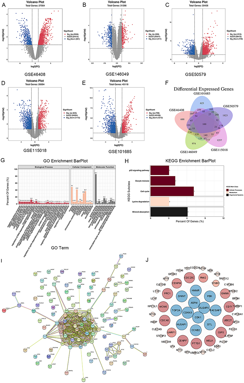 |
Figure 1 Identification of key genes in Hepatocellular carcinoma. (A–E) Volcano plot of DGEs in GSE46408, GSE146049, GSE115018, GSE50579 and GSE101685 datasets, the red dots represent up-regulated genes, the green dots represent down-regulated genes, and the grey dots represent genes with no significant difference in expression. For a–e, the screening criteria are set to P < 0.05, |log2FC| ≥0.6. (F) Venn diagram demonstrates the intersection of DGEs and positively correlated module genes of HCC in five GSEs data sets. (G and H) GO and KEGG pathway enrichment analysis of 100 DGEs, biological processes (BP) enrichment analysis (red bars); cellular components (CC) enrichment analysis (Orange bars); and molecular functions (MF) enrichment analysis (black bars). (I) PPI of 100 pivotal genes. (J) Visualized PPI analysis of the pivotal genes. |
The underlying mechanisms of action of these common crossover genes GO enrichment analysis revealed that the main biological processes (BP) mainly include cell division, cell cycle, negative regulation of growth DNA repair signaling pathway, etc. Cellular components (CC) mainly involve nucleus, cytoplasm, cytosol, nucleoplasm, and centrosome. Molecular functions (MF) involved in protein binding, DNA binding (Figure 1G); KEGG pathway analysis showed that the cross-targets mainly involved mineral absorption, cell cycle, oocyte meiosis and p53 signaling pathway (Figure 1H). Subsequently, we queried and screened the interaction relationships between indicated target proteins by the STRING online database to obtain a protein interaction network (Figure 1I). Besides, we visualized PPI analysis of the pivotal genes by the Cytoscape (Figure 1J) and obtained a core target interaction network consisting of 12 core genes (Supporting Table S3): Aspm, Ccnb2, Cdk1, Cdkn3, Dlgap5, Dtl, Hmmr, Nusap1, Pbk, Racgap1, Top2a and Zwint. Taken together, our joint analysis reveals key genomic alterations in HCC.
Expression of Key Genes in HCC from TCGA
Through the analysis of the GSE dataset, we identified a set of DEGs that exhibited significant differential expression in HCC. To further confirm the importance of these indicated genes, a total of 12 DEGs were screened according to stromal scores in HCC from TCGA dataset, which show the same expression patterns as in the GSE database (Figure 2). To substantiate the significance of the identified genes, we investigated their expression in Cholangiocarcinoma (CHOL) from TCGA. The study revealed a high expression level of these genes in CHOL (Supporting Figure S1), providing additional evidence for the accuracy of our screening results and suggesting their crucial roles in liver tumorigenesis, which strengthen the reliability of GEO data.
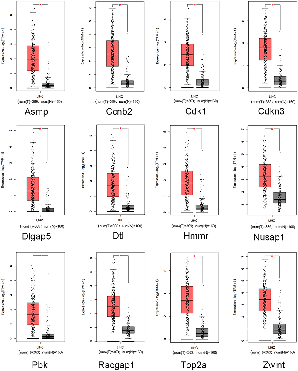 |
Figure 2 Expression of key genes in HCC from TCGA. mRNA expression analysis of Aspm, Ccnb2, Cdk1, Cdkn3, Dlgap5, Dtl, Hmmr, Nusap1, Pbk, Racgap1, Top2a, Zwint in LICH from TCGA. Red represents tumor tissue, and gray represents normal tissue. *P<0.01. |
CDK1and DLGAP5 is Highly Expressed and Associated with Poor Prognosis in HCC
To further investigate the significance and identify the most crucial genes in HCC, we utilized RNA-Seq data from the TCGA database to analyze these DEGs expression levels in HCC samples along with survival analysis. Kaplan–Meier survival analysis demonstrated that high expression levels of Aspm, Ccnb2, Cdk1, Cdkn3, Dlgap5, Dtl, Hmmr, Nusap1, Pbk, Racgap1, Top2a and Zwint were significantly associated with poor overall survival in HCC patients, among them, CDK1 and DLGAP5 are the most significant (Figure 3A). Therefore, we focused on CDK1 and DLGAP5 to reveal its important role in HCC.
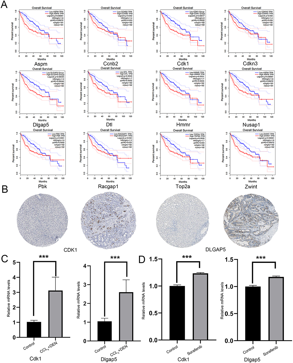 |
Figure 3 CDK1 and DLGAP5 is highly expressed and associated with poor survival. (A) Survival analyses of Aspm, Ccnb2, Cdk1, Cdkn3, Dlgap5, Dtl, Hmmr, Nusap1, Pbk, Racgap1, Top2a, Zwint in LICH from TCGA, Red: high-expression group; blue: low-expression group. p value was log-ranked. Auto-selected best cutoffs were used. (B) Immunohistochemical staining of CDK1 and DLGAP5 in normal liver tissue and HCC tissue. (C) Relative mRNA expression levels of CDK1 and DLGAP5 in control and HCC samples from CCl4+DEN treated mice. (D) Relative mRNA expression levels of CDK1 and DLGAP5 in hepG2 cells exposure to no sorafenib and 20% O2 with the presence of 6 µM sorafenib from GSE62813 datasets. ***P<0.001. |
Next, we validated the protein expression levels of CDK1 and DLGAP5 in HCC tissues using protein expression databases Human Protein Atlas. Our analysis revealed a significant upregulation of CDK1 and DLGAP5 expression in HCC tissues compared to normal liver tissues (Figure 3B). Additionally, CDK1 and DLGAP5 expressions were also significantly overexpressed in HCC mice model (Figure 3C). More importantly, these two genes were also significantly changed under the treatment of sorafenib, suggesting that they also play an important role in patients with HCC and sorafenib tolerance (Figure 3D). These findings suggested that CDK1 and DLGAP5 may play crucial roles in HCC progression and serve as potential prognostic indicators.
CDK1 and DLGAP5 are Influenced by Individual Cancer Types, Patient Characteristics, and TP53 Mutation Status
Subsequently, we conducted a correlation and expression pattern in different tumors analysis of these two genes, and the results suggest CDK1/DLGAP5 can be used as dual targets for treatment (Supporting Figure S2A and B). The above results indicated the expression of CDK1 and DLGAP5 may play a crucial role in the development and progression of HCC. Understanding the factors that influence their expression, such as individual cancer type, patient characteristics (age, weight, gender, race), and TP53 mutation status, can provide valuable insights into disease mechanisms and potential therapeutic targets. Expression of CDK1 and DLGAP5 in LICH based on individual cancer, patient’s age, patient’s weight, patient’s gender, patient’s race, and TP53 mutation status were conducted. The expression patterns of CDK1 and DLGAP5 in LICH are influenced by individual cancer types, patient characteristics, and TP53 mutation status (Figure 4A and B).
 |
Figure 4 CDK1 and DLGAP5 are influenced by individual cancer types, patient characteristics, and TP53 mutation status. (A and B) The relative expression of CDK1 and DLGAP5 in LIHC based on individual cancer type, age, weight, gender, race and TP53 mutation status from TCGA. |
Additionally, investigating the RNA expression of CDK1 and DLGAP5 at different periods and times during the cell cycle can shed light on their regulatory roles in cell division and proliferation. RNA expression of CDK1 and DLGAP5 in different periods of the cell cycle (G1, S, G2/M). Furthermore, their expression levels vary across different periods of the cell cycle (Supporting Figure S2C). These findings contribute to our understanding of HCC biology and may aid in the development of personalized treatment strategies targeting CDK1 and DLGAP5.
CDK1 and DLGAP5 is Associated with Tumor Immune Cell Infiltration
Next, we aim to find out the mechanisms by which CDK1 and DLGAP5 potentially regulate the occurrence of HCC. Understanding the interaction between tumor immune cells and key genes involved in cancer progression is crucial for improving cancer treatment strategies. To explore whether these two genes are related to tumor immunity in the development of HCC. In this study, we conducted a correlation analysis to explore the relationship between CDK1 and DLGAP5 in tumor immune cells.
We utilized the GSE166635 dataset, which provides single-cell transcriptomic data, our analysis revealed the distribution of CDK1 and DLGAP5 in different TME cell types at single-cell resolution. The cells with significant expression of the prognostic genes, indicated by the red box, suggest the presence of a potential correlation between CDK1 and DLGAP5 in specific tumor immune cell populations (Figure 5A). Conversely, the cells with trace expression of the prognostic genes, indicated by the green box, might represent cells where CDK1 and DLGAP5 are not significantly expressed. Specifically, we investigated the distribution of these prognostic genes across different types of tumor microenvironment (TME) cells, and found that CDK1 and DLGAP5 are associated with tumor immune cell infiltration (Figure 5B). Taken together, these data indicated that CDK1 and DLGAP5 may promote HCC through tumor immune cell infiltration.
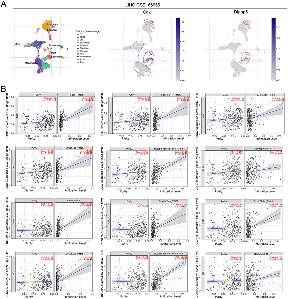 |
Figure 5 The expression of among CDK1 and DLGAP5 is associated with tumor immune cell infiltration. (A) The distribution of 2 prognostic genes in different TME cell types was explored with single-cell resolution in the GSE166635 dataset. The red box indicates the cells with significant expression of the prognostic genes, while the green box indicates the cells with trace expression of the prognostic genes. (B) Correlation analysis of CDK1 and DLGAP5 in tumor immune cells. |
Discussion
The identification of novel drug candidate target genes is paramount to advancing effective therapeutic strategies for hepatocellular carcinoma (HCC), an aggressive malignancy with limited treatment options.18 Firstly, we identified the gene expression profile in HCC. Additionally, of which, CDK1 and DLGAP5 were highly expressed and associated with poor prognosis. Lastly, the mechanism of CDK1 and DLGAP5 were found to be associated with tumor immune cell infiltration. These findings provide valuable insights into the development of novel therapeutic approaches for HCC (Figure 6).
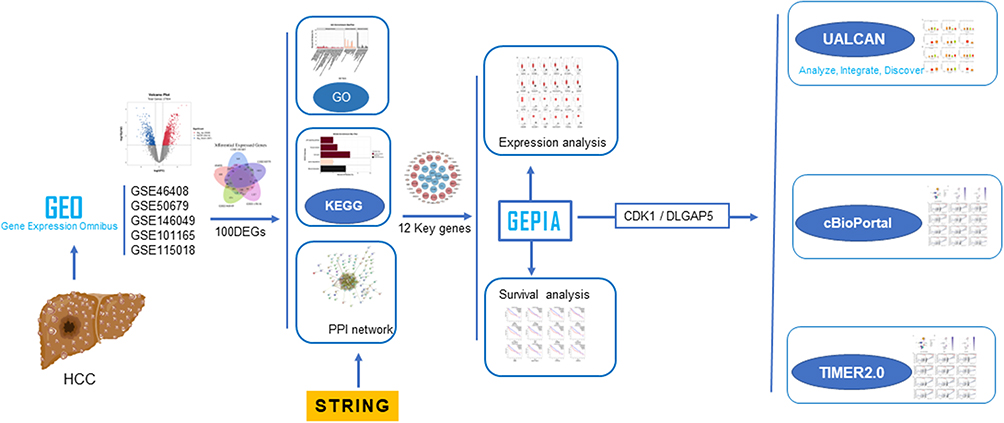 |
Figure 6 Key Modulators role of CDK1 and DLGAP5 in Hepatocellular Carcinoma. DLGAP5 and CDK1 were highly expressed in HCC and associated with poor prognosis. CDK1 and DLGAP5 are influenced by individual cancer types, patient characteristics, and TP53 mutation status. CDK1 and DLGAP5 is associated with tumor immune cell infiltration. |
HCC is characterized by its aggressive nature and limited treatment options, making it one of the leading causes of cancer-related deaths worldwide.19,20 Multi-omics bioinformatics analysis has revolutionized the landscape of cancer research by offering a comprehensive understanding of the molecular underpinnings of tumor diseases.21–23 To address this critical need, we employed bioinformatics analysis and identify CDK1 and DLGAP5 as potential targets.
Previous study indicated that CDK1 involved in cell proliferation,24 epigenetic regulation,25 gastrointestinal stromal tumor26 and mitochondrial metabolism.27 Our findings suggest that CDK1 and DLGAP5 are key genes that are highly expressed in HCC and are associated with poor patient prognosis. The association of these genes with tumor immune cell infiltration highlights their potential role in the tumor microenvironment, which has become an important focus in cancer research.28,29 This finding underscores the importance of targeting these genes not only for direct antitumor effects but also for modulating the immune response against HCC.
In conclusion, our study presents a novel strategy for identifying potential therapeutic targets for HCC. The integration of bioinformatics approaches offers a holistic view of the molecular landscape of HCC and paves the way for the development of targeted therapies. Future research should focus on validating these findings in experimental models and clinical trials to confirm their therapeutic potential.
Ethics Statement
Our organization supports the relevant provisions of the “Ethical Review Measures for Life Science and Medical Research Involving Human Beings” and the use of legally obtained public data can be exempted from ethical review.
The details contents are as follows:
Using human information data or biological samples to carry out life science and medical research involving humans in the following circumstances, which does not cause harm to the human body, does not involve sensitive personal information or commercial interests, can be exempted from ethical review to reduce unnecessary burdens on scientific researchers and promote the development of life science and medical research involving humans.
Consent for Publication
All authors agree to publish the final manuscript.
Acknowledgments
We would like to acknowledge the Gene Expression Omnibus (GEO) database and its contributors for providing valuable data resources for this study. This paper has been uploaded to ResearchGate as a preprint: https://www.researchgate.net/publication/378216585.
Author Contributions
All authors made a significant contribution to the work reported, whether that is in the conception, study design, execution, acquisition of data, analysis and interpretation, or in all these areas; took part in drafting, revising or critically reviewing the article; gave final approval of the version to be published; have agreed on the journal to which the article has been submitted; and agree to be accountable for all aspects of the work.
Funding
This research was supported by the Science and Technology Plan Project of Guizhou Province (QIAN KE HE JI CHU-ZK(2023)YI BAN556 and QIAN KE HE JI CHU-ZK(2023)YI BAN 229).
Disclosure
The authors declare no conflicts of interest.
References
1. Kulik L, El-Serag HB. Epidemiology and management of Hepatocellular Carcinoma. Gastroenterology. 2019;156(2):477–491.e1. doi:10.1053/j.gastro.2018.08.065
2. Yang JD, Hainaut P, Gores GJ, et al. A global view of hepatocellular carcinoma: trends, risk, prevention and management. Nat Rev Gastroenterol Hepatol. 2019;16(10):589–604. doi:10.1038/s41575-019-0186-y
3. Johnson P, Zhou Q, Dao DY, et al. Circulating biomarkers in the diagnosis and management of hepatocellular carcinoma. Nat Rev Gastroenterol Hepatol. 2022;19(10):670–681. doi:10.1038/s41575-022-00620-y
4. Caglar HO, Duzgun Z. Identification of upregulated genes in glioblastoma and glioblastoma cancer stem cells using bioinformatics analysis. Gene. 2023;848:146895. doi:10.1016/j.gene.2022.146895
5. Liu H, Dilger JP, Lin J. A pan-cancer-bioinformatic-based literature review of TRPM7 in cancers. Pharmacol Ther. 2022;240:108302. doi:10.1016/j.pharmthera.2022.108302
6. Hassan W, Zafar M, Duarte AE, Kamdem JP, da Rocha JB. Pharmacological research: a bibliometric analysis from 1989 to 2019. Pharmacol Res. 2021;169:105645. doi:10.1016/j.phrs.2021.105645
7. Foerster F, Gairing SJ, Müller L, et al. NAFLD-driven HCC: safety and efficacy of current and emerging treatment options. J Hepatol. 2022;76(2):446–457. doi:10.1016/j.jhep.2021.09.007
8. Toh MR, Wong EYT, Wong SH, et al. Global epidemiology and genetics of Hepatocellular Carcinoma. Gastroenterology. 2023;164(5):766–782. doi:10.1053/j.gastro.2023.01.033
9. Lu C, Rong D, Zhang B, et al. Current perspectives on the immunosuppressive tumor microenvironment in hepatocellular carcinoma: challenges and opportunities. Mol Cancer. 2019;18(1):130. doi:10.1186/s12943-019-1047-6
10. Granito A, Forgione A, Marinelli S, et al. Experience with regorafenib in the treatment of hepatocellular carcinoma. Therap Adv Gastroenterol. 2021;14:17562848211016959. doi:10.1177/17562848211016959
11. Stefanini B, Ielasi L, Chen R, et al. TKIs in combination with immunotherapy for hepatocellular carcinoma. Expert Rev Anticancer Ther. 2023;23(3):279–291. doi:10.1080/14737140.2023.2181162
12. Li L, Wang H. Heterogeneity of liver cancer and personalized therapy. Cancer Lett. 2016;379(2):191–197. doi:10.1016/j.canlet.2015.07.018
13. Williams HL, Dias Costa A, Zhang J, et al. Spatially resolved single-cell assessment of pancreatic cancer expression subtypes reveals co-expressor phenotypes and extensive intratumoral heterogeneity. Cancer Res. 2023;83(3):441–455. doi:10.1158/0008-5472.CAN-22-3050
14. Malumbres M, Barbacid M. Cell cycle, CDKs and cancer: a changing paradigm. Nat Rev Cancer. 2009;9(3):153–166. doi:10.1038/nrc2602
15. Xie B, Wang S, Jiang N, et al. Cyclin B1/CDK1-regulated mitochondrial bioenergetics in cell cycle progression and tumor resistance. Cancer Lett. 2019;443:56–66. doi:10.1016/j.canlet.2018.11.019
16. Chen R, Liu J, Hu J, et al. DLGAP5 knockdown inactivates the Wnt/β-catenin signal to repress endometrial cancer cell malignant activities. Environ Toxicol. 2023;38(3):685–693. doi:10.1002/tox.23720
17. Xu T, Dong M, Li H, et al. Elevated mRNA expression levels of DLGAP5 are associated with poor prognosis in breast cancer. Oncol Lett. 2020;19(6):4053–4065. doi:10.3892/ol.2020.11533
18. Vogel A, Meyer T, Sapisochin G, et al. Hepatocellular carcinoma. Lancet. 2022;400(10360):1345–1362. doi:10.1016/S0140-6736(22)01200-4
19. Chakraborty E, Sarkar D. Emerging therapies for Hepatocellular Carcinoma (HCC). Cancers. 2022;14(11):2798. doi:10.3390/cancers14112798
20. Zhao P, Malik S, Xing S. Epigenetic mechanisms involved in HCV-Induced Hepatocellular Carcinoma (HCC). Front Oncol. 2021;11:677926. doi:10.3389/fonc.2021.677926
21. He X, Liu X, Zuo F, et al. Artificial intelligence-based multi-omics analysis fuels cancer precision medicine. Semin Cancer Biol. 2023;88:187–200. doi:10.1016/j.semcancer.2022.12.009
22. Reel PS, Reel S, Pearson E, et al. Using machine learning approaches for multi-omics data analysis: a review. Biotechnol Adv. 2021;49:107739. doi:10.1016/j.biotechadv.2021.107739
23. Vandereyken K, Sifrim A, Thienpont B, et al. Methods and applications for single-cell and spatial multi-omics. Nat Rev Genet. 2023;24(8):494–515. doi:10.1038/s41576-023-00580-2
24. Haneke K, Schott J, Lindner D, et al. CDK1 couples proliferation with protein synthesis. J Cell Biol. 2020;219(3). doi:10.1083/jcb.201906147
25. Michowski W, Chick JM, Chu C, et al. Cdk1 controls global epigenetic landscape in embryonic stem cells. Mol Cell. 2020;78(3):459–476.e13. doi:10.1016/j.molcel.2020.03.010
26. Lu X, Pang Y, Cao H, et al. Integrated screens identify CDK1 as a therapeutic target in advanced gastrointestinal stromal tumors. Cancer Res. 2021;81(9):2481–2494. doi:10.1158/0008-5472.CAN-20-3580
27. Liu L, Xie B, Fan M, et al. Low-level saturated fatty acid palmitate benefits liver cells by boosting mitochondrial metabolism via CDK1-SIRT3-CPT2 cascade. Dev Cell. 2020;52(2):196–209.e9. doi:10.1016/j.devcel.2019.11.012
28. Gajewski TF, Schreiber H, Fu Y-X. Innate and adaptive immune cells in the tumor microenvironment. Nat Immunol. 2013;14(10):1014–1022. doi:10.1038/ni.2703
29. Mao X, Xu J, Wang W, et al. Crosstalk between cancer-associated fibroblasts and immune cells in the tumor microenvironment: new findings and future perspectives. Mol Cancer. 2021;20(1):131. doi:10.1186/s12943-021-01428-1
 © 2024 The Author(s). This work is published and licensed by Dove Medical Press Limited. The
full terms of this license are available at https://www.dovepress.com/terms.php
and incorporate the Creative Commons Attribution
- Non Commercial (unported, 3.0) License.
By accessing the work you hereby accept the Terms. Non-commercial uses of the work are permitted
without any further permission from Dove Medical Press Limited, provided the work is properly
attributed. For permission for commercial use of this work, please see paragraphs 4.2 and 5 of our Terms.
© 2024 The Author(s). This work is published and licensed by Dove Medical Press Limited. The
full terms of this license are available at https://www.dovepress.com/terms.php
and incorporate the Creative Commons Attribution
- Non Commercial (unported, 3.0) License.
By accessing the work you hereby accept the Terms. Non-commercial uses of the work are permitted
without any further permission from Dove Medical Press Limited, provided the work is properly
attributed. For permission for commercial use of this work, please see paragraphs 4.2 and 5 of our Terms.