")
Back to Journals » International Journal of Nanomedicine » Volume 20
Carbon Dot-Based Nanoparticles: A Promising Therapeutic Approach for Glioblastoma
Authors Wang Y, Wu H, Guo Y , Li F, Zhang H
Received 7 February 2025
Accepted for publication 7 May 2025
Published 31 May 2025 Volume 2025:20 Pages 7061—7092
DOI https://doi.org/10.2147/IJN.S519733
Checked for plagiarism Yes
Review by Single anonymous peer review
Peer reviewer comments 2
Editor who approved publication: Dr Kamakhya Misra
Yongzhi Wang,1,2,* Hao Wu,3,* Yu Guo,4 Fangbao Li,4 Hengzhu Zhang4
1Department of Neurosurgery, Fu Yang People’s Hospital, FuYang, People’s Republic of China; 2Department of Neurosurgery,The Second Affiliated Hospital of Soochow University, Suzhou, People’s Republic of China; 3Department of Neurosurgery, The Second Affiliated Hospital of Xi’an Medical University, Xi’an, People’s Republic of China; 4Department of Neurosurgery, Northern Jiangsu People’s Hospital Affiliated to Yangzhou University, Yangzhou, People’s Republic of China
*These authors contributed equally to this work
Correspondence: Hengzhu Zhang, Department of Neurosurgery, Northern Jiangsu People’s Hospital Affiliated to Yangzhou University, No. 98 Nantong West Road, Yangzhou City, Jiangsu Province, People’s Republic of China, Email [email protected] Yongzhi Wang, Department of Neurosurgery, Fu Yang People’s Hospital, No. 501 Sanqing Road, Yingzhou District, Fuyang City, Anhui Province, People’s Republic of China, Email [email protected]
Abstract: Glioblastoma (GBM) is a malignant tumor that currently still faces challenges for a complete cure. Although GBM treatment has made great progress, the prognosis of patients is still poor due to interference of various factors in treatment such as the blood-brain barrier (BBB), grade malignancy, intra- and intertumor heterogeneity, drug resistance, and poor targeting of anti-tumor drugs. In recent years, with marked advances in nanotechnology, different types of nanodrug delivery systems have been developed and have been considered as a promising therapeutic measure to gradually overcome chemotherapy resistance and improve tumor targeting. Carbon dots (CDs), as a new type of therapeutic NP, have become a research hotspot of concern for many researchers in recent years. NPs based on CDs have high modifiability and functionalization, allowing for covalent binding with chemotherapy drugs, genes, immune cells and photosensitizers, effectively targeting tumor cells and reducing peripheral cytotoxicity. However, at present, CDs are still in the basic research stage or the preclinical exploratory research stage, and has not yet entered the clinical trial stage or the implementation and application stage. Here, we review the fundamental principles of CDs in the broader field of nanotechnology, their development history, classification, synthesis, and potential for tumor treatment. Especially in the treatment of cancer, CDs can not only participate in photodynamic therapy, photothermal therapy, sonodynamic therapy, chemodynamic therapy, and chemotherapy, but also in multi-modal combination therapy. Here, we hope to provide some insights for further research.
Keywords: glioblastoma, cancer treatment, carbon dots, therapeutic approach
Introduction
GBM is the primary brain tumor with the highest degree of malignancy. GBM is not only difficult to treat but also has a poor prognosis and a higher recurrence rate, accounting for approximately 70% of malignant brain tumors.1,2 Based on the latest statistics from the World Health Organization, the median overall survival (OS) of patients with GBM is 12 to 16 months, and the 5-year OS rate is only approximately 5.6%.3 Current treatment for GBM usually relies on standard combination therapy, including surgical resection, postoperative radiotherapy, and chemotherapy.4 However, even with combination therapy, the median postoperative OS is only 10 to 16 months,5 as surgery cannot completely eradicate tumor cells, and GBM cells have a strong ability for invasion, migration, and infiltration, leading to recurrence of GBM.6 Therefore, in addition to traditional treatment methods, it is extremely urgent to explore several feasible alternative treatment strategies to effectively overcome the above issues, to improve postoperative quality of life, and prolong patient survival time. In recent years, with the rapid advancement of nanotechnology, nano drug delivery systems (NDDSs) have been extensively researched, repeatedly verified and ultimately proposed for application in the treatment of GBM in different species.7
In recent years, carbon dots (CDs) have been favored by researchers due to their low-cost synthesis and raw materials, good solubility and stability, excellent biocompatibility, low toxicity and degradability. Nowadays, they have been widely applied in various fields, such as drug sensing, biomarker detection and cell biological imaging8–13 Today, as a novel treatment method, CDs based nanoparticles (NPs) offer a feasible direction and approach for anti-tumor therapy.14–18 In this review, we summarize the development, characteristics, and anti-tumor mechanism of CDs NDDS proposed for the treatment of GBM.
Research and Development History of Carbon Dots
Looking back on the development history of CDs, they have gone through a long process. With the advancement of technology and the efforts of generations of scientists, they have gradually evolved from fluorescent carbon dots to new nano-drug delivery systems based on carbon dots with multiple functions and application characteristics. CDs were first surprisingly discovered in 2004. It is a new type of coupled material with a diameter of only approximately 10 nm. With the technological advancements in functionalization and modifiers, CDs now have the advantages and potential to be developed into nanomaterial carriers with anti-tumor activity.18 The development of CD has undergone three stages, namely, a discovery stage; an initial development stage; and a rapid development stage. The specific development process is as follows: Isolated fluorescent carbon in 2004; Surface passivation CDs in 2006; Fluorescent CQDs in 2010; Fluorescent GQDs in 2012; Fluorescent CPDs in 2013; Fluorescent β-C3N4 Nanocrystals, Fluorescent chiral CDs, and Fluorescent C3N QDs in 2017; Fluorescent triangle CDs in 2018. Then CDs entered a rapid stage of development, and go deep into various fields (Figure 1).15,19,20
 |
Figure 1 Research and Development History of CDs. |
 |
Figure 2 Classification of CDs. |
Classification of Carbon Dots
Due to the multiple functions and roles of CDs, interestingly, they possess diverse appellations, such as carbon nanoparticles (CNPs), carbon dots (CDs), and carbon nanodots (CNDs). Multitudinous carbon-based nanomaterials have been delved and applied, such as carbon nanotubes (CNTs),15,20 fullerenes,21 graphene,22 graphene quantum dots (GQDs),23 CDs,24 carbon nanocorners (CNHs),25 carbon nanoonions (CNOs)26 and so on. Among these, CDs have a huge potential development trend due to their unique biological properties.18,27–34 In particular, CDs are more advantageous in medical applications.35,36 The classification of CDs is shown in Figure 2.37 Table 1 offers a comparison of CDs and other carbon-based nanomaterials.
 |
Table 1 Comparison of CDs and Other Carbon-Based Nanomaterials |
Design of Carbon Dots
Due to the issue of the emission wavelength of CDs itself, its penetration rate in biological tissues is relatively poor, which thoroughly impedes their application domains. Because of the objective existence of the above-mentioned issues, multifarious endeavors have been carry out to enhance and improve CDs functionality, for instance, methods such as doping with heteroatoms and surface passivation of materials and so on.13 Most researches indicated that hetero-atom doping could be utilized to regulate and control the inherent traits of CDs. In addition, CDs are usually spherical in shape and have abundant functional groups on their surface, such as -OH, -COOH and -NH2 (amine) moieties, which equip CDs with good bio-compatibility and good water solubility, and the ability to form new conjugations with different inorganic and organic substances.45,48 Two types of CDs have been developed namely amorphous C-dots and G-Q-dots. The amorphous C dots are formed by simultaneous sp2 and sp3 hybridization of the carbon probes, which have the ability to undergo further surface modification. The G-Q dots possess hybrid sp2 nanocrystalline carbon nuclei, an asymmetric structure due to abundant N and O groups that perturb the carbon structure (Figure 3).49
 |
Figure 3 Design of CDs with different surface functional groups. |
Emerging Applications of Carbon Dots
In recent decades, research regarding the synthesis, characteristics and biological applications of CDs possess grown in a spiral. In terms of size and photoluminescence performance, CDs have a high degree of similarity to quantum dots (QDs). However, compared to QDs, CDs are relatively innocuous.50 CDs achieve relatively uniform dispersion, better stability, and biocompatibility.51 In addition, the fluorescence characteristics and plentiful surface functional groups of CDs allows them to be relatively easily modified or further functionalized.52 Combined with relatively better biocompatibility and excellent stability, CDs have been involved in many research fields and have received much attention, for example, in optical, energy, and biological applications, especially for the treatment and diagnosis of cancer, such as analysis of pharmaceuticals and biomarkers, cellular bio-imaging (in-vitro and in-vivo bio-imaging),13 medical imageology, nanomedicine, drug/gene delivery, gene-targeted combined therapy, PDT, PTT, and as antibacterial agents53–56 (Figure 4)
 |
Figure 4 Emerging applications of CDs. |
CDs Synthesis
Under normal conditions, the synthesis of CDs requires multifarious chemical substances and biological groups, such as –COOH (carboxyl groups). Because of the existence of carboxyl group, CDs have good solubility. The presence of functional groups gives CDs features that can be further modified or reassembled. Two common synthetic methods for the preparation of CDs8 are illustrated in Figure 5. That are top-down and bottom-up means. The first technique mainly include laser ablation, arc discharge and high energy ball milling. But above ways utilize strong chemical reagents and tedious conditions for synthesis.42
 |
Figure 5 Two synthetic methods “top-down” and “bottom-up” are common for the preparation of CDs. |
The second technique primarily contains hydrothermal/solvothermal synthesis, pyrolysis and electrochemical methods. However, the most crucial deficiencies of the bottom-up synthesis technique is its relative complexity, as special experimental instruments are required during the synthesis process and it takes a relatively long time.57 The formation of amide bonds is generally accomplished by covalent modification between the CD carboxyl group and the primary amine by EDC/NGS coupling chemistry (Figure 6).57–59
 |
Figure 6 Simplified scheme depicting the conjugation of CDs to a chemotherapeutic agent. |
CDs-Relevant Toxicity and Remedial Methods
Before the application of CDs, its toxic effects in organisms must be considered and verified. Table 2 summarizes the toxicity verification of CDs in vitro. Studies have confirmed that PEG-modified CDs can significantly reduce their own toxic effects and thus can be used in the fields of biological imaging and medical exploration. Although in vitro studies have shown that CDs is non-toxic or low-toxic, we still cannot be blindly optimistic. Because, up to now, there are still relatively few experiments on in vivo toxicity researches (Table 3), lacking reliable data to support and verify the toxic effects of CDs. However, we firmly believe that with technological innovation and the improvement of functionalization, more experiments will focus on toxicity verification in organisms.
 |
Table 2 In Vitro Toxicity Researches of CDs |
 |
Table 3 In Vivo Toxicity Researches of CDs |
How to Enforcing Remedies for Weakening Toxicity
It is well known that high molecular polymers such as polyacrylic acid (PAA) and branched-chain polyvinylimide (BPEI) themselves have certain toxic effects on living organisms. For instance, the exposure time of free PAA to CDs is at least about 10 hours or more, or even longer. Due to the inevitable toxic effect, it will cause a certain degree of damage to cells. But how to reduce this toxic and side effect? This is a fatal problem. Experimental research has proved that the remedial measures for toxicity mainly involve shortening the cell incubation time and exposure time, using the minimum concentration of drugs or materials, and innovating technologies, including the modification of physical and chemical properties and surface functionalization techniques. Although the toxicity of CDs is relatively low, it can still be further reduced through the intervention of the techniques mentioned above. At the same time, it can also be fully modified by using polymers PEG (polyethylene glycol) with good bio-compatibility and amino acids, etc.
Carbon Dots Based Nanoparticles for Photothermal Therapy of Tumors
In recent years, PTT has been widely applied in the research of cancer treatment. Its principle is to convert light energy into heat energy to deal a devastating blow to tumors. However, a crucial factor remains to be determined, that is, how to ingeniously design synergistic NPs as potential photothermal transducers that can truly and effectually improve the efficacy of PTT in killing tumor cells. On this basis, some researchers have strategically designed, prepared, and characterized hollow Cu NPs (CuSCDs). These NPs displayed excellent photothermal conversion efficiency, enhanced biocompatibility, and reduced toxicity under 808 nm laser irradiation. After coating a CuSCD-loaded proteasome inhibitor with the hybridized macrophage cell membrane of the T7 peptide, CuSCDB@MMT7 achieved specific targeting of cancer cells, immune escape, and boosted endocytosis. In general, CuSCDB@MMT7-triggered PTT displayed the accumulation of polyubiquitinized tumor suppressor protein, which was thermally steadied at a high temperature induced by near-infrared light, enhancing apoptosis and decreasing tumor cell metastasis. Through a series of characterization monitoring, the results indicated that CuSCDB@MMT7 possessed high stability, with relatively considerable loading and encapsulation efficiency of 20.5% and 81.9%, respectively. Meanwhile, the monitoring manifested that under laser irradiation, the temperature of CuSCD can rapidly accomplish above 90°C without obvious attenuation, and the heat generation capacity can be maintained at a certain steady-state level. More importantly, the research suggested that CuSCDB@MMT7 can specifically target tumor cells and has the ability to evade immunity. Laboratory monitoring demonstrated no obvious signs of damage to the liver and kidneys, which provided strong evidence for clinical transformation. It is gratifying that CuSCDB@MMT7 can accumulate effectively at tumor sites and manifest an increasing trend over time, further confirming that CuSCDB@MMT7 can accumulate at tumor sites via EPR and active targeting.
The above studies provide strong evidence for the PTT strategy of Cu/CDs nanocomposites loaded with proteasome inhibitors and highlight a promising therapeutic strategy that can achieve effective clinical cancer treatment (Figure 7). 66
 |
Figure 7 (A) Schematic illustration of the generation of proteasome inhibitor-encapsulated CuS/CDs nanocomposites (CuSCDB@MMT7). (B) Schematic illustration of the application of CuSCDB@MMT7 for enhanced PTT via heat-stabilization of various substrates in the ubiquitin-dependent proteasomal degradation pathway. |
In summary, the evidence to date strongly supports nanodrug delivery system designed based on CDs has a very good prospect for the destruction of tumor cells.
Therapy and the Principle of Carbon Dots in Photodynamic Therapy
The characteristics of PDT are determined by three key elements: light source, PS, and O2. The principle of the reaction is that when the light source is irradiated, PS absorbs energy from the ground phase to the excited phase and reacts to further produce reactive oxygen species (ROS), which in turn destroy cancer cells.67 When PS receives a certain amount of light and absorbs a certain amount of energy, it changes directly or indirectly from the ground state to the excited state, and then two types of reactions can occur. The first reaction, the Type I reaction, involves the generation of hydrogen peroxide, superoxide anion, and hydroxyl radical.68 The second reaction, type II, differs from the first by producing 1O2 directly through an “intersystem crossing” process (Figure 8).69 When the two types of reactions occur simultaneously, the resulting H2O2, O2−, •OH, and 1O2 are defined as ROS.69–73 The Type III reaction mechanism involves a direct and efficient destruction of biological target molecules.74 First, when CDs are activated by a light source, the absorbed electron energy is excited from S0 (ground state) to S1 (singlet state), and second, the energy is released through three routes and returned to a relatively lower energy level (Figure 9). The first route is nonradiative emission: that is, return from S1 to S0 as heat. The second route is radiative emission that involves phosphorescence and fluorescence, radiative processes in which energy relaxation occurs and in which energy is released in the form of electromagnetic radiation light. The third route is to transition from S1 to the excited state T1 via an intersystem crossing (ISC). The process in the third route is related to the ability of T1 to initiate a radical chain reaction (type I) after transferring electrons and O2 to the reaction substrate and further forming ROS such as H2O2, O2− and •OH, or it may acquire 1O2 by transferring energy to O2 and extracting electrons from some groups (such as aromatic rings and phenols) to produce a ground state via CDs (Type II). The three key free radicals (H2O2, O2−,•OH, and 1O2) generated by the two approaches in the third route above are the significant factors for CDs to implement the PDT effect.75–78 The 1O2 generated by type II reaction is generally considered the most important factor responsible for the efficacy of PDT.79 However, hypoxia that can occur in malignant neoplasms, such as in GBM, may reduce the antitumor effects of type II PDT. Hence, type II PDT is more suitable for tumor environments with O2.80,81 Although type I PDT is relatively better suited to anoxic environments, as it generates lower cytotoxicity due to O2− and H2O2.82,83 Hypericum perforatum has been applied to overcome these issues and achieve the control of Type I and Type II PDT. Researchers synthesized three red carbon dots (RCDs), which possess similar core structures and diverse surface states, resulting in various ΔES1-T1. However, a smallerΔES1-T1 is more favorable for energy transfer from S1 to T1, resulting in more 1O2 production. Moreover, since the three RCDS have the same REDOX potential, the same O2 is produced (Figure 10).84
 |
Figure 8 Schematic illustration of photodynamic reactions (either type I or type II) and cell death pathways in the process of PDT. |
 |
Figure 9 Description of mechanisms of CDs in PDT. |
 |
Figure 10 Schematic of RCDs as nano-PSs with tunable ROS generation. |
There are three key mechanisms by which PDT eliminates tumor cells: the first is to promote apoptosis and necrosis of tumor cells by inducing oxidative stress; the second is to cause vascular injury, and the final mechanism is to trigger an autoimmune and inflammatory response.85 In general, the key active treatment of PDT is to promote tumor cell apoptosis.86 To further evaluate the mechanism by which sulfur-doped carbon dots (S-CDs) conquer tumor cells, researchers took advantage of a Fluo-4 AM (Ca2+ probe) to calculate the effective concentration of Ca2+ entering tumor cells. The researchers found that under laser irradiation, the green fluorescence of the S-CDs treatment group was more significant than that of the 5-ALA group, indicating that the 1O2 generated by the S-CDs was traced in lysosomes and mitochondria, mediating the release of Ca2+ from internal storage into the cytoplasm, thereby triggering apoptosis.87
Pang and other researchers have developed a CD with bidirectional capabilities, that is, it not only has the feature of targeting but also the feature of ROS generation, which is more effective in the treatment of tumors and further improves the efficacy of PDT.88 Some researchers have also enhanced the PDT effect of CDs by functionalizing them to improve targeting. The new type of CDs developed by Jia et al can overcome hypoxia and greatly enhance the therapeutic effect of PDT.89 Similar studies also suggest, for instance, that the multifunctional CD nanocomposites designed by Zheng et al can significantly reduce the limitations of hypoxia on PDT in malignant tumors.90 In their research work, it was confirmed through cell viability experiments that functionalized CDs can eliminate the PDT resistance induced by hypoxia, and has good growth inhibition effect on tumor sites at 1% O2 concentration, showing superior ability to overcome cancer hypoxia.
Carbon Dots Based Nanoparticles for Photodynamic Therapy of Tumors
PDT is an activation therapy for antitumor activity. The rise of NDDS has made a tremendous advance in the domain of PDT.91–93 CDs are an ideal and novel nanomaterial, considered a perfect candidate for photosensitizer delivery systems because of their various excellent features. Researchers have developed a novel self-luminescent PDT system, which aims to improve the total PDT yield with efficient absorption by optimizing the CRET step between light and photosensitization. When coupled with yellow light-emitting CDs (y-CDs), the performance of the photosensitizer is extremely enhanced, and its Soret band and Q band can be motivated directly or indirectly by CL (Figure 11), and has potential to be implemented in tumor therapy. The study showed the potential treatment effect on cancer cells, by providing a feasible method to heighten photons while reducing the toxicity of CL. This CDs-based PDT has shown superiority in overcoming the deficiency of penetrating radiation and curing deep lesions that are difficult to solve with traditional PDT.94
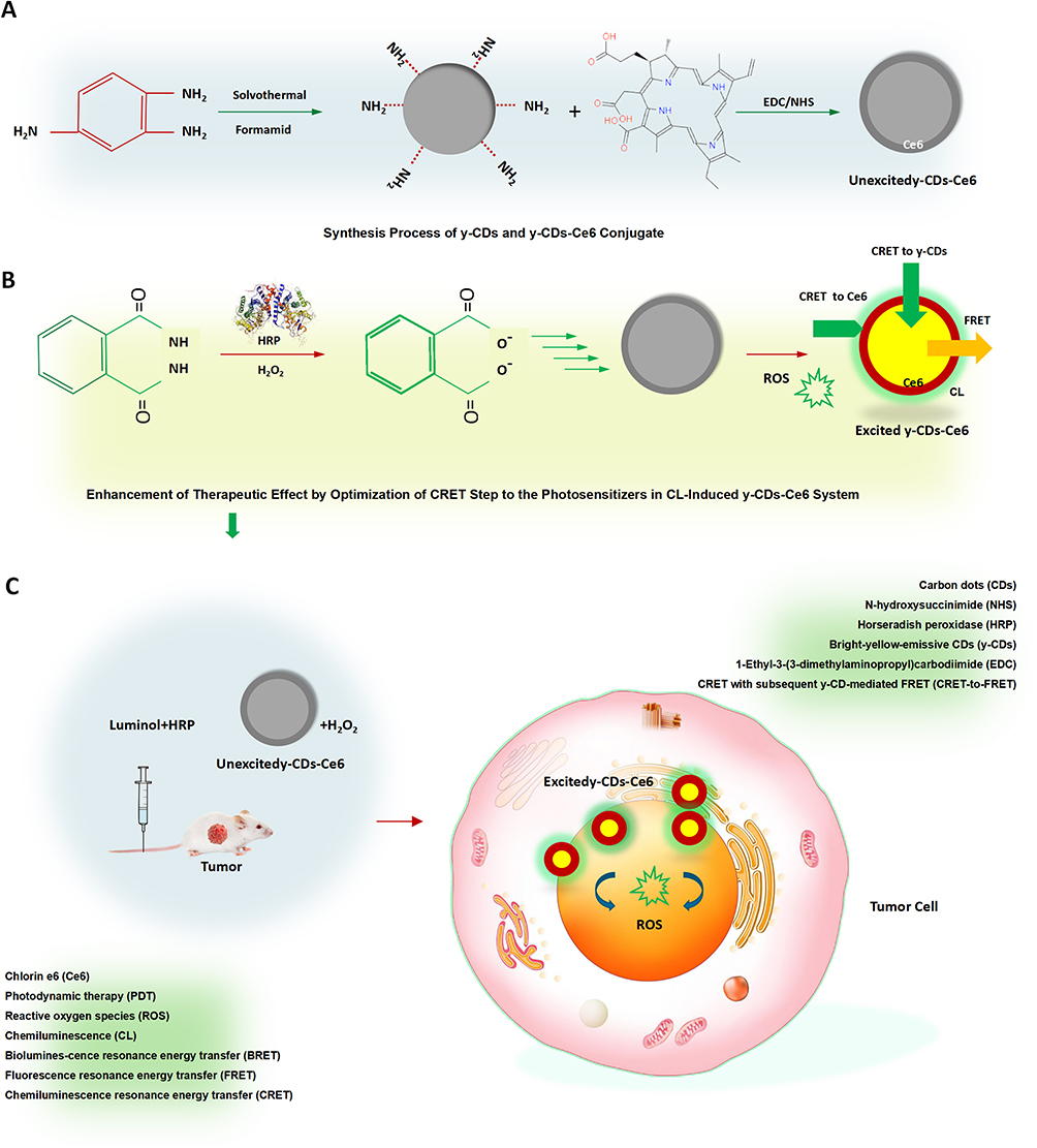 |
Figure 11 Schematic of the PDT System in vivo. (A) Synthesis process of y-CDs and y-CDs-Ce6 conjugate. (B) Enhancement of therapeutic effect by optimization of CRET step to the photosensitizers in CL-induced y-CDs-Ce6 system. (C) The mechanism of PDT. |
Overall, CDs-based NDDS has broad prospects for application in PDT therapy and has the potential for targeted GBM therapy. We hope to conduct further clinical studies in the future to benefit more patients with GBM.
Photodynamic Therapy and Photothermal Therapy Combination Therapy
Single treatment often has certain limitations on the killing of tumors and often requires combined treatment to further increase the efficacy of treatment. Because CDs have excellent PTT and PDT characteristics, PTT and PDT treatment in a single NDDS can afford a relatively better treatment effect against tumors than a either PTT or PDT alone. In general, PS acts as PDT agents in the tumor microenvironment (TME) and can induce generation of ROS under a certain amount of light, to achieve effective therapeutic purposes. However, to date, the ROS generation efficiency of PS from CD has been significantly restricted because of its poor solvability, light instability, and other factors. Accordingly, combination treatment of PDT with PTT may offer relatively excellent and unique antitumor activity. Due to increased tissue oxygen consumption and rupture of blood flow in tumor neovascularization, local anoxia of the tumor environment further interferes with and limits the curative effects of PDT. Therefore, combined therapy can overcome the problem of hypoxia and improve anticancer activity. In contrast, combination treatment could also address PS deficiencies based only on PDT or PTT. To date, researchers have continuously attempted to exploit light-triggered association treatments. For example, Ge et al developed CDs with both PTT and PDT traits using polythiophene benzoic acid as a carbon source (Figure 12A).95 In addition, neoteric nanomaterials have been designed to strengthen tumor therapy through the combined action of PDT and PTT by studying the uptake of polymer-polygamma-glutamate (gamma-gamma-glutamate) (gamma-PGA) by integrating gamma-glutamyltransferase (GGT) enzymes. γ-PGA@GOx@Mn, a Cu-CD preparation comprise a photosensitizer and self-supplying oxygen nanoparticle obtained by glucose metabolism reacting glucose oxidase (GOx), Mn, Cu-CDs. γ-PGA@GOx@Mn, Cu-CDs NPs remain in a slightly acidic environment of the tumor for a prolonged time and can further target cancer cells. Under 730 nm laser irradiation, NPs also achieve excellent photothermal and photodynamic effects and could significantly alleviate tumor hypoxia through endogenous formation of hydrogen peroxide (H2O2), further strengthening the curative effect. The combination treatment of PDT and PTT is effective and deserves further study to achieve tumor cure (Figure 12B and C).96
 |
Figure 12 (A) Synthetic route of PBA and CDs with simultaneous PTT and PDT capability by polymerization to carbonization. (B) Schematic illustration of starving and phototherapy mediated by γ-PGA@GOx@Mn, Cu-CDs NPs. (C) Schematic illustration of starving-like therapy, phototherapy, and immunotherapy mediated by γ-PGA@GOx@Mn, Cu-CDs NPs. |
Carbon Dots Based Drug/Gene Delivery
Chemotherapy (CT) is the most routine treatment for all kinds of malignant tumors. However, many factors, such as the drug resistance mechanism of the tumor, poor targeting of the drug and the tumor barrier, seriously limit their therapeutic effects in vivo. Thus, various NDDS have been developed to achieve enhanced targeted therapy against tumors and reduced drug resistance. In this field, CDs are also used as nanocarriers to encapsulate drugs, proteins, or cellular transmembrane peptides that target the TME through their unique surface structure to trigger targeted-release capabilities for precise drug delivery. In addition to better optical imaging and efficient drug encapsulation rates, these CD nanocarriers have shown excellent biocompatibility, safety, and low or no toxicity. The maximum drug loading of CDs is close to 96%, and there is no obvious inhibition of cell growth and inflammatory reaction. For example, Feng et al developed CDs-Pt(IV)@PEG-(PAH/DMMA), a drug nanocellular carrier with tumor extracellular microenvironment responsiveness based on preloading of the CT drug cisplatin (IV), as a bioimage-guided drug delivery system (Figure 13). This research offers a viable alternative to enhance the potential clinical application of CDs in antineoplastic protocols.97
 |
Figure 13 Schematic illustration for the drug delivery process of CDs-Pt(IV)@PEG-(PAH/DMMA). (A) Synthetic process of CDs-Pt(IV)@PEG-(PAH/DMMA). (B) The Drug release mechanism CDs-Pt(IV)@PEG-(PAH/DMMA). |
Gene therapy is a viable potential strategy for the cure of multiple tumors but demands a secure and effective vector for delivery. The unique properties of CDs make them an efficient novel gene carrier. Liu and his research team successfully developed a high-efficient dual functional nano gene vector based on polyethylenimine-passivated CDs as transfection agents via one-step strategy technique. Their research results indicated distinct fluorescence and better solubility with low cytotoxicity (The cell survival rate is approximately over 80%) and efficient in vitro DNA transfection).98
Kim et al contrasted the mechanism of PEI-functionalized C-dots (PEI-CDs), PEI-functionalized gold colloids (PEI-AU), and plasmid DNA (pDNA) in gene delivery through DNA transfection, and the results manifested that PEI-CDs and PEI-AU achieved poor fluorescence efficiency during transfection (The transfection efficiency was almost PEI25k close to approximately 1.0×10⁷ RLU/mg protein). After the PEI-C-dots were separated from the PEI-AU, it achieved a higher concentration of pDNA and high fluorescence, which promotes gene transfer and transport.99 Recently, Zhang et al have also exploited hyaluronic acid (HA)/PEI modified C-dots (HP-CDs) for tumor targeting and gene therapy. HP-CDs are easily implanted into cancer cells by endocytosis. The results suggest that HP-CDs are a potential vector for gene delivery.100
To sum up, CDs possess prominent development superiority in terms of transmission, especially for drug/gene delivery and deserve to conduct in-depth research and delve into anti-tumor applications.
CDs-Mediated Activation of the mTOR Signaling Pathway
Reduced glutathione (GSH) is a strong antioxidant, and ROS can be eliminated by it. When cells are damaged and in an oxidative stress state, GSH can afford the speedy metabolic response in the first instance.101,102 In view of the excellent bio-safety and compatibility of CDs, CD-based NPs have found prominent application in the monitoring and cure of malignant tumors. However, at present, the effects of CDs on tumor proliferation and cell metabolism are not fully understood, especially with regard to the regulation of tumor signaling pathways. The fundamental researches have manifested that CDs can increase ROS levels in UM cells in a dose-dependent manner. When the CDs concentration was <100 µg/mL, CDS-induced ROS could further increase the proliferation of UM cells and enhance cell aggressiveness. At 200 µg/mL, UM cells underwent further apoptosis. The addition of antioxidants reversed the proteinogenic effect of CDs. A concentration of 25–100 µg/mL CDs stimulate Akt/mTOR signaling, promote glutamine metabolism, and generate a cascade that accelerates UM cell proliferation. These data suggest that moderate subapoptotic doses of CDs facilitate tumorigenicity in UM cells. This feasibility study provides strong support for the combined effectiveness of ROS NPs in future cancer therapy (Figure 14).103
 |
Figure 14 Schematic of the opposing CDs-concentration-dependent effects on tumor cell progression and metastasis. |
The results of the above studies provide a very useful strategy for exploring CD-mediated signaling pathways and provide new insight and reference for further treatment of tumors.
Tumor Microenvironment-Responsive Imaging and Synergistic Tumor Treatment by CDs-Based NPs
The TME is characterized by angiogenesis, acidosis, and hypoxia, and provides a suitable environment for the growth and erosion of tumor cells, plays a crucial role in tumor deterioration, aggressive metastasis, and drug resistance, and thus, offers a breakthrough for exploring new anticancer strategies.104,105
The unique properties of the TME can provide a therapeutic target available for tumor cells.106 For purpose of achieving precise the cure of tumors, the use of smart nanomaterials with therapeutic capabilities responsive to TME stimulation is currently one of the most effective methods, with visible strengths in increasing therapeutic effect and lessening side effects.107 Although a more substantial study has been conducted on the design and development of TME-triggered nanotherapy platforms, it is still highly desirable to develop an easy way to manufacture such nanotherapy-borne systems with satisfactory results.108 Some researchers have modified CDs (CDs-Ce6) and Cu2+ by assembling a photosensitizer (chlorine e6, Ce6) to prepare TME stimulus-responsive NDDS, which can not only be used for fluorescence imaging, but also can be applied for collaborative tumor treatment. Due to the assembly of CDs-Ce6, the obtained nanocomponents (called Cu/CC, NPs) reveal quenched FL and photosensitivity. FL imaging and PDT function are effectively restored after penetration of the tumor under TME stimulation. The introduction of Cu2+ not only provides an attached CDT impact by reacting with H2O2 but also eliminates GSH in tumors through REDOX reactions; therefore, oxidative stress can be improved and the effect of ROS treatment can be enhanced. Cu/CC NPs can be used as a triad of FL-image-guided anti-tumor properties by PTT, PDT, and CDT (Figure 15).109
 |
Figure 15 Schematic of (A) the synthesis process of Cu/CC nanoassemblies and (B) their features for enhancing tumor accumulation, TME stimuli-responses and synergistic therapy. |
In summary, using CDs, we can design a nanoplatform with remarkable characteristics of TME stimulating response fluorescence imaging and PTT, PDT, and heat-enhanced CDT synergistic therapy, which can improve the therapeutic efficiency related to ROS and take advantage of the virtues of TME to effectively heighten curative effects and reduction of toxic and side effects.
CDs Penetrate the BBB
One of the largest impediments to the efficient remedy of gliomas is that most drugs cannot smoothly cross the blood-brain barrier (BBB). The unique microvascular system is a natural block that strictly limits the flow of molecules into and out of brain tissue. Although, tight connections between endothelial cells allow certain substances to cross the BBB. According to statistics, approximately 98% of small molecules and almost 100% of macromolecules cannot easily pass through the BBB, which strictly limits the effective concentration of glioma treatment drugs, such as temozolomide (TMZ).110 Nevertheless, through the ingenious encapsulation or modification of NDDS, researchers have conducted experiments to verify that both surface-modified or functionalized CDs and naked CDs can traverse the BBB. Adopting a zebrafish experiment, Li et al and Vallejo et al believed that CDs synthesized with carbon powder could not traverse the BBB by traditional synthesis methods, such as top-down methods. However, when modified with the iron transport protein, these CDs easily crossed the BBB.19,111
The presence of highly expressed transferrin receptors (TfRs) in brain endothelial cells makes them ideal targets for transferrin-binding CDs and other NDDSs to cross the BBB through receptor-mediated endocytosis. Although TfRs are generally used to promote BBB traversing, other receptors present in the BBB can also be used for delivery. These include angioendotheliin-2, which can penetrate the BBB and can serve to transport cargo to brain tissue or cells. Liu et al also confirmed that endothelin-2-modified CDs can fully penetrate the BBB of mouse C6 gliomas.112 Similarly, CDs can also be synthesized using the glucose transporter GLUT1 or the L-amino acid transporter 1 (LAT1), which are expressed at high levels throughout the BBB. Kirbas Cilinger et al proposed that fluorescein-coupled CDs prepared by glucose easily cross the BBB.113
Initially, CDs could be uniformly dispersed and permeated in all cells. Research has found that CDs can not only be taken up and accumulated in normal cells, but also be taken up and accumulated in malignant tumor cells, which will further confirm that CDs have good dispersibility and penetrability.114 However, the enhanced permeability and retention (EPR) effect has led miscellaneous researches to observe characteristic of the distinctive accumulation of CDs in cancer cells. The research of Su et al manifested that CDs firstly accumulated at the tumor microenvironment and were efficiently eliminated by circulatory system or metabolic system (for example, the kidney organ).115 Their research found that Hf-CDs have advantages such as the ability to target tumor accumulation preferentially. Meanwhile, it was also found that the reason why Hf-CDs can achieve rapid imaging is that CDs are highly likely to accumulate at the tumor site. To take advantage of the targeted imaging of CDs, researchers improved the cell uptake function enhanced by magnetic field to promote unique accumulation. Their research indicates that CDs NPs can target tumors by EPR.116. In conclusion, the effect regulation of BBB penetrability, EPR effect, endocytosis and excellent penetration power jointly contribute to the high targeting ability of CDs for brain tumors.
The penetration ability of CPDs was evaluated by co-establishing BBB model using human endothelial and C6 cells. The results suggested that CPDs had a good capacity to cross the BBB. Therefore, it can be used for the diagnosis and treatment of brain cancer, such as GBM. In vivo and in vitro imaging of rats with orthotopic glioma indicated that the targeted potential of CPDs for brain tumors was further confirmed. And it is suggested that CPDs may target gliomas rather than abnormal brain tissues. Furthermore, CPDs can clearly depict the boundaries of brain tumors, demonstrating the feasibility of CPDs for the visualization and localization of brain tumor surgeries.112,117 Similarly, Mintz et al used a zebrafish model to demonstrate that tryptophan cd can also easily cross the blood–brain barrier. Another way for CD to cross the blood–brain barrier involves adsorpt-mediated cell transport, which relies on the electrostatic interaction between brain capillary endothelial cells and CD. The common methods for delivering cargo through BBB are displayed in Figure 16.
 |
Figure 16 Schematic of the BBB and mechanisms by which CDs may cross. |
Through proper modification, functionalization, or covalent coupling, CDs can transport clinical chemotherapy drugs break through the BBB, providing new hope for the cure of various malignant tumors in the future. However, additional trials are needed to verify whether CDs can pierce through the BBTB, and more trials are needed to further probe and investigate the mechanism of penetration.
In fact, certain properties of NPs have been revealed to allow drugs to enter the central nervous system by conquering the BBB.118 Compared with metal-based NPs, CDs have lower cytotoxicity and superior bio-compatibility, so scholars have focused on it. In addition, the synthesis method of CDs is easy and the surface to volume ratio is high, which makes it possess a high drug encapsulation and loading rate. Due to its superior PL properties, the biological distribution of CDs can be monitored in vitro and in vivo studies. In addition, some CDs may be capable of piercing the BBB due to a number of beneficial surface properties, including low charge and amphiphilicity. In vitro and in vivo models have been established to verify whether CD and CDs covalent conjugate can cross the BBB. Lu et al prepared nitrogen-doped CDs (N-CDs) in a one-pot hydrothermal process and verified their competence to penetrate the BBB using an in vitro test model composed of endothelial cells and astrocytes, with satisfactory results.119 At the same time, the study also demonstrated that N-CDs are capable of crossing the BBB in the form of concentration and time dependence, which can put down to their small particle diameter ratio. In addition, the cation PEI on the surface of N-CDs also facilitates the infiltration of BBB.119,120
Table 4 provides research reports of functionalized CDs penetrating the BBB.
 |
Table 4 Research Reports of Functionalized CDs Penetrating the BBB |
Active Targeting of Carbon Dots Based Nanoparticles
Brain tumors range from benign to malignant. Two tumors that receive a similar pathological diagnosis could differ enormously in their susceptibility to the treatment. Furthermore, especially in malignant tumors such as GBM, individual cell populations within the same tumor tissue can also present differences in the expression of gene, various proteins, and also may regulate a variety of signaling pathways, and may present different sensitivities to certain therapies. Effective treatments should consider the intertumor and intratumor heterogeneity, which highlights the importance of targeted specific tumor tissue interventions. To date, different types of NDDS used in diverse trials have relied on reinforced EPR. This mechanism depends on the passive accumulation of NPs within the tumor in that reduced vascular leakage and lymphatic drainage, which is a significant reason in the development of NDDS.126,127 However, the trial data showed that only a small quantity of the drug can accumulate in tumor tissue or cells.128 In the process of uptake of cellular CDs, two main uptake mechanisms are involved. The first is a passive pathway, which refers to non-receptor-mediated uptake of CDs through diffusion or endocytosis, whereas the other is an active pathway, which refers to transporter- and receptor-ligand-mediated uptake of CDS in cells.121,129,130 CDs can be covalently coupled to ligands and drug cargoes by a link-agent or electrostatic coupling, resulting in the release of drugs in a particular environment through a specific pH microenvironment or through reductase-dependent bonds. For example, disulfide bonds. To efficiently deliver drugs to specific targets, the enzyme and cascade of signaling pathways that target the tumor, the TME, and the cell must be considered. Understanding the expression of glioma subtypes will especially help researchers design CD-based drug delivery targets. Approaches to build up tumor cell selectivity mainly contain the use of membrane receptors, transporters, regulation of signaling pathways, and alteration of tumor cell metabolism and migration. Thus, tumor cells exhibit strong glycolysis and amino acid metabolism activity. By exploiting nanospecific biomorphology, CDs can generate targeted glycolysis. Furthermore, due to the presence of the glucose transporter GLUT-1 and the AA transporter LAT1 on the BBB, these CDs can penetrate the BBB for imaging and drug delivery, thus achieving effective treatment of glioma. Studies of T cells for the cure of cell surface antigens in aggressive brain tumors have identified several promising drugs targeting NP-mediated NDDS.131 The most common targeted antigens are shown in Table 5. Most antigen-targeted functionalized NDDS have been shown to have anticancer effects and may also be used for NP-mediated drug delivery. Another option for CDs-mediated brain tumor cure is to target the TME. Using bevacizumab to target blood vessel growth, VEGF inhibitors have been shown to prolong the life of patients with GBM and have achieved a cure for recurrent GBM.132 In the early years, scholars confirmed the antiangiogenic effects of anti-VEGF aptamer modification of CDs in animal experiments.133
 |
Table 5 Potential Antigens and Targeted Ligands for CD-Mediated Drug Delivery to GBM |
Targeting immune cells is also currently considered a new treatment option for GBM. At the same time, integrin families that target transmembrane proteins are also a potential target for targeted drug delivery systems against GBM. A pH-dependent cisplatin drug has been developed that releases integrins to target CDs and shows significant tumor cell apoptosis, and even complete decomposition or death in a lower pH environment, thus targeting numerous levels of the TME.137
CDs have the potential for broad application in active targeting strategies, but the specific potential targeting mechanism and regulation of signaling pathways are not currently very clear, which is also a basic research issue that we need to explore and elucidate in the future.
In-Brain Multiphoton Imaging
Biocompatible fluorescent agents are a key factor in real-time in vivo imaging. The optical features of CDs support their feasibility for fluorescence imaging of the brain microstructure. Mesoporous vaterite NPs have been encapsulated with CDs to investigate the potential of a biocompatible image and the imaging efficacy of CD-vaterite compounds absorbed by different cells has been demonstrated. The CD-vaterite compound injected into mice allowed the hemodynamics within brain blood vessels to be monitored through cranial windows. These experimental results manifest that the platform possesses the capacity to achieve high-resolution biocompatibility imaging with both sensing and drug delivery capabilities (Figure 17). Finally, the interaction between CD-vaterite composites with MDM-DA-231 and C6 glioma cell lines using single-photon and two-photon confocal microscopy was evaluated. Injections of the CD-vaterite complex allowed visualization for the first time of the brain vessels of mice. In this way, particles in the blood can be tracked, accelerating the tracing of blood vessels and the BBB. Furthermore, with further improvement, the method can investigate drug–cell interactions in real time.138
 |
Figure 17 Experimental concept. |
In summary, CDs and vaterite coupling have strong potential as a multifunctional tool for glioma research and treatment, and can distinguish tumor tissue from normal brain tissue through intraoperative fluorescence agent tracking, and can further evaluate the heterogeneity of the neovasculature in tumor tissue. However, further research is needed before entering preliminary clinical trials.
Carbon Dots Based Nanoparticles for Treatment and Diagnostics of Glioblastoma
GBM is the most difficult tumor to treat and eradicate. In addition to PDT and PTT, SDT is a novel therapeutic measure, whose principle is to activate an acoustic sensitizer through low-intensity ultrasound to destroy the tumor. A new type of acoustic sensitizer was developed and delved adopting Cu-doped carbon dots (Cu-CDs) for the sonodynamic therapy of GBM. Cu-CDs were skillfully synthesized adopting one-step hydrothermal method using IR775 (I) and copper acetylacetonate. To assess the capability of Cu-CD, the therapeutic effects of Cu-CD were tested on glioma by establishing U87 an in situ tumor. Surprisingly, Cu-CDs crossed the BBB and enter glioma tissue, not only in that their small controllable particle size but also the negative charge of Cu-CDs helped to precisely permeate the BBB and enter brain tumors. During the whole treatment period, no death events were observed in Cu-CD+US group, indicating that SDT has a powerful anti-tumor effect. The weight of the experimental mice did not change significantly, confirming the bio-safety of SDT in the cure of brain tumors in animal models. With such a pronounced therapeutic effect, the researchers speculated that Cu-CD+US may initiate Cu-induced apoptosis, known as cuproptosis, in tumor cells during therapy, hence enhancing the total therapeutic effect of a synergistic anti-tumor way that combines SDT with the coordinated process of Cu-induced cell death. In addition, immunofluorescence analysis showed that the Cu-CD+US group achieved a considerable tumor inhibiting effect, and this phenomenon ascribe copper doping, which helps to enhance the sonodynamic therapy of Cu-CDs and the powerful biological influence of copper precipitation, further triggering tumor cell apoptosis (Figure 18).139
 |
Figure 18 Schematic diagram of Cu-CDs with a long-lived triple excited state (T1) with p-n type heterojunction for brain imaging and SDT. |
The marked BBB permeability and potent anti-tumor effect of Cu-CDs highlight their potential capacity in the targeted therapy of GBM. In the future, multi-modal schemes can be attempted to conquer GBM, such as PDT+PTT+SDT. The progress of CDs nanotechnology not only brings new hope for cancer treatment but also provides new ideas and breakthroughs for future basic and clinical research.
Early diagnosis of GBM is crucial, and early treatment is also a feasible method to heighten the prognosis of patients. A strategic approach involves the use of antibodies (Abs) to monitor gliofibrillary protein (GFAP) in samples. GFAP is specific to the brain tissue and does not exist in normal peripheral blood. Therefore, anti-GFAP antibodies can be used for the early diagnosis of GBM and provide some indications for early treatment. Currently, researchers have used CD-based fluorescent antibody nanoprobes for the early diagnosis of GBM. Proteins and antibodies labeled with fluorescent CDs produce a robust, light-stable diagnostic probe suitable for clinical diagnosis. The scheme relies on the coupling of dibenzocyclooctyl (DBCO)-functionalized CD with azide protein and promotes the linking chemical reaction of acetylene-azide cycloaddition (SPAAC) by binding amide coupling and strain. The new antibody CD conjugate developed by this strategy can be used for the early diagnosis of patients with GBM by experimental verification (Figure 19).140 The diagnostic value of CDs is compared with existing MRI (Table 6).
 |
Table 6 Differences Between CDs Imaging and MRI |
 |
Figure 19 General Antibody-CD conjugation strategy. (A) Synthetic process of antibody-CD. (B) Reagents and conditions (From I to (V). |
CD-based NDDS is a key component in the treatment of GBM, and if it is diagnosed early, it may further improve the prognosis of the patient and improve the quality of life of patients. The researches manifest that CDs can be used not only for the multi-synergistic treatment of GBM, but also for the early diagnosis of GBM via proper identification. Given the greater confidence in CD materials, one day CDs may serve clinical patients and improve their survival.
Summary of Drug Loading Rate of CDs
The drug loading rate of nanocarriers is crucial to the therapeutic effect, and we have summarized it as shown in Table 7 in order to provide useful information.
 |
Table 7 Summary of Drug Loading Rate of CDs |
Challenges and Opportunities of CDs-Based NPs Against GBM
Currently, CDs are continually served as sensors, probes, drug/gene carrier and imaging devices in every field.158,159 Bio-modification of CDs is also broadly used for therapy of malignant diseases, such as brain cancer, especially GBM.160 However, bio-modification of CDs could make some cells delivery infeasible because of the toxicity of the raw materials used to synthesize CDs.161 The mechanism of CDs metabolism and body circulation are not well understood, which induce toxicity in the animal or cell model.162 Some vital and critical limitations impeding its further utilization in clinical medicine field include toxic effect, bio-compatibility, side effect, bio-availability, immune inflammatory response and biodegradability and so on.163,164 The synthesis of nontoxic CDs such as bio-waste-based CDs and so on may solve above issues, especially toxic reaction.165,166 Recently, the broader focus in adopting natural resources in CDs synthesis utilizing hydrothermal carbonization (HTC) method demands a minimal experimental equipment.167–169 Furthermore, the approval of the regulation of CDs in the published academic papers is relatively low. At least, it has not been clinically approved and accepted yet, and needed to make unremitting efforts to achieve by every researcher and explorer.
According to the plasticity and modification of nanomaterials, the functionalization and covalent attachment of the surface could modify the physical characteristics, which could have extremely influence on their photoluminescence characteristics. The in vitro results indicated the strong anti-tumor features, showing them as potential replacements for imaging and medical applications because of the impacts of these nano-CDs purposes. CDs-based NPs have undergone three technological change. The first time is that they experience a relatively short circulation and accumulate in the spleen. This is because they lack a certain specificity and are instead more easily swallowed by the immune system. The second time is that CDs-based NPs are protected from the immune system, possess better solubility, and are less easy to gather when modified with polymer chains, such as PEG. However, the EPR effect could trigger these NPs to stay in particular position of the tumor microenvironment, such as tumor imaging.170 Due to a leaky, increased vascular permeability vascular system and the deficiency of lymphatic drainage. NPs can concentrate a specific target or tissue of tumor. Regrettably, this influence is multifactorial hinging on tumor characteristics, the patients, and the early stage or later stage of tumor. Third-time NPs were further modified with all kinds of conjugates, for instance, proteins, peptides, DNA or RNA and gene and so on. However, these adjustments could trigger fluorescence quenching, and thereby require to be strictly evaluated.
NIR emission of CDs are of extraordinary interest. In fact, the less near-infrared light is absorbed, the deeper it penetrates the tissue, up to a depth of about a few centimeters. However, due to reduced absorption coefficients of scalp, subcutaneous adipose tissue and skull, as well as oxygenated and hypoxic blood, tissue self-fluorescence is significantly reduced in the near-infrared window, especially between 650 and 950nm. Therefore, NIR CDs are attractive for biological imaging, especially for tumor imaging during surgical removal of GBM.170 At present, researchers are actively exploring and studying.
In addition, one of the biggest concerns for researchers is security, especially in medical applications. First of all, it is necessary to go through large-scale basic tests to verify the toxicity of CDs, and how to reduce the potential toxicity of CDs by physical or chemical modification; Secondly, after modification, CDs-based NPs need to be evaluated again whether it has life-threatening toxicity, whether it has inevitable side effects, even if it has certain side effects, once the toxic side effects occur, how can we use other drugs to rescue or combat? This is also a key issue that we cannot avoid; Similarly, ethical review, inspection by quality inspection department, approval by health department, and various problems in the production process all require strict control by the administrative department, and no errors are allowed. Finally, entering the clinical stage, it is more necessary to recruit more patients to participate in the evaluation of the safety of CDs-based nanodrug delivery system, so that it is possible to truly enter the clinical treatment stage. For the above problems, we need to work hard to conquer and verify step by step. However, we firmly believe that the future of CDs must be bright.
Conclusions and Future Perspectives
GBM remains a malignant tumor that cannot be completely conquered in the world and with high morbidity and mortality rates worldwide that currently can not be completely conquered. Recently, the introduction of NDDS, as a novel treatment method, has been favored by researchers, especially with regard to CDs because of their strengths of cheap cost, high repeatability, better bio-compatibility, low toxicity, high-definition imaging, and powerful modification of tumor-specific molecular targets. This area of continues to be a research hot spot and is garnering increasing interest. The excellent characteristics of CDs make it a suitable treatment strategy for GBM, especially when compared with other NDDS, for instance, exosomes, liposomes, polydopamine, and gold NPs, nowadays, which are widely used for drug delivery vectors in animal models.171 Compared with the drug delivery systems mentioned above, a notable property of CDs are their smaller size, which helps to penetrate the BBB and even the BBTB more rapidly and efficiently; thus allowing easier access to tumor cells for effective killing.172,173 The size of the NPs is of particular concern to researchers because larger NPs may accumulate in important off-target tissues or organs, such as the spleen and liver, increasing their toxicity. Although, smaller NPs can be cleared more efficiently by the kidneys.173 In addition, CDs can be organically modified and readily modified with tumor-targeting ligands and varieties of CT drugs, genes, or immune cells57,115 leading to specific, receptor- or transporter-mediated targeted therapy with more efficient tumor cell uptake and lowered off-target effects, which are critical for the treatment of GBM. In future, the characterization and synthesis of CDs and the availability of selective treatment options will favor the practical application of CDs in clinical efforts against GBM.
Abbreviations
BBB, Blood-brain barrier; BBTB, Blood–brain tumor barrier; CD, Carbon dots; EPR, Enhanced permeability and retention; NDDS, Nano drug delivery systems; OS, Overall survival; QD, Quantum dots; RCD, Red carbon dots; ROS, Reactive oxygen species; TfR, Transferrin receptors.
Acknowledgments
We thank our Teacher-Professor Zhang Hengzhu.
Author Contributions
All authors made a significant contribution to the work reported, whether that is in the conception, study design, execution, acquisition of data, analysis and interpretation, or in all these areas; took part in drafting, revising or critically reviewing the article; gave final approval of the version to be published; have agreed on the journal to which the article has been submitted; and agree to be accountable for all aspects of the work.
Funding
1.Hengrui Special Project of Health Research of Fuyang City (No. FYHR2024025). 2.Doctor Foundation of the Second Affiliated Hospital of Xi ‘an Medical University (No. X2Y-R11).
Disclosure
The authors report no conflicts of interest in this work.
References
1. Schaff LR, Mellinghoff IK. Glioblastoma and other primary brain malignancies in adults: a review. JAMA. 2023;329(7):574–587. doi:10.1001/jama.2023.0023
2. Roda D, Veiga P, Melo JB, Carreira IM, Ribeiro IP. Principles in the management of glioblastoma. Genes. 2024;15(4):501. doi:10.3390/genes15040501
3. Ostrom QT, Price M, Neff C, et al. CBTRUS statistical report: primary brain and other central nervous system tumors diagnosed in the United States in 2016-2020. Neuro-Oncol. 2023;25(12 suppl 2):iv1–iv99. doi:10.1093/neuonc/noad149
4. Xu C, Xiao M, Li X, et al. Origin, activation, and targeted therapy of glioma-associated macrophages. Front Immunol. 2022;13:974996. doi:10.3389/fimmu.2022.974996
5. Czarnywojtek A, Borowska M, Dyrka K, et al. Glioblastoma multiforme: the latest diagnostics and treatment techniques. Pharmacology. 2023;108(5):423–431. doi:10.1159/000531319
6. Whitehead CA, Fang H, Su H, et al. Small extracellular vesicles promote invadopodia activity in glioblastoma cells in a therapy-dependent manner. Cell Oncol. 2023;46(4):909–931. doi:10.1007/s13402-023-00786-w
7. Quinlan JA, Inglut CT, Srivastava P, et al. Carrier-Free, amorphous verteporfin nanodrug for enhanced photodynamic cancer therapy and brain drug delivery. Adv Sci. 2024;11(17):e2302872. doi:10.1002/advs.202302872
8. Magdy G, Elmansi H, Belal F, El-Deen AK. Doped carbon dots as promising fluorescent nanosensors: synthesis, characterization, and recent applications. Curr Pharm Des. 2023;29(6):415–444. doi:10.2174/1381612829666221103124856
9. Magdy G, Said N, El-Domany RA. Novel fluorescent probes based on sulfur and nitrogen co-doped carbon dots for determination of three N-substituted phenothiazine derivatives in dosage forms. Spectrochimica Acta Part A. 2024;314:124207. doi:10.1016/j.saa.2024.124207
10. Magdy G, Aboelkassim E, El-Domany RA, Belal F. A novel ultrafast synthesis of N, S-doped carbon quantum dots as a fluorescent nanoprobe for entacapone and clonazepam estimation in tablets and human plasma: compliance with greenness metrics and content uniformity testing, Sustainable. Chem Pharm. 2024;38:101488.ISSN 2352-5541. doi:10.1016/j.scp.2024.101488
11. El Hamd A, El-Maghrabey M, Almawash S, Radwan AS, El-Shaheny R, Magdy G. Citrus/urea nitrogen-doped carbon quantum dots as nanosensors for vanillin determination in infant formula and food products via factorial experimental design fluorimetry and smartphone. Luminescence. 2024;39:e4643. doi:10.1002/bio.4643
12. El-Maghrabey M, El Hamd MA, Al-Khateeb LA, et al. Design and synthesis of high quantum yield doped carbon nano probe derived from household sources for sensing of the anti-GERD drug pantoprazole. Spectrochimica Acta Part A. 2025;325:125067.ISSN 1386-1425. doi:10.1016/j.saa.2024.125067
13. El-libeshy A, Abdel-Megied A, Rashed N, Nasr Z, Magdy G. Nitrogen and sulfur co-doped carbon quantum dots as fluorescent nanosensors for determination of two tetracycline antibiotics in dosage forms and human plasma: compliance with greenness metrics. Luminescence. 2024;39:e4921. doi:10.1002/bio.4921
14. Mishra V, Patil A, Thakur S, Kesharwani P. Carbon dots: emerging theranostic nanoarchitectures. Drug Discov Today. 2018;23(6):1219–1232. doi:10.1016/j.drudis.2018.01.006
15. Xu J, Ning J, Wang Y, Xu M, Yi C, Yan F. Carbon dots as a promising therapeutic approach for combating cancer. Bioorg Med Chem. 2022;72:116987. doi:10.1016/j.bmc.2022.116987
16. Li J, Gong X. The emerging development of multicolor carbon dots. Small. 2022;18(51):e2205099. doi:10.1002/smll.202205099
17. Sun L, Zhao Y, Peng H, et al. Carbon dots as a novel photosensitizer for photodynamic therapy of cancer and bacterial infectious diseases: recent advances. J Nanobiotechnol. 2024;22(1):210. doi:10.1186/s12951-024-02479-4
18. Xu X, Ray R, Gu Y, et al. Electrophoretic analysis and purification of fluorescent single-walled carbon nanotube fragments. J Am Chem Soc. 2004;126(40):12736–12737. doi:10.1021/ja040082h
19. Vallejo FA, Sigdel G, Veliz EA, Leblanc RM, Vanni S, Graham RM. Carbon dots in treatment of pediatric brain tumors: past, present, and future directions. Int J Mol Sci. 2023;24(11):9562. doi:10.3390/ijms24119562
20. Ackermann J, Metternich JT, Herbertz S, Kruss S. Biosensing with fluorescent carbon nanotubes. Angew Chem Int Ed Engl. 2022;61(18):e202112372. doi:10.1002/anie.202112372
21. Ye L, Kollie L, Liu X, et al. Antitumor activity and potential mechanism of novel fullerene derivative nanoparticles. Molecules. 2021;26(11):3252. doi:10.3390/molecules26113252
22. Srimaneepong V, Skallevold HE, Khurshid Z, Zafar MS, Rokaya D, Sapkota J. Graphene for antimicrobial and coating application. Int J Mol Sci. 2022;23(1):499. doi:10.3390/ijms23010499
23. Barati F, Avatefi M, Moghadam NB, Asghari S, Ekrami E, Mahmoudifard M. A review of graphene quantum dots and their potential biomedical applications. J Biomater Appl. 2023;37(7):1137–1158. doi:10.1177/08853282221125311
24. Mp A, Pardhiya S, Rajamani P. Carbon dots: an excellent fluorescent probe for contaminant sensing and remediation. Small. 2022;18(15):e2105579. doi:10.1002/smll.202105579
25. Moreno-Lanceta A, Medrano-Bosch M, Melgar-Lesmes P. Single-walled carbon nanohorns as promising nanotube-derived delivery systems to treat cancer. Pharmaceutics. 2020;12(9):850. doi:10.3390/pharmaceutics12090850
26. Mohan H, Bincoletto V, Arpicco S, Giordani S. Supramolecular functionalisation of B/N co-doped carbon nano-onions for novel nanocarrier systems. Materials. 2022;15(17):5987. doi:10.3390/ma15175987
27. Chahal S, Macairan JR, Yousefi N, Tufenkji N, Naccache R. Green synthesis of carbon dots and their applications. RSC Adv. 2021;11(41):25354–25363. doi:10.1039/d1ra04718c
28. Janus Ł, Piątkowski M, Radwan-Pragłowska J, Bogdał D, Matysek D. Chitosan-based carbon quantum dots for biomedical applications: synthesis and characterization. Nanomaterials. 2019;9(2):274. doi:10.3390/nano9020274
29. Jing HH, Bardakci F, Akgöl S, et al. Green carbon dots: synthesis, characterization, properties and biomedical applications. J Funct Biomater. 2023;14(1):27. doi:10.3390/jfb14010027
30. Kanwal A, Bibi N, Hyder S, et al. Recent advances in green carbon dots (2015-2022): synthesis, metal ion sensing, and biological applications. Beilstein J Nanotechnol. 2022;13:1068–1107. doi:10.3762/bjnano.13.93
31. Mintz KJ, Zhou Y, Leblanc RM. Recent development of carbon quantum dots regarding their optical properties, photoluminescence mechanism, and core structure. Nanoscale. 2019;11(11):4634–4652. doi:10.1039/c8nr10059d
32. Dk K, Si PV, Panigrahi S, Mishra H. Carbon dots and their polymeric nanocomposites: insight into their synthesis, photoluminescence mechanisms, and recent trends in sensing applications. ACS Omega. 2024;9(10):11050–11080. doi:10.1021/acsomega.3c07612
33. Wang Y, Gu Z, Dong J, et al. Correction: green synthesis of chlorella-derived carbon dots and their fluorescence imaging in zebrafish. RSC Adv. 2024;14(21):14505. doi:10.1039/d4ra90048k
34. Khan R, Qureshi A, Azhar M, Zu H, Gul S, Ahmad S. Recent progress of fluorescent carbon dots and graphene quantum dots for biosensors: synthesis of solution methods and their medical applications. J Fluoresc. 2024. doi:10.1007/s10895-024-03809-3
35. Sun YP, Zhou B, Lin Y, et al. Quantum-sized carbon dots for bright and colorful photoluminescence. J Am Chem Soc. 2006;128(24):7756–7757. doi:10.1021/ja062677d
36. Kaczmarek A, Hoffman J, Morgiel J, et al. Luminescent carbon dots synthesized by the laser ablation of graphite in polyethylenimine and ethylenediamine. Materials. 2021;14(4):729. doi:10.3390/ma14040729
37. Cutroneo M, Silipigni L, Malinsky P, Slepicka P, Franco D, Torrisi L. Polyvinyl-alcohol composite filled with carbon dots produced by laser ablation in liquids. Polymers. 2024;16(10):1390. doi:10.3390/polym16101390
38. Xu J, Zhou Y, Cheng G, Dong M, Liu S, Huang C. Carbon dots as a luminescence sensor for ultrasensitive detection of phosphate and their bioimaging properties. Luminescence. 2015;30(4):411–415. doi:10.1002/bio.2752
39. Dong X, Shi Y, Huang W, Chen P, Li LJ. Electrical detection of DNA hybridization with single-base specificity using transistors based on CVD-grown graphene sheets. Adv Mater. 2010;22(14):1649–1653. doi:10.1002/adma.200903645
40. Zeni O, Palumbo R, Bernini R, Zeni L, Sarti M, Scarfì MR. Cytotoxicity investigation on cultured human blood cells treated with single-wall carbon nanotubes. Sensors. 2008;8(1):488–499. doi:10.3390/s8010488
41. Bernal-Ortega P, Bernal MM, Blume A, et al. Sulfur-modified carbon nanotubes for the development of advanced elastomeric materials. Polymers. 2021;13(5):821. doi:10.3390/polym13050821
42. Costa PM, Bourgognon M, Wang JT, Al-Jamal KT. Functionalised carbon nanotubes: from intracellular uptake and cell-related toxicity to systemic brain delivery. J Control Release. 2016;241:200–219. doi:10.1016/j.jconrel.2016.09.033
43. Chung S, Revia RA, Zhang M. Graphene quantum dots and their applications in bioimaging, biosensing, and therapy. Adv Mater. 2021;33(22):e1904362. doi:10.1002/adma.201904362
44. Osorio HM, Castillo-Solís F, Barragán SY, Rodríguez-Pólit C, Gonzalez-Pastor R. Graphene quantum dots from natural carbon sources for drug and gene delivery in cancer treatment. Int J Mol Sci. 2024;25(19):10539. doi:10.3390/ijms251910539
45. Park SY, Lee HU, Park ES, et al. Photoluminescent green carbon nanodots from food-waste-derived sources: large-scale synthesis, properties, and biomedical applications. ACS Appl Mater Interfaces. 2014;6(5):3365–3370. doi:10.1021/am500159p
46. Tang S, Li G, Zhang H, et al. Organic disulfide-modified folate carbon dots for tumor-targeted synergistic chemodynamic/photodynamic therapy. Biomater Sci. 2023;11(9):3128–3143. doi:10.1039/d3bm00124e
47. Zheng M, Ruan S, Liu S, et al. Self-targeting fluorescent carbon dots for diagnosis of brain cancer cells. ACS nano. 2015;9(11):11455–11461. doi:10.1021/acsnano.5b05575
48. Bao YW, Hua XW, Li YH, Jia HR, Wu FG. Endoplasmic reticulum-targeted phototherapy using one-step synthesized trace metal-doped carbon-dominated nanoparticles: laser-triggered nucleolar delivery and increased tumor accumulation. Acta Biomater. 2019;88:462–476. doi:10.1016/j.actbio.2019.02.005
49. Saleh Mohammadnia M, Roghani-Mamaqani H, Mardani H, Rezvani-Moghaddam A, Hemmati S, Salami-Kalajahi M. Fluorescent cellulosic composites based on carbon dots: recent advances, developments, and applications. Carbohydr Polym. 2022;294:119768. doi:10.1016/j.carbpol.2022.119768
50. Liu J, Li R, Yang B. Carbon dots: a new type of carbon-based nanomaterial with wide applications. ACS Cent Sci. 2020;6(12):2179–2195. doi:10.1021/acscentsci.0c01306
51. Zhang Z, Yi G, Li P, et al. A minireview on doped carbon dots for photocatalytic and electrocatalytic applications. Nanoscale. 2020;12(26):13899–13906. doi:10.1039/d0nr03163a
52. Giordano MG, Seganti G, Bartoli M, Tagliaferro A. An overview on carbon quantum dots optical and chemical features. Molecules. 2023;28(6):2772. doi:10.3390/molecules28062772
53. Pan Y, Yang J, Fang Y, Zheng J, Song R, Yi C. One-pot synthesis of gadolinium-doped carbon quantum dots for high-performance multimodal bioimaging. J Mater Chem B. 2017;5(1):92–101. doi:10.1039/c6tb02115h
54. Gong N, Wang H, Li S, et al. Microwave-assisted polyol synthesis of gadolinium-doped green luminescent carbon dots as a bimodal nanoprobe. Langmuir. 2014;30(36):10933–10939. doi:10.1021/la502705g
55. Jovanović SP, Syrgiannis Z, Marković ZM, et al. Modification of structural and luminescence properties of graphene quantum dots by gamma irradiation and their application in a photodynamic therapy. ACS Appl Mater Interfaces. 2015;7(46):25865–25874. doi:10.1021/acsami.5b08226
56. Jin H, Gui R, Sun J, Wang Y. Ratiometric two-photon excited photoluminescence of quantum dots triggered by near-infrared-light for real-time detection of nitric oxide release in situ. Anal Chim Acta. 2016;922:48–54. doi:10.1016/j.aca.2016.03.052
57. Li S, Amat D, Peng Z, et al. Transferrin conjugated nontoxic carbon dots for doxorubicin delivery to target pediatric brain tumor cells. Nanoscale. 2016;8(37):16662–16669. doi:10.1039/c6nr05055g
58. Liyanage PY, Zhou Y, Al-Youbi AO, et al. Pediatric glioblastoma target-specific efficient delivery of gemcitabine across the blood-brain barrier via carbon nitride dots. Nanoscale. 2020;12(14):7927–7938. doi:10.1039/d0nr01647k
59. Yan F, Jiang Y, Sun X, Bai Z, Zhang Y, Zhou X. Surface modification and chemical functionalization of carbon dots: a review. Mikrochim Acta. 2018;185(9):424. doi:10.1007/s00604-018-2953-9
60. Yang ST, Wang X, Wang H, et al. Carbon dots as nontoxic and high-performance fluorescence imaging agents. The journal of physical chemistry. C Nanomat Interfaces. 2009;113(42):18110–18114. doi:10.1021/jp9085969
61. Fan RJ, Sun Q, Zhang L, Zhang Y, Lu AH. Photoluminescent carbon dots directly derived from polyethylene glycol and their application for cellular imaging. Carbon. 2014;71:87–93.
62. Wang Z, Liao H, Wu H, Wang B, Zhao H, Tan M. Fluorescent carbon dots from beer for breast cancer cell imaging and drug delivery. Anal Meth. 2015;7(20):8911–8917.
63. Fu H, Ji Z, Chen X, et al. A versatile ratiometric nanosensing approach for sensitive and accurate detection of Hg2+ and biological thiols based on new fluorescent carbon quantum dots. Anal Bioanal Chem. 2017;409(9):2373–2382. doi:10.1007/s00216-017-0183-3
64. Tao H, Yang K, Ma Z, et al. In vivo NIR fluorescence imaging, biodistribution, and toxicology of photoluminescent carbon dots produced from carbon nanotubes and graphite. Small. 2012;8(2):281–290. doi:10.1002/smll.201101706
65. Wang K, Gao Z, Gao G, et al. Systematic safety evaluation on photoluminescent carbon dots. Nanoscale Res Lett. 2013;8(1):122. doi:10.1186/1556-276X-8-122
66. Yu Y, Song M, Chen C, et al. Bortezomib-encapsulated CuS/carbon dot nanocomposites for enhanced photothermal therapy via stabilization of polyubiquitinated substrates in the proteasomal degradation pathway. ACS Nano. 2020;14(8):10688–10703. doi:10.1021/acsnano.0c05332
67. Cramer GM, Cengel KA, Busch TM. Forging forward in photodynamic therapy. Cancer Res. 2022;82(4):534–536. doi:10.1158/0008-5472.CAN-21-4122
68. Ochsner M. Photophysical and photobiological processes in the photodynamic therapy of tumours. J Photochem Photobiol B. 1997;39(1):1–18. doi:10.1016/s1011-1344(96)07428-3
69. Abrahamse H, Hamblin MR. New photosensitizers for photodynamic therapy. Biochem J. 2016;473(4):347–364. doi:10.1042/BJ20150942
70. Ding H, Yu H, Dong Y, et al. Photoactivation switch from type II to type I reactions by electron-rich micelles for improved photodynamic therapy of cancer cells under hypoxia. J Control Release off J Control Release Soc. 2011;156(3):276–280. doi:10.1016/j.jconrel.2011.08.019
71. Sharman WM, Allen CM, van Lier JE. Role of activated oxygen species in photodynamic therapy. Methods Enzymol. 2000;319:376–400. doi:10.1016/s0076-6879(00)19037-8
72. Zhang ZJ, Wang KP, Mo JG, Xiong L, Wen Y. Photodynamic therapy regulates fate of cancer stem cells through reactive oxygen species. World J Stem Cells. 2020;12(7):562–584. doi:10.4252/wjsc.v12.i7.562
73. Zhou Z, Song J, Nie L, Chen X. Reactive oxygen species generating systems meeting challenges of photodynamic cancer therapy. Chem Soc Rev. 2016;45(23):6597–6626. doi:10.1039/c6cs00271d
74. Yao Q, Fan J, Long S, et al. The concept and examples of type-III photosensitizers for cancer photodynamic therapy. Chem. 2022;8:197–209. doi:10.1016/j.chempr.2021
75. Chepurna OM, Yakovliev A, Ziniuk R, et al. Core-shell polymeric nanoparticles co-loaded with photosensitizer and organic dye for photodynamic therapy guided by fluorescence imaging in near and short-wave infrared spectral regions. J Nanobiotechnol. 2020;18(1):19. doi:10.1186/s12951-020-0572-1
76. Sekar R, Basavegowda N, Jena S, et al. Recent developments in heteroatom/metal-doped carbon dot-based image-guided photodynamic therapy for cancer. Pharmaceutics. 2022;14(9):1869. doi:10.3390/pharmaceutics14091869
77. Di Y, Deng R, Liu Z, et al. Optimized strategies of ROS-based nanodynamic therapies for tumor theranostics. Biomaterials. 2023;303:122391. doi:10.1016/j.biomaterials.2023.122391
78. Chung YJ, Kim J, Park CB. Photonic carbon dots as an emerging nanoagent for biomedical and healthcare applications. ACS Nano. 2020;14(6):6470–6497. doi:10.1021/acsnano.0c02114
79. Farajzadeh N, Çelik Ç, Atmaca GY, et al. Photophysicochemical, sonochemical, and biological properties of novel hexadeca-substituted phthalocyanines bearing fluorinated groups. Dalton Trans. 2022;51(2):478–490. doi:10.1039/d1dt02919c
80. Teng KX, Chen WK, Niu LY, Fang WH, Cui G, Yang QZ. BODIPY-based photodynamic agents for exclusively generating superoxide radical over singlet oxygen. Angew Chem Int Ed Engl. 2021;60(36):19912–19920. doi:10.1002/anie.202106748
81. Jiang M, Liu Y, Dong Y, Wang K, Yuan Y. Bioorthogonal chemistry and illumination controlled programmed size-changeable nanomedicine for synergistic photodynamic and hypoxia-activated therapy. Biomaterials. 2022;284:121480. doi:10.1016/j.biomaterials.2022.121480
82. Ghaemi B, Moshiri A, Herrmann IK, et al. Supramolecular insights into domino effects of Ag@ZnO-Induced oxidative stress in melanoma cancer cells. ACS Appl Mater Interfaces. 2019;11(50):46408–46418. doi:10.1021/acsami.9b13420
83. Xuan W, Xia Y, Li T, Wang L, Liu Y, Tan W. Molecular self-assembly of bioorthogonal aptamer-prodrug conjugate micelles for hydrogen peroxide and pH-independent cancer chemodynamic therapy. J Am Chem Soc. 2020;142(2):937–944. doi:10.1021/jacs.9b10755
84. Zhang Y, Jia Q, Nan F, et al. Carbon dots nanophotosensitizers with tunable reactive oxygen species generation for mitochondrion-targeted type I/II photodynamic therapy. Biomaterials. 2023;293:121953. doi:10.1016/j.biomaterials.2022.121953
85. Lagos KJ, García D, Cuadrado CF, et al. Carbon dots: types, preparation, and their boosted antibacterial activity by photoactivation. Current Status future Perspectives Wiley Interdiscip Rev Nanomed Nanobiotechnol. 2023;15(4):e1887. doi:10.1002/wnan.1887
86. Juarranz A, Jaén P, Sanz-Rodríguez F, Cuevas J, González S. Photodynamic therapy of cancer. Basic principles and applications. Clin Transl Oncol. 2008;10(3):148–154. doi:10.1007/s12094-008-0172-2
87. Barbosa HFG, Piva HL, Matsuo FS, et al. Hybrid lipid-biopolymer nanocarrier as a strategy for GBM photodynamic therapy (PDT). Int J Biol Macromol. 2023;242(1):124647. doi:10.1016/j.ijbiomac.2023.124647
88. Pang W, Jiang P, Ding S, et al. Nucleolus-targeted photodynamic anticancer therapy using renal-clearable carbon dots. Adv Healthcare Mater. 2020;9(16):e2000607. doi:10.1002/adhm.202000607
89. Jia Q, Ge J, Liu W, et al. A magnetofluorescent carbon dot assembly as an acidic H2 O2 -driven oxygenerator to regulate tumor hypoxia for simultaneous bimodal imaging and enhanced photodynamic therapy. Adv Mater. 2018;30(13):e1706090. doi:10.1002/adma.201706090
90. Zheng DW, Li B, Li CX, et al. Carbon-dot-decorated carbon nitride nanoparticles for enhanced photodynamic therapy against hypoxic tumor via water splitting. ACS nano. 2016;10(9):8715–8722. doi:10.1021/acsnano.6b04156
91. Candido NM, de Melo MT, Franchi LP, et al. Combining photodynamic therapy and chemotherapy: improving breast cancer treatment with nanotechnology. J Biomed Nanotechnol. 2018;14(5):994–1008. doi:10.1166/jbn.2018.2558
92. He H, Zheng X, Liu S, et al. Diketopyrrolopyrrole-based carbon dots for photodynamic therapy. Nanoscale. 2018;10(23):10991–10998. doi:10.1039/c8nr02643b
93. Zhang W, Lu J, Gao X, et al. Enhanced photodynamic therapy by reduced levels of intracellular glutathione obtained by employing a nano-MOF with CuII as the active center. Angew Chem Int Ed Engl. 2018;57(18):4891–4896. doi:10.1002/anie.201710800
94. Yang K, Wang C, Wei X, et al. Self-illuminating photodynamic therapy with enhanced therapeutic effect by optimization of the chemiluminescence resonance energy transfer step to the photosensitizer. Bioconjug Chem. 2020;31(3):595–604. doi:10.1021/acs.bioconjchem.9b00740
95. Ge J, Jia Q, Liu W, et al. Carbon dots with intrinsic theranostic properties for bioimaging, red-light-triggered photodynamic/photothermal simultaneous therapy in vitro and in vivo. Adv Healthc Mater. 2016;5(6):665–675. doi:10.1002/adhm.201500720
96. Zhang M, Wang W, Wu F, et al. Biodegradable poly(γ-glutamic acid)@glucose oxidase@carbon dot nanoparticles for simultaneous multimodal imaging and synergetic cancer therapy. Biomaterials. 2020;252:120106. doi:10.1016/j.biomaterials.2020.120106
97. Feng T, Ai X, An G, Yang P, Zhao Y. Correction to charge-convertible carbon dots for imaging-guided drug delivery with enhanced in vivo cancer therapeutic efficiency. ACS Nano. 2016;10(5):5587. doi:10.1021/acsnano.6b02794
98. Liu C, Zhang P, Zhai X, et al. Nano-carrier for gene delivery and bioimaging based on carbon dots with PEI-passivation enhanced fluorescence. Biomaterials. 2012;33(13):3604–3613. doi:10.1016/j.biomaterials.2012.01.052
99. Kim J, Park J, Kim H, Singha K, Kim WJ. Transfection and intracellular trafficking properties of carbon dot-gold nanoparticle molecular assembly conjugated with PEI-pDNA. Biomaterials. 2013;34(29):7168–7180. doi:10.1016/j.biomaterials.2013.05.072
100. Zhang M, Zhao X, Fang Z, et al. Fabrication of HA/PEI-functionalized carbon dots for tumor targeting, intracellular imaging and gene delivery. RSC Adv. 2017;7(6):3369–3375. doi:10.1039/C6RA26048A
101. Lushchak VI. Glutathione homeostasis and functions: potential targets for medical interventions. J Amino Acids. 2012;2012:736837. doi:10.1155/2012/736837
102. Bansal A, Simon MC. Glutathione metabolism in cancer progression and treatment resistance. J Cell Biol. 2018;217(7):2291–2298. doi:10.1083/jcb.201804161
103. Ding Y, Yu J, Chen X, et al. Dose-dependent carbon-dot-induced ROS promote Uveal melanoma cell tumorigenicity via activation of mTOR signaling and glutamine metabolism. Adv Sci. 2021;8(8):2002404. doi:10.1002/advs.202002404
104. Peng C, Xu Y, Wu J, Wu D, Zhou L, Xia X. TME-related biomimetic strategies against cancer. Int J Nanomed. 2024;19:109–135. doi:10.2147/IJN.S441135
105. Bejarano L, Jordāo MJC, Joyce JA. Therapeutic targeting of the tumor microenvironment. Cancer Discov. 2021;11(4):933–959. doi:10.1158/2159-8290.CD-20-1808
106. Bilotta MT, Antignani A, Fitzgerald DJ. Managing the TME to improve the efficacy of cancer therapy. Front Immunol. 2022;13:954992. doi:10.3389/fimmu.2022.954992
107. Jin Y, Huang Y, Ren H, et al. Nano-enhanced immunotherapy: targeting the immunosuppressive tumor microenvironment. Biomaterials. 2024;305:122463. doi:10.1016/j.biomaterials.2023.122463
108. Cao F, Jin L, Zhang C, et al. Engineering clinically relevant probiotics with switchable “nano-promoter” and “nano-effector” for precision tumor therapy. Adv Mater. 2024;36(5):e2304257. doi:10.1002/adma.202304257
109. Sun S, Chen Q, Tang Z, et al. Tumor microenvironment stimuli-responsive fluorescence imaging and synergistic cancer therapy by carbon-dot-Cu2+ nanoassemblies. Angew Chem Int Ed Engl. 2020;59(47):21041–21048. doi:10.1002/anie.202007786
110. Deeken JF, Löscher W. The blood-brain barrier and cancer: transporters, treatment, and Trojan horses. Clin Cancer Res off J Am Assoc Cancer Res. 2007;13(6):1663–1674. doi:10.1158/1078-0432.CCR-06-2854
111. Li S, Peng Z, Dallman J, et al. Crossing the blood-brain-barrier with transferrin conjugated carbon dots: a zebrafish model study. Colloids Surf B Biointerfaces. 2016;145:251–256. doi:10.1016/j.colsurfb.2016.05.007
112. Liu Y, Liu J, Zhang J, et al. Noninvasive brain tumor imaging using red emissive carbonized polymer dots across the blood-brain barrier. ACS Omega. 2018;3(7):7888–7896. doi:10.1021/acsomega.8b01169
113. Kirbas Cilingir E, Sankaran M, Garber JM, et al. Surface modification of carbon nitride dots by nanoarchitectonics for better drug loading and higher cancer selectivity. Nanoscale. 2022;14(27):9686–9701. doi:10.1039/d2nr02063g
114. Sengar P, Chauhan K, Hirata GA. Progress on carbon dots and hydroxyapatite based biocompatible luminescent nanomaterials for cancer theranostics. Transl Oncol. 2022;24:101482. doi:10.1016/j.tranon.2022.101482
115. Hettiarachchi SD, Graham RM, Mintz KJ, et al. Triple conjugated carbon dots as a nano-drug delivery model for glioblastoma brain tumors. Nanoscale. 2019;11(13):6192–6205. doi:10.1039/c8nr08970a
116. Zhang Y, Shen Y, Teng X, Yan M, Bi H, Morais PC. Mitochondria-targeting nanoplatform with fluorescent carbon dots for long time imaging and magnetic field-enhanced cellular uptake. ACS Appl Mater Interfaces. 2015;7(19):10201–10212. doi:10.1021/acsami.5b00405
117. Seven ES, Seven YB, Zhou Y, et al. Crossing the blood-brain barrier with carbon dots: uptake mechanism and in vivo cargo delivery. Nanoscale Adv. 2021;3(13):3942–3953. doi:10.1039/d1na00145k
118. Zhang W, Sigdel G, Mintz KJ, et al. Carbon Dots: a Future Blood-Brain Barrier Penetrating Nanomedicine and Drug Nanocarrier. Int J Nanomed. 2021;16:5003–5016. doi:10.2147/IJN.S318732
119. Lu S, Guo S, Xu P, et al. Hydrothermal synthesis of nitrogen-doped carbon dots with real-time live-cell imaging and blood-brain barrier penetration capabilities. Int J Nanomed. 2016;11:6325–6336. doi:10.2147/IJN.S119252
120. Yang Y, Bai L, Li X, et al. Transport of active flavonoids, based on cytotoxicity and lipophilicity: an evaluation using the blood-brain barrier cell and Caco-2 cell models. Toxicol In Vitro. 2014;28(3):388–396. doi:10.1016/j.tiv.2013.12.00
121. Zhou Y, Liyanage PY, Devadoss D, et al. Nontoxic amphiphilic carbon dots as promising drug nanocarriers across the blood-brain barrier and inhibitors of β-amyloid. Nanoscale. 2019;11(46):22387–22397. doi:10.1039/c9nr08194a
122. Mintz KJ, Mercado G, Zhou Y, et al. Tryptophan carbon dots and their ability to cross the blood-brain barrier. Colloids Surf B Biointerfaces. 2019;176:488–493. doi:10.1016/j.colsurfb.2019.01.031
123. Niu Y, Tan H, Li X, et al. Protein-Carbon Dot Nanohybrid-Based Early Blood-Brain Barrier Damage Theranostics. ACS Appl Mater Interfaces. 2020;12(3):3445–3452. doi:10.1021/acsami.9b19378
124. Ji Z, Ai P, Shao C, et al. Manganese-Doped Carbon Dots for Magnetic Resonance/Optical Dual-Modal Imaging of Tiny Brain Glioma. ACS Biomater Sci Eng. 2018;4(6):2089–2094. doi:10.1021/acsbiomaterials.7b01008
125. Wang S, Li C, Qian M, et al. Augmented glioma-targeted theranostics using multifunctional polymer-coated carbon nanodots. Biomaterials. 2017;141:29–39. doi:10.1016/j.biomaterials.2017.05.040
126. Maeda H, Wu J, Sawa T, Matsumura Y, Hori K. Tumor vascular permeability and the EPR effect in macromolecular therapeutics: a review. J Control Release. 2000;65(1–2):271–284. doi:10.1016/s0168-3659(99)00248-5
127. Matsumura Y, Maeda H. A new concept for macromolecular therapeutics in cancer chemotherapy: mechanism of tumoritropic accumulation of proteins and the antitumor agent smancs. Cancer Res. 1986;46(12 Pt 1):6387–6392.
128. Muresan P, McCrorie P, Smith F, et al. Development of nanoparticle loaded microneedles for drug delivery to a brain tumour resection site. Eur J Pharm Biopharm Off J Arbeitsgem Pharm Verfahrenstech V. 2023;182:53–61. doi:10.1016/j.ejpb.2022.11.016
129. Saadat M, Zahednezhad F, Zakeri-Milani P, Reza Heidari H, Shahbazi-Mojarrad J, Valizadeh H. Drug targeting strategies based on charge dependent uptake of nanoparticles into cancer cells. J Pharm Sci. 2019;22(1):191–220. doi:10.18433/jpps30318
130. Futaki S, Suzuki T, Ohashi W, et al. Arginine-rich peptides. An abundant source of membrane-permeable peptides having potential as carriers for intracellular protein delivery. J Biol Chem. 2001;276(8):5836–5840. doi:10.1074/jbc.M007540200
131. Bandekar A, Zhu C, Gomez A, Menzenski MZ, Sempkowski M, Sofou S. Masking and triggered unmasking of targeting ligands on liposomal chemotherapy selectively suppress tumor growth in vivo. Mol Pharm. 2013;10(1):152–160. doi:10.1021/mp3002717
132. Cohen MH, Shen YL, Keegan P, Pazdur R. FDA drug approval summary: bevacizumab (Avastin) as treatment of recurrent glioblastoma multiforme. Oncologist. 2009;14(11):1131–1138. doi:10.1634/theoncologist.2009-0121
133. Shoval A, Markus A, Zhou Z, et al. Anti-VEGF-aptamer modified C-dots-A hybrid nanocomposite for topical treatment of ocular vascular disorders. Small. 2019;15(40):e1902776. doi:10.1002/smll.201902776
134. Jiang Y, Wang X, Liu X, et al. Enhanced antiglioma efficacy of ultrahigh loading capacity paclitaxel prodrug conjugate self-assembled targeted nanoparticles. ACS Appl Mater Interfaces. 2017;9(1):211–217. doi:10.1021/acsami.6b13805
135. Genta I, Chiesa E, Colzani B, Modena T, Conti B, Dorati R. GE11 peptide as an active targeting agent in antitumor therapy: a minireview. Pharmaceutics. 2017;10(1):2. doi:10.3390/pharmaceutics10010002
136. Tivnan A, Heilinger T, Ramsey JM, et al. Anti-GD2-ch14. 18/CHO coated nanoparticles mediate glioblastoma (GBM)-specific delivery of the aromatase inhibitor, letrozole, reducing proliferation, migration and chemoresistance in patient-derived GBM tumor cells. Oncotarget. 2017;8(10):16605–16620. doi:10.18632/oncotarget.15073
137. Feng T, Ai X, Ong H, Zhao Y. Dual-responsive carbon dots for tumor extracellular microenvironment triggered targeting and enhanced anticancer drug delivery. ACS Appl Mater Interfaces. 2016;8(29):18732–18740. doi:10.1021/acsami.6b06695
138. Barhum H, McDonnell C, Peltek O, et al. In-brain multiphoton imaging of vaterite cargoes loaded with carbon dots. Nano Lett. 2024;24(27):8232–8239. doi:10.1021/acs.nanolett.4c00325
139. Cheng M, Liu Y, You Q, et al. Metal-doping strategy for carbon-based Sonosensitizer in Sonodynamic therapy of glioblastoma. Adv Sci. 2024;11(34):e2404230. doi:10.1002/advs.202404230
140. Ghirardello M, Shyam R, Liu X, et al. Carbon dot-based fluorescent antibody Nanoprobes as brain tumour glioblastoma diagnostics. Nanoscale Adv. 2022;4(7):1770–1778. doi:10.1039/d2na00060a
141. Zhu P, Wang S, Zhang Y, et al. Carbon Dots in Biomedicine: a Review. ACS Appl Bio Mater. 2022;5(5):2031–2045. doi:10.1021/acsabm.1c01215
142. Davis CP, McKinnon GC, Debatin JF, von Schulthess GK. 2.Ultra-high-speed MR imaging. Eur Radiol. 1996;6(3):297–311. doi:10.1007/BF00180599
143. Karthik S, Saha B, Ghosh SK, Pradeep Singh ND. Photoresponsive quinoline tethered fluorescent carbon dots for regulated anticancer drug delivery. Chem Commun. 2013;49(89):10471–10473. doi:10.1039/c3cc46078a
144. Wang Q, Huang X, Long Y, et al. Hollow luminescent carbon dots for drug delivery. Carbon. 2013. doi:10.1016/j.carbon.2013.03.009
145. Gogoi N, Chowdhury D. Novel carbon dot coated alginate beads with superior stability, swelling and pH responsive drug delivery. J Mat Chem B. 2014;2(26):4089–4099. doi:10.1039/c3tb21835j
146. Zheng M, Liu S, Li J, et al. Integrating oxaliplatin with highly luminescent carbon dots: an unprecedented theranostic agent for personalized medicine. Adv Mater. 2014;26(21):3554–3560. doi:10.1002/adma.201306192
147. Feng T, Ai X, An G, Yang P, Zhao Y. Charge-Convertible Carbon Dots for Imaging-Guided Drug Delivery with Enhanced in Vivo Cancer Therapeutic Efficiency. ACS nano. 2016;10(4):4410–4420. doi:10.1021/acsnano.6b00043
148. Gong X, Zhang Q, Gao Y, Shuang S, Choi MM, Dong C. Phosphorus and Nitrogen Dual-Doped Hollow Carbon Dot as a Nanocarrier for Doxorubicin Delivery and Biological Imaging. ACS Appl Mater Interfaces. 2016;8(18):11288–11297. doi:10.1021/acsami.6b01577
149. Zeng Q, Shao D, He X, et al. Carbon dots as a trackable drug delivery carrier for localized cancer therapy in vivo. J Mat Chem B. 2016;4(30):5119–5126. doi:10.1039/c6tb01259k
150. Gao N, Yang W, Nie H, et al. Turn-on theranostic fluorescent nanoprobe by electrostatic self-assembly of carbon dots with doxorubicin for targeted cancer cell imaging, in vivo hyaluronidase analysis, and targeted drug delivery. Biosens Bioelectron. 2017;96:300–307. doi:10.1016/j.bios.2017.05.019
151. Mao, Quan-Xing, Wang, et al. Ionic liquid mediated organophilic carbon dots for drug delivery and bioimaging. Carbon Int J Sponsored Ame Carbon Soc. 2017;114:324–33.
152. Wang HJ, He X, Luo TY, Zhang J, Liu YH, Yu XQ. Amphiphilic carbon dots as versatile vectors for nucleic acid and drug delivery. Nanoscale. 2017;9(18):5935–5947. doi:10.1039/c7nr01029j
153. Yao YY, Gedda G, Girma WM, Yen CL, Ling YC, Chang JY. Magneto fluorescent Carbon Dots Derived from Crab Shell for Targeted Dual-Modality Bioimaging and Drug Delivery. ACS Appl Mater Interfaces. 2017;9(16):13887–13899. doi:10.1021/acsami.7b01599
154. Ma J, Kang K, Zhang Y, Yi Q, Gu Z. Detachable polyzwitterion-coated ternary nanoparticles based on peptide dendritic carbon dots for efficient drug delivery in cancer therapy. ACS Appl Mater Interfaces. 2018;10(50):43923–43935. doi:10.1021/acsami.8b17041
155. Wu R, Liu J, Chen D, et al. Carbon nanodots modified with catechol–borane moieties for ph-stimulated doxorubicin delivery: toward nuclear targeting[J]. ACS Appl Nano Mater. 2019. doi:10.1021/acsanm.9b00779
156. Hailing Y, Xiufang L, Lili W, et al. Doxorubicin-loaded fluorescent carbon dots with PEI passivation as a drug delivery system for cancer therapy. Nanoscale. 2020;12(33):17222–17237. doi:10.1039/d0nr01236j
157. Liang YC, Gou SS, Liu KK, et al. Ultralong and efficient phosphorescence from silica confined carbon nanodots in aqueous solution. Nano Today. 2020;34:100900. doi:10.1016/j.nantod.2020.100900
158. Wang Y, Chen J, Tian J, et al. Tryptophan-sorbitol based carbon quantum dots for theranostics against hepatocellular carcinoma. J Nanobiotechnol. 2022;20(1):78. doi:10.1186/s12951-022-01275-2
159. Lesani P, Mohamad Hadi AH, Lu Z, Palomba S, New EJ, Zreiqat H. Design principles and biological applications of red-emissive two-photon carbon dots. Communicat Mater. 2021;2(1):108.
160. Wang X, Sun X, Lao J, et al. Multifunctional graphene quantum dots for simultaneous targeted cellular imaging and drug delivery. Colloids Surf B. 2014;122:638–644. doi:10.1016/j.colsurfb.2014.07.043
161. Smith AM, Duan H, Mohs AM, Nie S. Bioconjugated quantum dots for in vivo molecular and cellular imaging. Adv Drug Delivery Rev. 2008;60(11):1226–1240. doi:10.1016/j.addr.2008.03.015
162. Pi QM, Zhang WJ, Zhou GD, Liu W, Cao Y. Degradation or excretion of quantum dots in mouse embryonic stem cells. BMC Biotech. 2010;10:36. doi:10.1186/1472-6750-10-36
163. Wang X, He K, Hu Y, Tang M. A review of pulmonary toxicity of different types of quantum dots in environmental and biological systems. Chem Biol Interact. 2022;368:110247. doi:10.1016/j.cbi.2022.110247
164. Liang Y, Zhang T, Tang M. Toxicity of quantum dots on target organs and immune system. J appl toxicol. 2022;42(1):17–40. doi:10.1002/jat.4180
165. He X, Ma N. An overview of recent advances in quantum dots for biomedical applications. Colloids Surf B. 2014;124:118–131. doi:10.1016/j.colsurfb.2014.06.002
166. Atchudan R, Chandra Kishore S, Gangadaran P, et al. Tunable fluorescent carbon dots from biowaste as fluorescence ink and imaging human normal and cancer cells. Environ Res. 2022;204(Pt D):112365. doi:10.1016/j.envres.2021.112365
167. Shabbir H, Wojtaszek K, Rutkowski B, et al. Milk-Derived Carbon Quantum Dots: study of Biological and Chemical Properties Provides Evidence of Toxicity. Molecules. 2022;8728. doi:10.3390/molecules27248728
168. Malavika JP, Shobana C, Sundarraj S, Ganeshbabu M, Kumar P, Selvan RK. Green synthesis of multifunctional carbon quantum dots: an approach in cancer theranostics. Biomat adv. 2022;136:212756. doi:10.1016/j.bioadv.2022.212756
169. Barman MK, Patra A. Current status and prospects on chemical structure driven photoluminescence behaviour of carbon dots. J Photochem Photobiol C Photochem Rev. 2018;37:1–22.
170. Dirheimer L, Pons T, Marchal F, Bezdetnaya L. Quantum Dots Mediated Imaging and Phototherapy in Cancer Spheroid Models: state of the Art and Perspectives. Pharmaceutics. 2022;14(10):2136. doi:10.3390/pharmaceutics14102136
171. Etheridge ML, Campbell SA, Erdman AG, Haynes CL, Wolf SM, McCullough J. The big picture on nanomedicine: the state of investigational and approved nanomedicine products. Nanomed Nanotechnol Biol Med. 2013;9(1):1–14. doi:10.1016/j.nano.2012.05.013
172. Kulkarni SA, Feng SS. Effects of particle size and surface modification on cellular uptake and biodistribution of polymeric nanoparticles for drug delivery. Pharm Res. 2013;30(10):2512–2522. doi:10.1007/s11095-012-0958-3
173. Chauhan VP, Stylianopoulos T, Martin JD, et al. Normalization of tumour blood vessels improves the delivery of nanomedicines in a size-dependent manner. Nat Nanotechnol. 2012;7(6):383–388. doi:10.1038/nnano.2012.45
174. Namdari P, Negahdari B, Eatemadi A. Synthesis, properties and biomedical applications of carbon-based quantum dots: an updated review. Biomed Pharmacothe. 2017;87:209–222. doi:10.1016/j.biopha.2016.12.108
175. Li Q, Zhou R, Xie Y, Li Y, Chen Y, Cai X. Sulphur-doped carbon dots as a highly efficient nano-photodynamic agent against oral squamous cell carcinoma. Cell Prolif. 2020;53(4):e12786. doi:10.1111/cpr.12786
 © 2025 The Author(s). This work is published and licensed by Dove Medical Press Limited. The
full terms of this license are available at https://www.dovepress.com/terms.php
and incorporate the Creative Commons Attribution
- Non Commercial (unported, 4.0) License.
By accessing the work you hereby accept the Terms. Non-commercial uses of the work are permitted
without any further permission from Dove Medical Press Limited, provided the work is properly
attributed. For permission for commercial use of this work, please see paragraphs 4.2 and 5 of our Terms.
© 2025 The Author(s). This work is published and licensed by Dove Medical Press Limited. The
full terms of this license are available at https://www.dovepress.com/terms.php
and incorporate the Creative Commons Attribution
- Non Commercial (unported, 4.0) License.
By accessing the work you hereby accept the Terms. Non-commercial uses of the work are permitted
without any further permission from Dove Medical Press Limited, provided the work is properly
attributed. For permission for commercial use of this work, please see paragraphs 4.2 and 5 of our Terms.