")
Back to Journals » Clinical Ophthalmology » Volume 19
Central Corneal Thickness Measurement in Patients with Glaucoma and Glaucoma Suspects by Spectral-Domain and Swept-Source Optical Coherence Tomography Compared with the Standard Ultrasonic Method
Authors Machado Luz P, Lubisco VM , Leivas Lindenmeyer R, Swarovsky AP, Back da Silva A, D´Azevedo Silveira V, Silva MO , Schopf C, Lavinsky J, Lavinsky D, Pakter HM, Lavinsky F
Received 13 December 2024
Accepted for publication 26 February 2025
Published 10 March 2025 Volume 2025:19 Pages 819—825
DOI https://doi.org/10.2147/OPTH.S512052
Checked for plagiarism Yes
Review by Single anonymous peer review
Peer reviewer comments 2
Editor who approved publication: Dr Scott Fraser
Pedro Machado Luz,1,* Valentina Mostardeiro Lubisco,1,* Rodrigo Leivas Lindenmeyer,2 Ariadne Peres Swarovsky,1 Anais Back da Silva,2 Victoria D´Azevedo Silveira,2 Monica Oliveira Silva,2 Carolina Schopf,1 Jaco Lavinsky,2– 4 Daniel Lavinsky,2– 4 Helena Messinger Pakter,2,4 Fabio Lavinsky1,3,5
1School of Medicine, Universidade do Vale do Rio dos Sinos, São Leopoldo, Brazil; 2Department of Ophthalmology, Hospital de Clínicas de Porto Alegre, Porto Alegre, Brazil; 3GEPO (Grupo de Estudos e Pesquisa em Oftalmologia); Lavinsky Eye Institute, Porto Alegre, Brazil; 4Department of Ophthalmology, Federal University of Rio Grande do Sul, Porto Alegre, Brazil; 5Wills Eye Hospital, Philadelphia, PA, USA
*These authors contributed equally to this work
Correspondence: Fabio Lavinsky, GEPO (Grupo de Estudos e Pesquisa em Oftalmologia) Lavinsky Eye Institute, Rua Quintino Bocaiuva 673, Porto Alegre, Brazil, Tel +55 51 33302444, Email [email protected]
Purpose: To evaluate the agreement of central corneal thickness (CCT) measurements between spectral-domain (SD) and swept-source (SS) optical coherence tomography (OCT) compared with the gold standard ultrasonic corneal pachymetry (US CP).
Methods: Adults with glaucoma and glaucoma suspects presenting with typical optic nerve head findings and high intraocular pressure with or without visual field damage were included. Patients underwent anterior segment (AS) SD-OCT (Spectralis, Heidelberg Engineering), AS SS-OCT (DRI-Triton, Topcon), and US CP. For both AS SD-OCT and AS SS-OCT, an 8.3 and a 3.0 mm single horizontal B-scan at the central cornea were performed and manually measured respectively. CCT was measured by three blinded different trained examiners. Intraclass correlation coefficient (ICC) and Bland-Altman plots were used for statistical analysis.
Results: Twenty-six eyes from 16 patients with glaucoma and glacuoma suspects were included. The mean (SD) age was 59.1 (10.5) years, and 87% were women. The mean (SD) CCT measurements were 548.04 (33.11) μm for AS SD-OCT, 538.88 (34.84) μm for AS SS-OCT, and 537.12 (29.20) μm for US CP. The overall agreement ICC was 0.915 (95% CI, 0.785– 0.964; p< 0.001). The best agreement was found between AS SS-OCT and US CP (ICC = 0.929; 95% CI, 0.849– 0.967; p< 0.001). When comparing AS-OCT methods with US CP, a difference of less than 10 μm was found in 53.8% of the eyes examined by SS-OCT, and in 50% of the eyes examined by SD-OCT.
Conclusion: OCT emerges as an effective modality for CCT measurements. This technology provides advantages such as patient comfort, B-scan trackability, and the possibility to conduct multiple glaucoma diagnostic modalities in a single device.
Keywords: optical coherence tomography, ultrasonic pachymetry, glaucoma, multimodal imaging
Introduction
Glaucoma is a worldwide leading cause of irreversible vision loss.1 As the global population ages, awareness, screening, and early detection of glaucoma will be crucial to mitigate its impact on eye health.2,3 Glaucomatous damage occurs due to a complex neuropathy with multiple contributing factors, marked by the gradual neurodegeneration of retinal ganglion cells and their interconnected axons.4 Because elevated intraocular pressure (IOP) is a major risk factor for the progression of glaucoma, early detection of risk and implementation of a strategy aimed at lowering IOP through targeted therapies can alleviate the mechanical stress exerted on retinal ganglion cells.5,6
Measurement of central corneal thickness (CCT), also called corneal pachymetry (CP), plays a pivotal role in risk assessment in ocular hypertension (Ocular Hypertension Treatment Study [OHTS]) and in the propaedeutics of glaucoma (Early Manifest Glaucoma Trial [EMGT]).5,7–9 CCT does indeed correlate positively with IOP as measured by applanation tonometry. Since CCT could be overestimated or underestimated in some cases, its effect on measured IOP may be clinically significant. In the OHTS, pachymetry helped identify individuals with ocular hypertension who are at higher risk to the development of glaucoma, enabling a more precise risk stratification.7,8 Similarly, the EMGT incorporated CP as a key component of its propaedeutic approach, contributing to a meticulous evaluation of glaucoma and its progression.5,9 In both settings, CP emerges as a valuable tool in clinical practice, providing essential information for informed and personalized clinical decision-making.
In the study of glaucoma, the use of advanced optical coherence tomography (OCT) as part of the structural work-up has become the standard of care.10 However, using anterior segment (AS) OCT for screening is not a strategy employed by all. Nevertheless, AS-OCT has greatly increased the possibility of a precise noncontact assessment of the central cornea. This innovation has been particularly showcased through different iterations of OCT instruments, such as spectral-domain (SD) and swept-source (SS), which have combined accuracy and noninvasiveness in the field of corneal assessment.11 In the context of CCT measurements, OCT’s inherent ability to discern minute structural variations and to perform CCT measurements without direct contact with the corneal surface has added value to clinical practice.
The OCT technology provides a potentially more accurate, trackable, noncontact method for measuring CCT in glaucoma that could be utilized with posterior segment OCT and fundus photographs in a multimodal fashion in devices that become an all-in-one tool for structural evaluation. The aim of this study was to evaluate the agreement of CCT measurements using 3 different devices: SS-OCT, SD-OCT, and the gold standard ultrasonic (US) CP.
Materials and Methods
This prospective longitudinal study enrolled patients with glaucoma who were examined by AS SD-OCT, AS SS-OCT, and US CP at the ophthalmology department of Hospital de Clínicas de Porto Alegre, Brazil. The ethics committee of Hospital de Clínicas de Porto Alegre approved the study, which was conducted in accordance with the Declaration of Helsinki. Informed consent was obtained from all participants.
Participants
Patients with glaucoma and glaucoma suspects presenting with typical optic nerve head findings and high IOP with or without visual field damage were included in the study if they were 18 years or older. To capture adequate quality images, the requirements were visual acuity equal to or greater than 20/60, spherical equivalent equal to or less than ±6 SD, and absence of media opacity.
Study Protocol
All participants underwent a complete ophthalmologic examination, including best-corrected visual acuity, biomicroscopy, applanation tonometry, and fundus examination. In addition, AS SD-OCT (Spectralis; Heidelberg Engineering), AS SS-OCT (DRI OCT Triton Plus; Topcon), and US CP (Ocuscan RXP Pachymeter; Alcon, USA) were performed. Only images of adequate quality score (>40 in SS-OCT and >20 in SD-OCT) were eligible for the study. Images with artifacts and/or poor-quality images were excluded. The US CP measurements were performed by a trained examiner at least 1 day prior to the OCT measurements, which were performed by a different trained examiner.
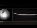
The scanning protocol consisted of an 8.3 mm single B-scan for AS SD-OCT (Figure 1A) and a 3.0 mm single horizontal B-scan at the central cornea for AS SS-OCT (Figure 1B). CCT was measured manually with the caliper available in the software by 2 different trained blinded examiners.
 |
Figure 1 (A) Central corneal thickness measured by anterior segment (AS) spectral-domain (SD) optical coherence tomography (OCT). The position of the B-scan is visible over the cornea, and the portion measured by the caliper is also visible. (B) Central corneal thickness measured by AS swept-source (SS) OCT. The same principles were applied. Note a difference of 11 µm between the methods. |
Statistical Analysis
Quantitative baseline data were described as mean (SD). Intraclass correlation coefficient (ICC) with 95% CI and a Bland-Altman assessment for agreement were used to compare CCT measurements among the 3 different methods. The data were analyzed using SPSS version 22.0 (IBM Corporation, Armonk, NY, USA).
Results
Twenty-six eyes from 16 patients were eligible for inclusion in the study. The mean (SD) age was 59.1 (10.5) years, and 87% were women. Average (SD) circumpapillary retinal nerve fiber layer (cpRNFL) was 95.73± 16.54. The mean (SD) CCT measurements were 548.04 (33.11) µm for AS SD-OCT, 538.88 (34.84) µm for AS SS-OCT, and 537.12 (29.20) µm for US CP. The overall agreement ICC was 0.915 (95% CI, 0.785–0.964; p<0.001). The best agreement between AS-OCT methods and US CP was found for SS-OCT and US CP (ICC = 0.929; 95% CI, 0.849–0.967; p<0.001). The ICC was 0.871 (95% CI, 0.459–0.956; p<0.001) between SD-OCT and US CP, and 0.942 (95% CI, 0.435–0.984; p<0.001) between SD-OCT and SS-OCT. When comparing AS-OCT methods with US CP, a difference of less than 10 µm was found in 53.8% of the eyes examined by SS-OCT, and in 50% of the eyes examined by SD-OCT. The Bland-Altman plots depicting the differences between the methods and the average CCT between the methods are shown in Figure 2A–C.
 |
Figure 2 Bland-Altman plots showing the differences in central corneal thickness (CCT) as measured by swept-source (SS) optical coherence tomography (OCT), spectral-domain (SD) OCT, and ultrasonic corneal pachymetry (US CP). (A) SS-OCT vs SD-OCT (in µm); (B) US CP vs SS-OCT (in µm); and (C) US CP vs SD-OCT (in µm). |
Discussion
Our study demonstrated that both SD-OCT and SS-OCT were comparable to the gold standard US CP for CCT measurement in a group of patients with glaucoma. The overall agreement was robust, as indicated by an ICC of 0.915 (95% CI, 0.785–0.964). Notably, the most substantial agreement was observed between AS SS-OCT and US CP (ICC = 0.929; 95% CI, 0.849–0.967). The Bland-Altman analysis supported the results, revealing that more than 50% of the eyes displayed CCT measurement differences of less than 10 µm when comparing AS SS-OCT with US CP, with AS SD-OCT yielding a similar level of agreement when compared with the gold standard US CP.
The clinical equivalence of AS SD-OCT and AS SS-OCT with US CP for obtaining precise CCT measurements provides clinicians with a potentially more accurate, noninvasive, noncontact alternative to the standard US CP. Also, AS SD-OCT and AS SS-OCT provide precise measurements that allow examiners to target the specific corneal location for the assessment of CCT since the location of the B-scan remains trackable, which can be seen as an advantage over traditional US measurements. Furthermore, US is a technique that relies substantially on the expertise of the examiner, whereas OCT methods can reduce this bias by offering a more precise and more operator-independent assessment. Coupled with the noninvasive, noncontact, and eye drop-free nature of AS SD-OCT and AS SS-OCT, these capabilities render both techniques highly appealing alternatives in clinical practice for comprehensively assessing both the structure and thickness of the cornea and its particularities.11 In our study, the average CCT measured by the US CP was the lowest and the repercussion of these differences should be considered in IOP measurements, especially in the extreme ends of the pachymetry spectrum.
Several studies have compared CCT measurements between different brands of optical imaging devices and US pachymeters. A study by Vonor et al demonstrated that AS- OCT (Topcon 2000) was an alternative to US CP showing a strong correlation between them. The authors also presented OCT as an alternative for performing CCT at the same time and device as the posterior segment OCT.12 Scotto et al measured CCT using AS-OCT, noncontact specular microscopy (SM), and US CP in healthy eyes. The mean (SD) CCT readings were 535.8 (35.5) μm, 547.7 (38.2) μm, and 537.4 (37.5) μm, respectively. The mean difference between US CP and OCT measurements was small, yet significant: 1.6 (8.6) μm (p=0.02).13 Differently from studies evaluating healthy individuals, we included a population with glaucoma or glaucoma suspects, and we employed ICC and Bland-Altman plots to access the interchangeability of the technologies.
There were studies comparing multiple technologies. Beutelspacher et al measured CCT using 4 different devices: Orbscan II (Bausch & Lomb, Germany), a scanning-slit Scheimpflug-based corneal analysis system; IOPac (Reichert/Heidelberg Engineering, Germany), a conventional US pachymeter; SL-OCT (Heidelberg Engineering, Germany), a slit-lamp-mounted AS-OCT-based analysis system; and an optical low coherence reflectometry (OLCR) pachymeter (Haag Streit, Switzerland). While the repeatability of each device was high, the authors advised exercising caution when utilizing the Orbscan device interchangeably with US CP, since it has the potential to overestimate the results.14
Gokcinar et al examined the agreement of CCT measurements using SD-OCT (Nidek RS-3000 advance OCT), corneal topography (CoT) with a combined Scheimpflug-Placido system (CSO Sirius), optical biometry (OB) (Nidek AL-scan partial coherence interferometry-based), specular microscopy (SM), corrected SM (Tomey EM-3000), and US CP (Reichert iPac). Except for the differences between OCT and US CP and between CoT and corrected SM, there was a significant difference in mean CCT between the devices (p<0.05), with mean paired differences ranging from 0.68 to 20.41 μm.15
Other studies comparing previous iterations of OCT with US CP also showed good association between the measurements.16–20 Rao et al compared two iterations of OCT and demonstrated that the CCT measurements using SD OCT RTVue had exceptional repeatability and were comparable to those using US CP, Orbscan, and Visante AS-OCT. However, there was a wide range of 95% limits of agreement among the CCT measurements using multiple devices, which might have a substantial impact depending on the specific clinical scenario.21 Unlike the studies mentioned above, ours compared two newer iterations of OCT (SD and SS OCT) with US CP.
Both AS SD-OCT and AS SS-OCT confer distinct advantages over traditional US CP measurements because they offer precise manual measurements that allow examiners to target a specific and trackable corneal location for the assessment of CCT, with the B-scan showing its exact location on the cornea. Additionally, AS-OCT methods provide clinicians with the invaluable opportunity to observe other morphological features of the cornea, allowing them to extend their valuable clinical insights beyond CCT measurement alone. The AS-OCT also allows evaluation of the angle structures, thus being helpful in assessing angle-closure glaucoma and other clinical subsets. The device used for SS-OCT in our study has the capability of taking fundus photographs, rendering it a potential all-in-one multimodal structural assessment tool for the propaedeutics of glaucoma.
Limitations of our study include the small sample size and the examiner-determined B-scans for SD-OCT and SS-OCT, which were measured manually. However, the measurements were performed by 2 different trained examiners. Another limitation is that the study population included only patients with glaucoma and glaucoma suspects.
In conclusion, OCT emerges as an effective modality for CCT measurements. Patient comfort, B-scan trackability on the cornea, and the possibility to perform multiple glaucoma examinations in a single multimodal device translate into a potential advantage of OCT for the comprehensive propaedeutics of glaucoma.
Statistical Analysis
Outsourced biostatistician.
Acknowledgments
Portions of this work were presented at the Brazilian Council of Ophthalmology (CBO) meeting, in Fortaleza, Brazil, 2023.
Administrative, technical, or material support supervision: Monica Oliveira, Jaco Lavinsky, Daniel Lavinsky. Research group leadership: Fabio Lavinsky, Helena Messinger Pakter, Rodrigo Leivas Lindenmeyer.
Author Contributions
All authors made a significant contribution to the work reported, whether that is in the conception, study design, execution, acquisition of data, analysis and interpretation, or in all these areas; took part in drafting, revising or critically reviewing the article; gave final approval of the version to be published; have agreed on the journal to which the article has been submitted; and agree to be accountable for all aspects of the work.
Funding
There is no funding to report.
Disclosure
The authors report no conflicts of interest in this work.
References
1. Jonas JB, Aung T, Bourne RR, Bron AM, Ritch R, Panda-Jonas S. Glaucoma. Lancet. 2017;390(10108):2183–2193. doi:10.1016/S0140-6736(17)31469-1
2. Quigley HA, Broman AT. The number of people with glaucoma worldwide in 2010 and 2020. Br J Ophthalmol. 2006;90(3):262–267. doi:10.1136/bjo.2005.081224
3. Tham YC, Li X, Wong TY, Quigley HA, Aung T, Cheng CY. Global prevalence of glaucoma and projections of glaucoma burden through 2040: a systematic review and meta-analysis. Ophthalmology. 2014;121(11):2081–2090. doi:10.1016/j.ophtha.2014.05.013
4. Weinreb RN, Aung T, Medeiros FA. The pathophysiology and treatment of glaucoma: a review. JAMA. 2014;311(18):1901–1911. doi:10.1001/jama.2014.3192
5. Heijl A, Leske MC, Bengtsson B, Hyman L, Bengtsson B, Hussein M; Early Manifest Glaucoma Trial Group. Reduction of intraocular pressure and glaucoma progression: results from the Early Manifest Glaucoma Trial. Arch Ophthalmol. 2002;120(10):1268–1279. doi:10.1001/archopht.120.10.1268
6. De Moraes CG, Liebmann JM, Levin LA. Detection and measurement of clinically meaningful visual field progression in clinical trials for glaucoma. Prog Retin Eye Res. 2017;56:107–147. doi:10.1016/j.preteyeres.2016.10.001
7. Brandt JD, Beiser JA, Gordon MO, Kass MA; Ocular Hypertension Treatment Study (OHTS) Group. Central corneal thickness and measured IOP response to topical ocular hypotensive medication in the Ocular Hypertension Treatment Study. Am J Ophthalmol. 2004;138(5):717–722. doi:10.1016/j.ajo.2004.07.036
8. Brandt JD, Beiser JA, Kass MA, Gordon MO. Central corneal thickness in the Ocular Hypertension Treatment Study (OHTS). Ophthalmology. 2001;108(10):1779–1788. doi:10.1016/S0161-6420(01)00760-6
9. Leske MC, Heijl A, Hyman L, Bengtsson B. Early Manifest Glaucoma Trial: design and baseline data. Ophthalmology. 1999;106(11):2144–2153. doi:10.1016/S0161-6420(99)90497-9
10. Dong ZM, Wollstein G, Schuman JS. Clinical Utility of Optical Coherence Tomography in Glaucoma. Invest Ophthalmol Vis Sci. 2016;57(9):OCT556–67. doi:10.1167/iovs.16-19933
11. Shan J, DeBoer C, Xu BY. Anterior segment optical coherence tomography: applications for clinical care and scientific research. Asia Pac J Ophthalmol. 2019;8(2):146–157. doi:10.22608/APO.201910
12. Vonor K, Amédomé KM, Santos MAK, et al. Fiabilité de la pachymétrie par OCT comparée à la pachymétrie par ultrasons [Accuracy of Optical Coherence Tomography versus Ultrasound in pachymetry]. J Fr Ophtalmol. 2021;44(7):1047–1051. doi:10.1016/j.jfo.2020.08.037
13. Scotto R, Bagnis A, Papadia M, Cutolo CA, Risso D, Traverso CE. Comparison of Central Corneal Thickness Measurements Using Ultrasonic Pachymetry, Anterior Segment OCT and Noncontact Specular Microscopy. J Glaucoma. 2017;26(10):860–865. doi:10.1097/IJG.0000000000000745
14. Beutelspacher SC, Serbecic N, Scheuerle AF. Assessment of central corneal thickness using OCT, ultrasound, optical low coherence reflectometry and Scheimpflug pachymetry. Eur J Ophthalmol. 2011;21(2):132–137. doi:10.5301/EJO.2010.1093
15. Gokcinar NB, Yumusak E, Ornek N, Yorubulut S, Onaran Z. Agreement and repeatability of central corneal thickness measurements by four different optical devices and an ultrasound pachymeter. Int Ophthalmol. 2019;39(7):1589–1598. doi:10.1007/s10792-018-0983-2
16. Leung DY, Lam DK, Yeung BY, Lam DS. Comparison between central corneal thickness measurements by ultrasound pachymetry and optical coherence tomography. Clin Exp Ophthalmol. 2006;34(8):751–754. doi:10.1111/j.1442-9071.2006.01343.x
17. Li H, Leung CK, Wong L, et al. Comparative study of central corneal thickness measurement with slit-lamp optical coherence tomography and visante optical coherence tomography. Ophthalmology. 2008;115(5):796–801.e2. doi:10.1016/j.ophtha.2007.07.006
18. Wong AC, Wong CC, Yuen NS, Hui SP. Correlational study of central corneal thickness measurements on Hong Kong Chinese using optical coherence tomography, Orbscan and ultrasound pachymetry. Eye. 2002;16(6):715–721. doi:10.1038/sj.eye.6700211
19. Zhao PS, Wong TY, Wong WL, Saw SM, Aung T. Comparison of central corneal thickness measurements by visante anterior segment optical coherence tomography with ultrasound pachymetry. Am J Ophthalmol. 2007;143(6):1047–1049. doi:10.1016/j.ajo.2007.01.050
20. Li EY, Mohamed S, Leung CK, et al. Agreement among 3 methods to measure corneal thickness: ultrasound pachymetry, Orbscan II, and Visante anterior segment optical coherence tomography. Ophthalmology. 2007;114(10):1842–1847. doi:10.1016/j.ophtha.2007.02.017
21. Rao HL, Kumar AU, Kumar A, et al. Evaluation of central corneal thickness measurement with RTVue spectral domain optical coherence tomography in normal subjects. Cornea. 2011;30(2):121–126. doi:10.1097/ICO.0b013e3181e16c65
 © 2025 The Author(s). This work is published and licensed by Dove Medical Press Limited. The
full terms of this license are available at https://www.dovepress.com/terms.php
and incorporate the Creative Commons Attribution
- Non Commercial (unported, 3.0) License.
By accessing the work you hereby accept the Terms. Non-commercial uses of the work are permitted
without any further permission from Dove Medical Press Limited, provided the work is properly
attributed. For permission for commercial use of this work, please see paragraphs 4.2 and 5 of our Terms.
© 2025 The Author(s). This work is published and licensed by Dove Medical Press Limited. The
full terms of this license are available at https://www.dovepress.com/terms.php
and incorporate the Creative Commons Attribution
- Non Commercial (unported, 3.0) License.
By accessing the work you hereby accept the Terms. Non-commercial uses of the work are permitted
without any further permission from Dove Medical Press Limited, provided the work is properly
attributed. For permission for commercial use of this work, please see paragraphs 4.2 and 5 of our Terms.
Recommended articles
Reproducibility of Neuroretinal Rim Measurements Obtained from High-Density Spectral Domain Optical Coherence Tomography Volume Scans
Kim J, Men CJ, Ratanawongphaibul K, Papadogeorgou G, Tsikata E, Ben-David GS, Antar H, Poon LYC, Freeman M, Park EA, Guzman Aparicio MA, de Boer JF, Chen TC
Clinical Ophthalmology 2022, 16:2595-2608
Published Date: 13 August 2022
Glaucoma Diagnosis Through the Integration of Optical Coherence Tomography/Angiography and Machine Learning Diagnostic Models
Kooner KS, Angirekula A, Treacher AH, Al-Humimat G, Marzban MF, Chen A, Pradhan R, Tunga N, Wang C, Ahuja P, Zuberi H, Montillo AA
Clinical Ophthalmology 2022, 16:2685-2697
Published Date: 18 August 2022
Spotlight on Lattice Degeneration Imaging Techniques
Maltsev DS, Kulikov AN, Shaimova VA, Burnasheva MA, Vasiliev AS
Clinical Ophthalmology 2023, 17:2383-2395
Published Date: 16 August 2023