Back to Journals » Journal of Inflammation Research » Volume 18
Cervical Conization and Systemic Inflammatory Markers: The Predictive Value of Neutrophil Percentage to Albumin Ratio (NPAR) to Identify High-Grade Cervical Intraepithelial Neoplasia (CIN)
Authors Özbilgeç S , Akkuş F
Received 18 December 2024
Accepted for publication 12 April 2025
Published 19 April 2025 Volume 2025:18 Pages 5343—5353
DOI https://doi.org/10.2147/JIR.S512991
Checked for plagiarism Yes
Review by Single anonymous peer review
Peer reviewer comments 2
Editor who approved publication: Professor Ning Quan
Sıtkı Özbilgeç,1 Fatih Akkuş2
1Department of Obstetrics and Gynecology, Gynecologic Oncology Clinic, Konya City Hospital, Konya, Turkey; 2Department of Obstetrics and Gynecology, Perinatology Clinic, Kütahya City Hospital, Kütahya, Turkey
Correspondence: Sıtkı Özbilgeç, Konya City Hospital, Konya, Turkey, Email [email protected]
Objective: This study aimed to evaluate the predictive value of systemic inflammatory markers, particularly the Neutrophil Percentage to Albumin Ratio (NPAR), in identifying high-grade cervical intraepithelial neoplasia (CIN) in patients undergoing colposcopy and cervical conization.
Materials and Methods: A retrospective analysis was conducted on 116 patients who underwent cervical conization between January 2020 and May 2024. Demographic, clinical, and laboratory data were collected, and inflammatory indices, including NPAR, NLR (Neutrophil-to-Lymphocyte Ratio), PLR (Platelet-to-Lymphocyte Ratio), and SII (Systemic Immune-Inflammation Index), were calculated. Sample size estimation was based on prior studies assessing inflammatory markers in CIN, ensuring adequate statistical power for detecting differences in biomarker levels. ROC curve analysis was performed to assess the diagnostic accuracy of these markers.
Results: NPAR demonstrated the highest predictive value for high-grade CIN, with an AUC of 0.893. Significant correlations were found between NPAR and other systemic inflammatory markers, such as NLR and SII. However, NLR and PLR showed lower predictive accuracy compared to NPAR.
Conclusion: NPAR is a valuable biomarker for predicting high-grade CIN and can aid in patient stratification and treatment planning. Integrating NPAR with other systemic markers may enhance the accuracy of clinical decisions. Further studies with larger cohorts are recommended to validate these findings and explore their clinical utility.
Keywords: cervical conization, systemic inflammatory markers, neutrophil percentage to albumin ratio, NPAR, cervical intraepithelial neoplasia, CIN, prognostic biomarkers, systemic immune-inflammation index, SII
Introduction
Cervical cancer remains a global health burden, particularly in regions with limited access to effective screening and vaccination programs.1 Cervical intraepithelial neoplasia (CIN), a precursor to cervical cancer, offers an opportunity for early detection and treatment through procedures such as colposcopy and cervical conization.2 However, identifying patients at high risk of progressing to severe lesions remains a challenge.3 Biomarkers that reflect systemic inflammation, such as the neutrophil-to-lymphocyte ratio (NLR) and platelet-to-lymphocyte ratio (PLR), are gaining attention for their predictive value in cancer prognosis.4,5
Chronic inflammation plays a central role in the development and progression of CIN. Persistent infection with high-risk human papillomavirus (HPV) induces an inflammatory microenvironment characterized by immune evasion, cytokine dysregulation, and oxidative stress, which contribute to the progression from low-grade lesions to high-grade CIN and invasive cancer.6,7 Inflammatory markers provide indirect insight into these biological processes. Elevated neutrophil levels are associated with enhanced tumor-promoting inflammation, while reduced albumin levels reflect a poor nutritional and immune status, both of which contribute to cancer progression.8 The ability of systemic inflammatory markers to capture these dynamic changes makes them valuable tools in predicting CIN progression.
In the context of cervical cancer, these inflammatory indices can provide insight into both the host immune response to HPV and the severity of cervical lesions.9 Xu and Song (2021) demonstrated that elevated NLR levels could predict the development of CIN, particularly in patients with persistent HPV infections.4 Such findings highlight the importance of systemic immune-inflammation markers in identifying patients who may benefit from closer surveillance or more aggressive treatment strategies.
In addition to NLR and PLR, other indices such as the neutrophil percentage to albumin ratio (NPAR) and systemic immune-inflammation index (SII) have been explored as potential biomarkers. NPAR, in particular, reflects both the inflammatory burden and the nutritional status of patients, making it a promising tool for risk stratification. Since CIN2-3 represents a critical threshold for intervention, evaluating NPAR’s role in distinguishing these lesions is essential.10 As current screening methods, including cytology and HPV testing, have limitations in predicting lesion progression, integrating systemic inflammatory markers like NPAR into clinical practice could enhance risk stratification and guide individualized patient management.11
This study aims to evaluate the predictive value of NPAR, along with other systemic inflammatory markers such as NLR, PLR, and SII, in patients undergoing cervical conization to identify CIN2-3. We hypothesize that these markers may help identify high-risk patients with CIN2-3 and guide clinical decisions regarding follow-up and treatment strategies.
Materials and Methods
Study Design and Participants
This retrospective study was conducted at a tertiary care hospital and included patients who underwent colposcopy and cervical conization between January 2020 and May 2024. This study was approved by the Necmettin Erbakan University Faculty of Medicine Ethics Committee (Approval No: [21165]) institutional review board, and informed consent was waived due to the retrospective nature of the study. The primary objective was to investigate the predictive value of systemic inflammatory markers, including the Neutrophil Percentage to Albumin Ratio (NPAR), in detecting high-grade cervical intraepithelial neoplasia (CIN).
Inclusion and Exclusion Criteria
Inclusion Criteria
- Patients aged 18 years or older.
- Patients who underwent colposcopy and cervical conization during the study period.
- Complete blood count (CBC) and albumin measurements taken within one week prior to the procedure.
Exclusion Criteria
- Patients with acute infections, chronic inflammatory diseases, or autoimmune conditions.
- Patients with ongoing malignancies or those receiving immunosuppressive therapy.
- Incomplete or missing laboratory or clinical data.
Data Collection
Demographic data (age) and clinical characteristics were collected from the hospital’s electronic medical records. Preoperative laboratory data included complete blood count parameters (neutrophil, lymphocyte, platelet counts) and serum albumin levels. Based on these values, the following systemic inflammatory indices were calculated:
- NPAR = Neutrophil % / Albumin
- NLR = Neutrophil / Lymphocyte
- PLR = Platelet / Lymphocyte
- SII = (Neutrophil × Platelet) / Lymphocyte
Histopathological Evaluation
Tissue specimens obtained during conization were evaluated by experienced pathologists. Patients were stratified into two groups based on their histopathological results:
- Low-grade lesions (CIN1)
- High-grade lesions (CIN2, CIN3, or carcinoma in situ)
Sample Size and Statistical Power The sample size was determined based on prior studies assessing inflammatory markers in CIN, ensuring adequate statistical power to detect significant differences in biomarker levels between low-grade and high-grade CIN groups. Post hoc power analysis confirmed that the study had sufficient power (≥80%) to evaluate the predictive performance of NPAR.
To control for potential confounding factors, patients with known comorbidities that could influence inflammatory markers (such as diabetes, cardiovascular diseases, or chronic infections) were excluded from the study. Additionally, information regarding medication use (such as corticosteroids or immunosuppressants) was reviewed, and patients on these treatments were not included. Lifestyle factors such as smoking and obesity were not systematically assessed due to retrospective data limitations but were acknowledged as potential limitations in the discussion.
HPV Genotyping HPV genotyping was performed using polymerase chain reaction (PCR)-based methods to identify high-risk HPV types. DNA was extracted from cervical tissue samples, and specific primers targeting high-risk HPV subtypes were used to determine genotype distributions.
Surgical Margin Assessment Histopathological examination of surgical margins was performed to classify them as positive or negative. A margin was considered positive if CIN2-3 or carcinoma in situ was present at the surgical edge, while a margin was classified as negative if no dysplastic cells were detected at the resection boundary.
Ethical approval was obtained from the Ethics Committee of the Necmettin Erbakan University Faculty of Medicine Ethics Committee and the study adhered to the principles outlined in the Declaration of Helsinki. All patient data were handled in strict confidentiality and in accordance with institutional regulations.
Statistical Analysis
All statistical analyses were performed with SPSS version 26.0. Descriptive statistics for continuous variables are presented as mean ± standard deviation (SD) and median with interquartile range (IQR). Categorical variables are presented as frequencies and percentages. Comparisons between HSIL and above and LSIL and below groups were made using independent samples t-test for normally distributed continuous variables and Mann–Whitney U-test for non-normally distributed variables. Data are presented as mean ± standard deviation (SD). Categorical data were analysed using the chi-squared test or Fisher’s exact test where appropriate. Correlation analyses between continuous variables were performed using Pearson’s correlation coefficient for normally distributed variables and Spearman correlation coefficient for non-normally distributed variables.
Receiver operating characteristic (ROC) curve analysis was performed to assess the predictive performance of the laboratory parameters in detecting lesions of HSIL and above. The area under the curve (AUC) was calculated, and optimal cut-off points were determined using the Youden index, which maximizes sensitivity and specificity. The rationale for using the Youden index is its ability to provide a balance between false positives and false negatives, making it a clinically relevant measure for identifying high-risk patients. Alternative methods, such as predefined clinical thresholds, were not available for NPAR, making statistical optimization essential. Sensitivity, specificity, positive predictive value (PPV), and negative predictive value (NPV) were also reported. A p-value of less than 0.05 was considered statistically significant for all tests.
Results
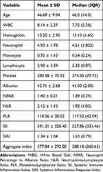
In this study, various hematological and biochemical parameters of patients who underwent colposcopy were analyzed. The mean age of the patients was 46.69 ± 9.94 years, with a median age of 46.0. The mean values for white blood cell count, hemoglobin, neutrophil, monocyte, lymphocyte, and platelet counts were 8.14 ± 2.27, 13.20 ± 2.95, 4.93 ± 1.78, 0.72 ± 1.10, 2.90 ± 3.39, and 280.58 ± 70.32, respectively. The mean values for NPAR, NLR, PLR, SII, and SIRI were 1.40 ± 0.21, 2.12 ± 1.10, 118.56 ± 38.02, 591.31 ± 320.40, and 1.34 ± 1.04, respectively. The detailed mean and median values of all parameters are presented in Table 1.
 |
Table 1 Descriptive Statistics of Haematological and Biochemical Parameters in Patients Undergoing Colposcopy |
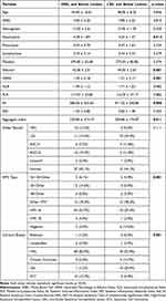
In the evaluation based on HPV genotypes and biopsy results, HSIL lesions were identified in 8.6%, LSIL lesions in 18.1%, ASC-H lesions in 8.6%, and ASCUS lesions in 17.2% of the cases. Normal smear results were observed in 44.8%. Regarding HPV genotype distribution, HPV 16 was detected in 46.6%, HPV 18 in 4.3%, and other HPV types in 24.1% of the cases. Biopsy results revealed HSIL in 74.1%, LSIL in 9.5%, chronic cervicitis in 4.3%, and SCC in 8.6%. According to cone biopsy results, HSIL was found in 57.8%, LSIL in 17.2%, and SCC in 9.5%. Surgical margin assessment indicated negative margins in 69.8% and positive margins in 21.6% of the cases, while follow-up cytology showed ASCUS in 2.6% and negative cytology in 16.4% (Table 2).
 |
Table 2 Cervical Lesions in Colposcopy Patients: HPV Genotypes and Biopsy Results |
In the comparison of clinical and laboratory results between HSIL and above lesions versus LSIL and below lesions, significant differences were observed in albumin levels (HSIL and above: 42.00 ± 2.51; LSIL and below: 44.00 ± 2.65, p=0.001), neutrophil count (HSIL and above: 4.78 ± 1.84; LSIL and below: 4.24 ± 1.47, p=0.014), NPAR values (HSIL and above: 1.45 ± 0.18; LSIL and below: 1.21 ± 0.17, p=0.001), and systemic inflammatory index (SII) values (HSIL and above: 586.26 ± 335.63; LSIL and below: 471.52 ± 240.86, p=0.006). However, no significant differences were found between the groups in terms of white blood cell (WBC) count, hemoglobin, monocyte, lymphocyte, or platelet counts. In the analysis of categorical data, significant differences were observed between the groups in terms of HPV genotypes (p=0.001) and cervical biopsy results (p=0.001), with a higher prevalence of HPV 16 and HSIL in the HSIL and above group. However, no significant differences were found in smear results (p=0.111) (Table 3).
 |
Table 3 Comparing Clinical and Laboratory Results of HSIL and Above vs, LSIL and Below Lesions |
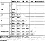
In the correlation analyses, a positive correlation was found between NPAR and NLR (r=0.408, p=0.001). A significant correlation was also observed between NLR and SIRI (r=0.276, p=0.003). Additionally, significant correlations were detected between SII and SIRI (r=0.497, p=0.001) and between PLR and SII (r=0.276, p=0.001) (Table 4). Furthermore, significant correlations were identified between the Aggregate index and all other parameters (r=0.477, p=0.001) (Table 4).
 |
Table 4 Correlations of NPAR, NLR, PLR, SII, SIRI and Aggregate Index |
In the evaluation of the performance of laboratory parameters in predicting HSIL and above lesions, the NPAR cutoff value was determined as 1.30, with a sensitivity of 87.8% and a specificity of 82.3% (AUC: 0.893, p=0.001). The NLR cutoff value was 1.47, with a sensitivity of 81.7% and a specificity of 41.1% (AUC: 0.621, p=0.040). The SII cutoff value was determined as 527.42, with a sensitivity of 62.2% and a specificity of 76.4% (AUC: 0.700, p=0.001). The SIRI cutoff value was 0.93, with a sensitivity of 71.9% and a specificity of 56.1% (AUC: 0.677, p=0.003). Lastly, the Aggregate index cutoff value was 194.70, with a sensitivity of 84.1% and a specificity of 54.4% (AUC: 0.668, p=0.004) (Figure 1 and Table 5).
 |
Table 5 Prediction Performance of Laboratory Parameters for Prediction of HSIL and Above Lesions |
Discussion
Our study demonstrates that systemic inflammatory markers, particularly NPAR and SII, are valuable tools for predicting the severity of cervical intraepithelial neoplasia (CIN). NPAR showed the highest predictive value, with an AUC of 0.893, making it a promising biomarker for identifying patients at risk for high-grade lesions. These results align with Xu and Song (2021), who reported that elevated NLR levels were associated with higher-grade CIN, especially in patients with persistent HPV infections.4 Chronic inflammation, as reflected in these systemic markers, plays a pivotal role in the development and progression of CIN, influencing both immune regulation and the tissue environment.12
The biological mechanisms linking NPAR to CIN severity likely involve a combination of chronic inflammation and immune system dysregulation. Persistent HPV infection induces an inflammatory microenvironment, leading to increased neutrophil activity and cytokine release, which promote tissue remodeling and carcinogenesis. Meanwhile, serum albumin levels, an indicator of nutritional and systemic health, tend to decrease in patients with chronic disease and malignancies, further reflecting immune dysfunction.13,14 This dual role of NPAR, capturing both inflammatory burden and nutritional status, may explain its strong predictive ability in CIN severity assessment.
The integration of NPAR with other markers, such as NLR and SII, offers a more comprehensive picture of the patient’s immune status and inflammatory burden. While NLR and PLR have been widely used as predictive markers in other cancers, including colorectal and lung cancer,15,16 our findings suggest that NPAR may provide additional insight by incorporating nutritional status. This aligns with earlier research showing that systemic immune-inflammation indices like NPAR and SII are relevant in capturing both immune suppression and nutritional deficiencies, which are closely tied to cancer progression and patient outcomes.17,18
In the context of cervical cancer, systemic inflammatory markers may also serve as indicators of the host immune response to HPV infection. Studies show that chronic immune activation and persistent HPV infections increase the risk of progression from low-grade to high-grade CIN.19 Our findings corroborate these observations, demonstrating that systemic inflammatory markers like NPAR and SII can help clinicians stratify patients based on their risk of developing severe lesions, guiding personalized treatment and follow-up strategies.20
Beyond its diagnostic value, the implementation of NPAR in clinical practice should consider its feasibility and cost-effectiveness. Compared to more advanced molecular diagnostic techniques, calculating NPAR is relatively simple and inexpensive, as it requires only standard laboratory tests that are routinely performed in clinical settings. This accessibility makes NPAR a practical tool for resource-limited settings where HPV testing and colposcopy may not be readily available.21 However, further cost-benefit analyses are needed to determine its true economic impact and potential role in routine screening algorithms.
However, certain limitations must be acknowledged. First, the retrospective design limits the ability to draw causal inferences. Additionally, the relatively small sample size may reduce the statistical power to detect subtle differences between groups. Further, confounding factors such as HPV genotype and viral load, which are known to influence inflammatory marker levels, were not evaluated in our study.11 Although we attempted to minimize confounding by excluding patients with known inflammatory diseases and malignancies, unmeasured factors such as smoking and obesity may still have influenced our results. Future prospective studies with larger sample sizes are needed to validate these findings and explore the interaction between systemic inflammation and HPV status in greater detail.22
Incorporating these markers into clinical practice has the potential to enhance patient care. As Xu and Song (2021) suggested, identifying patients with elevated inflammatory markers early can enable clinicians to initiate more aggressive interventions or closer monitoring to prevent disease progression.4 This approach may be particularly valuable for patients with persistent HPV infections or those with multiple risk factors for developing high-grade CIN. Given the promising findings of our study, future research should focus on establishing standardized cut-off values and assessing the longitudinal impact of NPAR in predicting disease outcomes.
Conclusion
Our study shows that systemic inflammatory markers, particularly NPAR and SII, are valuable in predicting the severity of CIN. NPAR demonstrated superior diagnostic accuracy in predicting CIN2-3 compared to other systemic inflammatory markers, highlighting its potential as a reliable biomarker. These findings align with previous research, highlighting the role of inflammatory markers in CIN progression.
The combined use of NPAR with other indices, such as NLR and PLR, provides complementary insights, enhancing patient stratification and follow-up strategies. Integrating these markers into clinical practice could refine risk assessment, allowing for more tailored follow-up and intervention approaches. Given its ease of measurement and cost-effectiveness, NPAR has the potential to be integrated into routine clinical practice. Future research should validate these findings in larger cohorts and explore the integration of inflammatory markers with HPV-related factors to optimize treatment decisions.
Data Sharing Statement
The datasets used and/or analyzed during the current study are available from the corresponding author on reasonable request.
Funding
No funding was received.
Disclosure
The authors declare that they have no competing interests in this work.
References
1. Massad LS, Einstein MH, Huh WK, et al. 2012 updated consensus guidelines for the management of abnormal cervical cancer screening tests and cancer precursors. J Low Genit Tract Dis. 2013;17(5 Suppl 1):S1–s27. doi:10.1097/LGT.0b013e318287d329
2. Hillemanns P, Soergel P, Hertel H, Jentschke M. Epidemiology and early detection of cervical cancer. Oncol Res Treat. 2016;39(9):501–506. doi:10.1159/000448385
3. Kalliala I, Athanasiou A, Veroniki AA, et al. Incidence and mortality from cervical cancer and other malignancies after treatment of cervical intraepithelial neoplasia: a systematic review and meta-analysis of the literature. Ann Oncol. 2020;31(2):213–227. doi:10.1016/j.annonc.2019.11.004
4. Xu L, Song J. Elevated neutrophil-lymphocyte ratio can be a biomarker for predicting the development of cervical intraepithelial neoplasia. Medicine. 2021;100(28):e26335. doi:10.1097/MD.0000000000026335
5. Liu Y, Fan P, Yang Y, et al. Human papillomavirus and human telomerase RNA component gene in cervical cancer progression. Sci Rep. 2019;9(1):15926. doi:10.1038/s41598-019-52195-5
6. Long T, Long L, Chen Y, et al. Severe cervical inflammation and high-grade squamous intraepithelial lesions: a cross-sectional study. Arch Gynecol Obstet. 2021;303(2):547–556. doi:10.1007/s00404-020-05804-y
7. Huang R, Liu Z, Sun T, Zhu L. Cervicovaginal microbiome, high-risk HPV infection and cervical cancer: mechanisms and therapeutic potential. Microbiol Res. 2024;287:127857. doi:10.1016/j.micres.2024.127857
8. Li X, Wu M, Chen M, et al. The association between Neutrophil-Percentage-to-Albumin Ratio (NPAR) and mortality among individuals with cancer: insights from national health and nutrition examination survey. Cancer Med. 2025;14(2):e70527. doi:10.1002/cam4.70527
9. Bruno M, Bizzarri N, Teodorico E, et al. The potential role of systemic inflammatory markers in predicting recurrence in early-stage cervical cancer. Eur J Surg Oncol. 2024;50(1):107311. doi:10.1016/j.ejso.2023.107311
10. Jiang Y, Yin F, Chen Y, Yue L, Li L. Discovery of microarray-identified genes associated with the progression of cervical intraepithelial neoplasia. Int J Clin Exp Pathol. 2018;11(12):5667–5681.
11. Naizhaer G, Yuan J, Mijiti P, Aierken K, Abulizi G, Qiao Y. Evaluation of multiple screening methods for cervical cancers in rural areas of Xinjiang, China. Medicine. 2020;99(6):e19135. doi:10.1097/MD.0000000000019135
12. Ayhan S, Akar S, Kar İ, et al. Prognostic value of systemic inflammatory response markers in cervical cancer. J Obstet Gynaecol. 2022;42(6):2411–2419. doi:10.1080/01443615.2022.2069482
13. Liu CF, Chien LW. Predictive role of Neutrophil-Percentage-to-Albumin Ratio (NPAR) in nonalcoholic fatty liver disease and advanced liver fibrosis in nondiabetic US adults: evidence from NHANES 2017–2018. Nutrients. 2023;15(8). doi:10.3390/nu15081892
14. Zhao M, Huang X, Zhang Y, Wang Z, Zhang S, Peng J. Predictive value of the neutrophil percentage-to-albumin ratio for coronary atherosclerosis severity in patients with CKD. BMC Cardiovasc Disord. 2024;24(1):277. doi:10.1186/s12872-024-03896-x
15. He W, Yin C, Guo G, et al. Initial neutrophil lymphocyte ratio is superior to platelet lymphocyte ratio as an adverse prognostic and predictive factor in metastatic colorectal cancer. Med Oncol. 2013;30(1):439. doi:10.1007/s12032-012-0439-x
16. Kobayashi N, Usui S, Kikuchi S, et al. Preoperative lymphocyte count is an independent prognostic factor in node-negative non-small cell lung cancer. Lung Cancer. 2012;75(2):223–227. doi:10.1016/j.lungcan.2011.06.009
17. Ferro M, Babă DF, de Cobelli O, et al. Neutrophil percentage-to-albumin ratio predicts mortality in bladder cancer patients treated with neoadjuvant chemotherapy followed by radical cystectomy. Future Sci OA. 2021;7(7):Fso709. doi:10.2144/fsoa-2021-0008
18. Liao YC, Ying HQ, Huang Y, et al. Role of chronic inflammatory ratios in predicting recurrence of resected patients with stage I-III mucinous colorectal adenocarcinoma. Cancer Manag Res. 2021;13:3455–3464. doi:10.2147/CMAR.S303758
19. Li N, Zhang Y, Qu W, et al. Analysis of systemic inflammatory and coagulation biomarkers in advanced cervical cancer: prognostic and predictive significance. Int J Biol Markers. 2023;38(2):133–138. doi:10.1177/03936155231163599
20. Santos Thuler LC, Reis Wariss B, Nogueira-Rodrigues A, de Melo AC, Bergmann A. The utility of pretreatment systemic inflammatory response biomarkers on overall survival of cervical cancer patients stratified by clinical staging. Eur J Obstet Gynecol Reprod Biol. 2021;264:281–288. doi:10.1016/j.ejogrb.2021.07.034
21. Bruni L, Serrano B, Roura E, et al. Cervical cancer screening programmes and age-specific coverage estimates for 202 countries and territories worldwide: a review and synthetic analysis. Lancet Glob Health. 2022;10(8):e1115–e27. doi:10.1016/S2214-109X(22)00241-8
22. Zhang W, Yin Y, Jiang Y, et al. Relationship between vaginal and oral microbiome in patients of human papillomavirus (HPV) infection and cervical cancer. J Transl Med. 2024;22(1):396. doi:10.1186/s12967-024-05124-8
 © 2025 The Author(s). This work is published and licensed by Dove Medical Press Limited. The
full terms of this license are available at https://www.dovepress.com/terms.php
and incorporate the Creative Commons Attribution
- Non Commercial (unported, 4.0) License.
By accessing the work you hereby accept the Terms. Non-commercial uses of the work are permitted
without any further permission from Dove Medical Press Limited, provided the work is properly
attributed. For permission for commercial use of this work, please see paragraphs 4.2 and 5 of our Terms.


