Back to Journals » Infection and Drug Resistance » Volume 17
Clinical Application of Metagenomic Next-Generation Sequencing (mNGS) in Patients with Early Pulmonary Infection After Liver Transplantation
Authors Peng HB , Liu Y , Hou F, Zhao S , Zhang YZ , He ZY, Liu JY, Xiong HF , Sun LY
Received 26 August 2024
Accepted for publication 1 December 2024
Published 19 December 2024 Volume 2024:17 Pages 5685—5698
DOI https://doi.org/10.2147/IDR.S483684
Checked for plagiarism Yes
Review by Single anonymous peer review
Peer reviewer comments 3
Editor who approved publication: Dr Zhi Ruan
Hua-Bin Peng,1 Ying Liu,1 Fei Hou,1 Shuang Zhao,1 Yi-Zhi Zhang,1 Zhi-Ying He,1 Jing-Yi Liu,1 Hao-Feng Xiong,1 Li-Ying Sun1– 4
1Department of Critical Liver Diseases, Beijing Friendship Hospital, Capital Medical University, Beijing, People’s Republic of China; 2Laboratory for Clinical Medicine, Capital Medical University, Beijing, People’s Republic of China; 3Liver Transplantation Center, National Clinical Research Center for Digestive Diseases, Beijing Friendship Hospital, Capital Medical University, Beijing, People’s Republic of China; 4Clinical Center for Pediatric Liver Transplantation, Capital Medical University, Beijing, People’s Republic of China
Correspondence: Li-Ying Sun; Hao-Feng Xiong, Department of Critical Liver Diseases, Beijing Friendship Hospital, Capital Medical University, Number 101, Luyuan East Road, Tongzhou District, Beijing, 101100, People’s Republic of China, Email [email protected]; [email protected]
Purpose: To examine the clinical utility of metagenomic next-generation sequencing (mNGS) in individuals with early pulmonary infection following liver transplantation.
Patients and Methods: mNGS and traditional detection results were retrospectively collected from 99 patients with pulmonary infection within one week following liver transplantation. These patients were admitted to the Department of Critical Liver Diseases at Beijing Friendship Hospital from February 2022 to February 2024, along with their general clinical data.
Results: mNGS exhibited a significantly higher detection rate than traditional methods (92.93% vs 54.55%, P < 0.05) and was more effective in identifying mixed infections (67.68% vs 14.81%, P < 0.05). mNGS identified 303 pathogens in 92 patients, with Enterococcus faecium, Pneumocystis jirovecii, and human herpesvirus types 5 and 7 being the most prevalent bacteria, fungi, and viruses. A total of 26 positive cases were identified through traditional culture methods (sputum and bronchoalveolar lavage fluid), with 18 cases consistent with mNGS detection results, representing 69.23% consistency. Among the three drug-resistant bacteria that showed positivity in mNGS and traditional culture, the presence of drug-resistance genes—mecA in Staphylococcus aureus; KPC-2, KPC-9, KPC-18, KPC-26, OXA27, OXA423 in Klebsiella pneumoniae; and OXA488 and NDM6 in Pseudomonas aeruginosa—reliably predicted drug-resistance phenotype. The treatment regimen for 76 of the 92 patients with positive mNGS relied on these results; 74 exhibited significant symptom improvement, yielding a 97.37% recovery rate. The overall prognosis was favorable.
Conclusion: mNGS offers rapid detection, a high positivity rate, insensitivity to antibiotics, and a superior ability to detect mixed infections in patients with early post-transplant pulmonary infections. Additionally, mNGS shows good consistency with traditional culture and can predict drug-resistant phenotypes to guide targeted antibiotic therapy for early-stage post-transplant pulmonary infection after liver transplantation. Patients whose antibiotic therapy is based on mNGS results have experienced decreased mortality rates and overall improved prognosis.
Keywords: Liver transplantation, Pulmonary infection, Metagenomic next-generation sequencing, Clinical value
Introduction
Liver transplantation has emerged as the sole efficacious intervention for individuals with end-stage liver disease.1 Recently, advancements in surgical techniques, the introduction of new immune agents, and ongoing enhancements in perioperative care have significantly increased the long-term survival rates of organ transplantation recipients and their grafts.2 Nevertheless, early post-operative infections significantly impact patient prognosis,3 with pulmonary infections being a prevalent complication and a leading cause of patient mortality.4 Despite the common practice of administering prophylactic antibiotics, the incidence of early post-operative infections remains as high as 71.4%.5 Meanwhile, as a result of the growing misuse of antibiotics, bacterial resistance has emerged as a significant concern, presenting challenges in clinical treatment and posing a serious threat to the life and health of patients.6 Hence, patients must receive early preventive measures and effective anti-infective treatment, along with timely and precise identification of the causative agent and selection of appropriate antibiotics to combat drug-resistant bacteria.
Recently, the clinical application of metagenomic next-generation sequencing (mNGS) has increased due to its heightened sensitivity and efficiency compared to conventional detection techniques, facilitated by advancements in genomics technology. Evidence indicates that mNGS effectively detects pathogens after liver transplantation.7–9 This method allows for swift and thorough identification of DNA and RNA sequences of pathogenic microorganisms from clinical samples, potentially revealing the presence of resistance genes.10
This study aims to demonstrate the advantages of mNGS in clinical applications through comparative analysis with traditional detection methods. By analyzing the distribution characteristics of pathogens in the lower respiratory tract of patients with early post-liver transplant pulmonary infections detected by mNGS, we aim to provide a reference basis for early and accurate pathogen diagnosis for such patients. Furthermore, by analyzing the value of mNGS in detecting drug-resistant genes in clinical settings, we aspire to formulate rational and effective anti-infective strategies, thereby further improving patient prognosis.
Material and Methods
Basic Information on the Research Object
This study retrospectively examined a cohort of 99 patients admitted to the Intensive Care Unit of Critical Liver Diseases in Beijing Friendship Hospital between February 2022 and February 2024 who experienced pulmonary infections within one-week post-liver transplantation.
Inclusion and Exclusion Criteria
Inclusion criteria were as follows: (1) a diagnosis of post-operative pulmonary infection within one week; (2) complete clinical data, including mNGS and traditional detection results; (3) informed consent obtained from the patient or their family for relevant examinations and inspections.
Exclusion criteria included the following: (1) pre-operative complications of pulmonary infection; (2) severe respiratory and circulatory diseases preventing relevant examinations; (3) Incomplete clinical data.
Post-Operative Empirical Anti-Infective Regimen
All patients routinely received prophylactic anti-infective treatment post-surgery. For recipients of cadaveric donor livers, the regimen typically included meropenem/imipenem, vancomycin, and micafungin combination to cover Gram-positive cocci, Gram-negative bacilli, atypical pathogenic bacteria, and fungi. For recipients of living donor livers without pre-operative infections, third-generation cephalosporin antibiotics or carbapenem antibiotics were used post-surgery. If pre-operative infection was detected in the recipient, the treatment was guided by the results of pre-operative pathogen culture and drug sensitivity.
Diagnostic Criteria for Pulmonary Infection
Diagnostic criteria for pulmonary infection were as follows: new or progressive patchy, infiltrating shadows or interstitial changes on post-operative chest imaging accompanied by fever (body temperature > 38.3 °C) or body temperature < 36 °C, abnormal white blood cell count (> 10 or < 4 × 10⁹/L), and clinical symptoms (new or worsening cough, dyspnea, and purulent discharge).11,12
Lower Respiratory Tract Samples Collected and Submitted for Examination
The patient underwent bedside bronchoscopy in the intensive care unit; aseptic tubes were used to collect bronchoalveolar lavage fluid (≥ 4 mL) or sputum (≥ 2 mL). The tubes were then sent to Jieyi Biotechnology (Hangzhou, China) for mNGS detection.All sputum samples for mNGS testing were obtained under aseptic conditions via bronchoscopy to minimize contamination from upper respiratory tract microorganisms and ensure accurate pathogen identification. For lower respiratory tract samples sent for conventional culture, the standard practice in our center is as follows: if a patient with suspected pulmonary infection has already undergone tracheal intubation, sputum is directly collected at the bedside using bronchoscopy or a sterile suction catheter. In case of insufficient sputum, bronchoalveolar lavage fluid is obtained through bronchoscopy and sent for testing. This two methods are the most common approaches for obtaining samples for conventional culture. If a patient with suspected pulmonary infection has not undergone tracheal intubation and has good respiratory muscle strength, sputum can be acquired through voluntary coughing. In our study, only a few patients had their sputum collected using this method.
Library Preparation and Metagenomic Sequencing
DNA libraries were prepared through automatic nucleic acid extraction, enzymatic fragmentation, end repair, terminal adenylation, and adaptor ligation according to a previous study.13 Finished libraries were quantified using real-time polymerase chain reaction (PCR) (KAPA) and pooled. Shotgun sequencing was performed on illumina Nextseq, generating approximately 20 million 50-bp single-end reads per library. Bioinformatic analysis was conducted as described in a previous report.14 Briefly, Low-quality sequences, human-derived sequences, reagent-engineered microbial sequences, and laboratory environmental contamination sequences were filtered (GRCh38.p13). The remaining reads were aligned to reference databases (NCBI nt, GenBank and an in-house curated genomic database) to identify microbial species and read counts. For each sequencing run, a negative control (NC) (culture medium containing 104 Jurkat cells/mL) was included.
MNGS Reporting Criteria
Microbial reads identified from a library were reported if 1) the sequencing data passed quality control filters (library concentration > 50 pM, Q20 > 85%, and Q30 > 80%); and 2) the NC in the same sequencing run did not contain the species or the reads per million (RPM) for the sample/RPM for the NC was ≥ 5, as determined empirically by previous studies as a cutoff for discriminating true-positives from background contaminations.13,15,16 In the final report, we evaluated whether the detected microorganisms were pathogens or commensal bacteria according to the pathogenicity of microorganism, specimen type, number of detected sequences, relative abundance, the rank of genus sequences among all genera in the specimen, and clinical information provided by the clinic.
Traditional Detection Methods
Routine samples, including nasopharyngeal swabs, sputum, bronchoalveolar lavage fluid, and blood, were collected. We employed the traditional detection methods, mainly smear-stained microscopy of sputum and bronchoalveolar lavage fluid. BASO (brand name) Gram stain solution was utilized for common nosocomial bacteria and most fungi, whereas BASO acid-fast stain solution was used for Mycobacterium tuberculosis. Additionally, bacteria, fungi, and mycobacteria in sputum and bronchoalveolar lavage fluid were cultured (MacConkey agar and Columbia agar, China),17 and the 1, 3-β-D-glucan test was performed on blood and bronchoalveolar lavage fluid (Jinshanchuan, China).18 Moreover, PCR testing was conducted on nasopharyngeal swabs for six respiratory viruses: influenza A/B, respiratory syncytial virus, adenovirus, and parainfluenza virus types I and III (Zhuochenghuisheng, China), as well as Mycoplasma pneumoniae (Baoruiyuan, China).19 Furthermore, antibody detection was performed for Epstein-Barr virus and cytomegalovirus (CMV) in the blood (Suoling, China).
Statistical Methods
RPM refers to the normalized sequence count of microorganisms detected in sequencing data. This index standardizes data, eliminating discrepancies in sequence numbers caused by varying data volumes, enabling researchers to compare loads of the same pathogen across different samples. In this study, microbial sequences from 99 samples were uniformly processed using RPM normalization for comparison. Statistical analysis was conducted using SPSS 27.0; GraphPad Prism 7 software was utilized for data visualization. Measurement data adhering to normal distribution and variance homogeneity were expressed as mean ± standard deviation and analyzed using the independent sample t-test. Non-normally distributed measurements were represented by the median or interquartile range and analyzed using the Mann–Whitney U-test. Categorical count data were presented as the number of cases (percentage) and tested using the X2 method. A significance level of P < 0.05 was employed in this study.
Results
General Characteristics of the Study Population
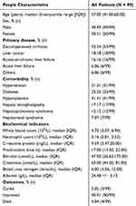
A total of 99 patients meeting the criteria were included in the study for analysis (Figure 1). Among these patients, there were 49 males (49.49%) and 50 females (50.51%), with a median age of 57 years. The majority of patients had decompensated cirrhosis as their primary disease (53.54%); common complications included hypersplenism (41.41%) and hepatorenal syndrome (17.17%). Additionally, some patients had comorbid chronic conditions such as hypertension (31.31%) and diabetes (25.25%). Laboratory tests revealed a median C-reactive protein level of 9.59, a median prothrombin time of 17.00, and a median total bilirubin level of 63.00, all of which exceeded the upper limit of the normal reference range (Table 1).
 |
Table 1 Baseline Characteristics of the Study Population |
Results of mNGS Detection in the Study Population
Sputum or alveolar lavage fluid from 99 patients was collected for mNGS detection. mNGS detected pathogens in 92 patients, with a positivity rate of 92.93% (92/99, 95% CI 87.80%–98.10%). According to the results of mNGS detection, bacterial (17.19%,16/92), bacterial + viral (21.74%, 20/92) and bacterial + viral + fungal infections (27.17%, 25/92) were the most common types of pulmonary infection. Mixed infections were detected in 67 patients, with a total detection rate of 67.68% (67/92) (Figure 2).
 |
Figure 2 Pathogen profiles detected via mNGS. |
All 99 patients were tested using traditional detection methods, with 54 patients testing positive for pathogens, yielding a positivity rate of 54.55% (54/99, 95% CI 44.60%–64.50%), which was significantly lower than that of mNGS (P < 0.001). Among patients with lung infections detected through conventional methods, bacterial infections were the most prevalent types at 44.44% (24/54), followed by viral infections at 20.37% (11/54) and fungal infections at 20.37% (11/54). Additionally, mixed infections were identified in eight patients, accounting for 14.81% (8/54), a rate significantly lower than that detected via mNGS (P < 0.05). mNGS exhibited superior performance in pathogen detection rates and the identification of mixed infections (Figure 3).
 |
Figure 3 Pathogen spectra detected via traditional detection methods. |
Given that culture is the standard method for detecting bacterial and fungal pathogens, we compared the diagnoses of mNGS and culture using bronchoalveolar lavage fluid and sputum samples. Among the 99 patients included in the study, 26 had a positive culture, yielding a positivity rate of 26.26% (95% CI 17.40%–35.10%), significantly lower than that obtained via mNGS (P < 0.05). In 18 patients (69.23%), the same pathogen was identified via mNGS and culture. These findings suggest a high concordance between the results of mNGS and those by traditional culture methods, indicating mNGS reliability in pathogen detection. In this research, all subjects received empirical anti-infective treatment post-surgery, resulting in a 92.93% positivity rate for mNGS compared to 26.26% for culture (bronchoalveolar lavage fluid and sputum). These findings suggest that culture exhibits greater susceptibility to anti-infective therapy, while mNGS remains relatively unaffected.
The Distribution Characteristics of Pathogens in Each Category Detected via mNGS
This study identified 303 pathogens in 92 samples that tested positive using mNGS. Bacterial infections were the most prevalent types, comprising 51.48% (156/303) of the cases, followed by viral infections at 28.71% (87/303) and fungal infections at 18.81% (57/303). Mycoplasma infections represented 0.99% (3/303) of the cases. Within the bacterial infections, Gram-negative bacteria accounted for the majority, comprising 55% (86/156) of the cases. We comprehensively analyzed the distribution characteristics of each type of pathogen within the identified population.
The distribution of bacterial species was limited to the 20 most frequently detected species (Table 2). Enterococcus faecium, Pseudomonas aeruginosa, and Klebsiella pneumoniae were the leading bacterial pathogens. Within the cohort of bacteria exhibiting a high frequency of detection, Pseudomonas aeruginosa and Klebsiella pneumoniae displayed relatively elevated average sequence numbers.
 |
Table 2 Bacterial Species Distribution in 92 Patients with Positive Sequencing Results |
The study identified nine fungal species, a significantly lower number than that of the bacterial species. The distribution of all detected fungi is presented in Table 3. Pneumocystis jirovecii, Candida albicans, and Aspergillus fumigatus were the most frequently detected fungi, with a significantly high mean sequence number.
 |
Table 3 Distribution of Fungal Species in 92 Patients with Positive Sequencing Results |
In our study, 15 species of viruses were classified at the species level. The distribution of all identified viruses is presented in Table 4. Human herpesvirus types 1,4,5, and 7 were the leading viral pathogens. The highest average sequence count was observed for human herpesvirus type 4, followed by type 1.
 |
Table 4 Distribution of Virus Species in 92 Patients with Positive Sequencing Results |
Moreover, three instances of mycoplasma infection were identified, including two cases of human Mycoplasma hominis and one case of Ureaplasma urealyticum. The mean sequence number for Mycoplasma hominis was greater than that for Ureaplasma urealyticum (Table 5).
 |
Table 5 Mycoplasma Species Distribution in 92 Patients with Positive Sequencing Results |
mNGS Identified Gram-Positive and Gram-Negative Bacteria Carrying Drug-Resistance Genes
An mNGS-based comprehensive analysis identified 10 drug-resistance genes within 21 Gram-positive strains representing seven distinct species, with a predominant presence of mec and van genes. Notably, mecA was the most commonly detected gene, followed by vanA and vanC, primarily observed in Staphylococcus and Enterococcus faecalis (faecium) (Table 6).
 |
Table 6 Identification Frequency of Drug-Resistance Genes in Gram-Positive Bacteria via mNGS |
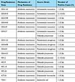
A total of 16 drug-resistance genes, predominantly OXA and KPC, were identified using mNGS in 19 Gram-negative bacterial isolates from various species. The majority of these genes were found in Acinetobacter baumannii, Klebsiella pneumoniae, and Pseudomonas aeruginosa. Notably, OXA-818 and KPC-2 were among the most frequently detected genes (Table 7).
 |
Table 7 Identification Frequency of Drug-Resistance Genes in Gram-Negative Bacteria Using mNGS |
Drug-Resistant Bacterial Genotypes and Phenotypes Identified by mNGS and Traditional Culture Methods
Five strains of drug-resistant bacteria were identified as positive via mNGS and traditional culture methods (sputum and bronchoalveolar lavage fluid). These strains included one strain of Staphylococcus aureus, two strains of Klebsiella pneumoniae, and two strains of Pseudomonas aeruginosa. The presence of drug-resistance genes mecA (in Staphylococcus aureus), KPC-2, KPC-9, KPC-18, KPC-26, OXA27, and OXA423 (in Klebsiella pneumoniae), as well as OXA488 and NDM6 (in Pseudomonas aeruginosa), shows strong concordance with the drug-resistance phenotypes predicted via traditional culture methods. Among the genes analyzed, tetM, a tetracycline-resistance gene identified in Staphylococcus aureus, exhibited limited reliability in forecasting drug-resistance phenotype when compared to conventional culture methods (Table 8).
 |
Table 8 Drug-Resistant Bacterial Genotypes and Phenotypes Identified by mNGS and Traditional Culture Methods |
Utilizing mNGS Findings to Tailor the Anti-Infective Therapeutic Regimen
Out of the 99 patients, 40 were identified as having the pathogen solely through mNGS, leading to the adjustment of their anti-infective regimen based on these results. An additional 52 patients were identified through mNGS and traditional detection methods, with 36 showing concordant pathogen detection between the two methods. A total of 76 patients had their anti-infective regimen guided exclusively by mNGS results, resulting in 76.32% (58/76) of these patients requiring modifications to their empirical antibiotic therapy. Conversely, 23.68% (18/76) of the patients did not necessitate adjustments to the anti-infective regimen as the pathogens identified by mNGS were effectively targeted by the prophylactic anti-infective regimen. Two out of 76 patients achieved complete resolution, while 72 demonstrated improvement, yielding an overall recovery rate of 97.37%. Tragically, the remaining two patients succumbed to septic shock following transplantation.
Discussion
Despite notable progress in the development of immunosuppressive drugs and surgical techniques, a considerable number of liver transplant recipients still encounter early post-operative infections, which have a significant impact on transplant outcomes.5,20 Research21–23 suggests that pulmonary infections are the most prevalent. The frequency of early post-operative infections among liver transplant recipients at our institution between 2022 and 2024 was 44.10%, aligning with Elif’s reported range of 13.7%–51%.24 Research indicates that around 70% of these infections manifest within the first week post-surgery, with mortality rates varying between 10% and 30%, significantly impacting patient outcomes.25 Hence, the timely detection of infections and the expeditious formulation of targeted anti-infective approaches directed toward pertinent pathogens, particularly drug-resistant bacteria, are imperative for mitigating patient mortality rates and enhancing prognostic outcomes.
Applying the traditional methods for detecting pathogens in the clinical diagnosis of pulmonary infection is restricted due to prolonged detection cycles, limited pathogen coverage, and susceptibility to various influencing factors that impact the positive detection rate.26,27 mNGS provides an impartial sequencing method with extensive pathogen inclusivity and heightened sensitivity, rendering it beneficial for detecting unidentified pathogens and mixed infections.28 Through the direct isolation of nucleic acid fragments from pathogens, mNGS enables the concurrent identification of pertinent resistant genes,29 impervious to the influence of antibacterial agents.28
We collected and analyzed the mNGS and traditional pathogen detection results of lower respiratory tract samples (sputum and bronchoalveolar lavage fluid) from 99 patients meeting the study criteria. We compared the differences in pathogen detection levels between the two methods. The results demonstrated that, despite all patients receiving prophylactic anti-infective drugs post-surgery, mNGS achieved a high pathogen detection rate of 92.93%, significantly surpassing that of the traditional method (54.55%, P < 0.05). Additionally, mNGS exhibited a significantly higher detection rate in mixed infection than traditional methods (67.68% vs 14.81%, P < 0.05), highlighting the substantial advantage of mNGS.
Moreover, the study included a comparative analysis of the diagnostic outcomes of mNGS and culture (bronchoalveolar lavage fluid and sputum), revealing a concordant rate of 69.23%. However, there were a few instances where samples were collected via voluntary coughing. Moreover, considering that many patients are elderly with compromised immune function, these samples may be susceptible to contamination by respiratory pathogens and colonization of oral bacteria. This could have caused contamination and oral bacterial colonization, and the limited sample size potentially influenced the outcomes. Despite these factors, mNGS and traditional cultivation demonstrated a consistency rate of 69.23%, indicating the high reliability of mNGS.
The statistical analysis of the pathogen species distribution in the 92 patients with positive sequencing shows that Enterococcus faecium, Pseudomonas aeruginosa, and Klebsiella pneumoniae were the leading bacterial pathogens, aligning with the study of Shen.30 Fungal infections were mainly represented by Pneumocystis jirovecii, Candida albicans, and Aspergillus fumigatus, which is consistent with the multiple reports31,32 on the common fungal infections in immunosuppressed patients. Human herpesvirus was the most frequently detected virus, especially CMV and HHV-7, which is consistent with the findings of Engelmann33 and Bermek.34 These results further demonstrate the consistent and credible utilization of MNGS for pathogen detection, aligning with the traditional methods employed in previous studies.
Infections following liver transplantation are characterized by the high incidence and mortality rate of drug-resistant bacterial infections, presenting a significant clinical challenge.35 The most common Gram-positive bacteria include methicillin-resistant Staphylococcus aureus, vancomycin-resistant Enterococcus faecium, and drug-resistant Streptococcus pneumoniae.36 Among the Gram-negative bacteria, the predominant strains are carbapenem-resistant Pseudomonas aeruginosa, carbapenem-resistant Acinetobacter baumannii, and carbapenem-resistant Klebsiella pneumoniae.37 In our study, all drug-resistant bacteria were identified using mNGS. This study revealed that mecA exhibited the highest detection frequency among the 10 antibiotic-resistance genes identified in Gram-positive bacteria. This gene encodes the synthesis of methicillin-resistant penicillin-binding protein 2a, serving as the primary mechanism of resistance in Staphylococcus aureus.38 The primary mechanism for conferring resistance to Gram-negative bacteria involves the production of carbapenemases.39 This study identified antibiotic-resistant genes in Gram-negative bacteria, with KPC and OXA being the predominant ones. KPC and OXA are classified as class A and class D carbapenemases, respectively.40,41 The distribution of drug-resistance genes among these pathogens offers a critical reference for clinical empirical therapy.
In this study, the drug-resistance phenotypes and genotypes of bacteria identified through mNGS and traditional culture methods were analyzed. The efficacy of mNGS in predicting bacterial drug resistance was also evaluated. The findings revealed that among the three drug-resistant bacterial strains that were double-positive for mNGS and traditional culture, the drug-resistance genes mecA (in Staphylococcus aureus), KPC-2, KPC-9, KPC-18, KPC-26, OXA27, OXA423 (in Klebsiella pneumoniae), and OXA488 and NDM6 (in Pseudomonas aeruginosa) could effectively predict drug-resistance phenotype. The detection of the tetM gene associated with tetracycline resistance in Staphylococcus aureus exhibited poor concordance with traditional culture methods. This observation suggests that the drug-resistance genes identified through mNGS generally demonstrate high predictive efficacy for bacterial drug-resistance phenotypes. Consequently, mNGS can be instrumental in aiding clinicians in developing timely, accurate, and effective anti-infective treatment strategies.
Finally, this study demonstrates the clinical utility of mNGS in diagnosing and treating early pulmonary infections in liver transplant recipients by evaluating patient prognoses in real-world clinical settings. A retrospective analysis of electronic medical records revealed that 76 patients received anti-infective treatment plans based on mNGS results. Among these patients, two were cured, 72 showed improvement, and two succumbed to their conditions. The overall recovery rate was 97.37%, suggesting a favorable prognosis. Nevertheless, it is essential to note that while the clinical superiority of mNGS is undeniable, patients with liver transplantation have increased susceptibility to post-operative secondary infections due to immune suppression. Consequently, majority of these patients receive prophylactic antibiotics that cover various pathogens after surgery, which might significantly contribute to favorable prognoses. Hence, in clinical practice, it is necessary to consider patient-specific factors and carefully develop anti-infectious plans to prevent antibiotic misuse.
This study has several limitations. The retrospective design may have introduced bias in the results. Consequently, the clinical utility of mNGS technology requires validation through prospective, large-sample, multi-center randomized controlled trials. Furthermore, mNGS detection technology faces several challenges. For example, in immunocompromised patients with liver transplants, many colonizing or opportunistic pathogens can cause infections. mNGS can detect these microorganisms, making it challenging to identify the true pathogen. Clinically, a comprehensive analysis of the infection site, symptoms, lab tests, and pathophysiology is needed to determine the cause.
Conclusion
In summary, this study assessed the clinical utility of mNGS in identifying lower respiratory tract pathogens and drug-resistance genes in patients experiencing early post-operative lung infections following liver transplantation. The findings indicate that mNGS offers significant advantages over traditional detection methods, demonstrating high concordance with conventional culture results and yielding reliable outcomes. Pathogen and drug-resistance gene distributions detected by mNGS can serve as a valuable reference for early and accurate etiological diagnosis, offering critical insights for clinical decision-making regarding antimicrobial therapy. mNGS has demonstrated significant predictive capability for bacterial drug-resistance phenotypes, aiding clinicians in timely adjustments to treatment regimens and ultimately enhancing patient prognosis.
Ethics Approval and Consent to Participate
This study was conducted in accordance with the Declaration of Helsinki and approved by the Research Ethics Committee of Beijing Friendship Hospital. In our study, all relevant examinations and inspections obtained informed consent from the patient or their family during the hospital stay. After they were discharged and when we collected their clinical data related to the study, informed consent is waived with the approval of Ethics Committee of Beijing Friendship Hospital due to the nature of retrospective study and anonymized patient’s information. All methods were carried out in accordance with relevant guidelines and regulations. At the same time, in our study, all organs were donated voluntarily with written informed consent, and that this was conducted in accordance with the Declaration with the Istanbul.
Acknowledgments
We thank the study stuff of Beijing Friendship Hospital affiliated with Capital Medical University, Department of Critical Liver Diseases and Liver Transplantation Center, who contributed to the study. All authors significantly contributed to various aspects of this study, encompassing clinical patient management, sample collection, study design, project implementation, data collection, and statistical analysis. Each author assumed responsibility for their respective tasks, and all authors reviewed and approved the final manuscript.
Disclosure
The authors report no conflicts of interest in this work.
References
1. Reardon LC, Lin JP, VanArsdell GS, et al. Orthotopic heart and combined heart liver transplantation: the ultimate treatment option for failing Fontan physiology. Curr Transplant Rep. 2021;8(1):9–20. doi:10.1007/s40472-021-00315-4
2. Shetty A, Kodali S. Long-term management of the successful adult liver transplant: a patient-friendly summary of the 2012 AASLD and AST practice guideline. Clin Liver Dis (Hoboken) Mar. 2022;19(3):83–88. doi:10.1002/cld.1179
3. Beam E, Razonable RR. A survey of infection prevention and control practices among solid organ transplantation centers. Am J Infect Control Jan. 2019;47(1):101–104. doi:10.1016/j.ajic.2018.06.020
4. Guo L, Peng P, Peng WT, Zhao J, Wan QQ. Klebsiella pneumoniae infections after liver transplantation: drug resistance and distribution of pathogens, risk factors, and influence on outcomes. World J Hepatol Apr. 2024;16(4):612–624. doi:10.4254/wjh.v16.i4.612
5. Wu X, Wu L, Wan Q. Pathogen distribution and risk factors of bacterial and fungal infections after liver transplantation. Zhong Nan Da Xue Xue Bao Yi Xue Ban. 2022;47(8):1120–1128. doi:10.11817/j.issn.1672-7347.2022.220054
6. GBD. Antimicrobial Resistance Collaborators. Global mortality associated with 33 bacterial pathogens in 2019: a systematic analysis for the Global Burden of Disease Study 2019. Lancet. 2022;400(10369):2221–2248. doi:10.1016/s0140-6736(22)02185-7
7. Zhao D, Guo L, Lian D, et al. Diagnostic value and clinical application of mNGS for post-liver transplantation infection: a cross-sectional study with case reports. Front Microbiol. 2022;13:919363. doi:10.3389/fmicb.2022.919363
8. Huang JF, Miao Q, Cheng JW, et al. Metagenomic next-generation sequencing versus traditional laboratory methods for the diagnosis and treatment of infection in liver transplantation. Front Cell Infect Microbiol. 2022;12:886359. doi:10.3389/fcimb.2022.886359
9. Zhuang L, Zhu C, Ma J, et al. Predictive performance of Metagenomic Next Generation Sequencing in early detection of post-liver transplantation infections. Heliyon. 2024;10(16):e36405. doi:10.1016/j.heliyon.2024.e36405
10. Han D, Li Z, Li R, Tan P, Zhang R, Li J. mNGS in clinical microbiology laboratories: on the road to maturity. Crit Rev Microbiol Sep-Nov. 2019;45(5–6):668–685. doi:10.1080/1040841x.2019.1681933
11. Sharma S, Maycher B, Eschun G. Radiological imaging in pneumonia: recent innovations. Curr Opin Pulm Med. 2007;13(3):159–169. doi:10.1097/MCP.0b013e3280f3bff4
12. Aydin C, Otan E, Akbulut S, et al. Postoperative pulmonary complications after liver transplantation: assessment of risk factors for mortality. Transplant Proc. 2015;47(5):1488–1494. doi:10.1016/j.transproceed.2015.04.058
13. Luan Y, Hu H, Liu C, et al. A proof-of-concept study of an automated solution for clinical metagenomic next-generation sequencing. J Appl Microbiol. 2021;131(2):1007–1016. doi:10.1111/jam.15003
14. Shen H, Shen D, Song H, et al. Clinical assessment of the utility of metagenomic next-generation sequencing in pediatric patients of hematology department. Int J Lab Hematol Apr. 2021;43(2):244–249. doi:10.1111/ijlh.13370
15. Schlaberg R, Chiu CY, Miller S, et al. Validation of metagenomic next-generation sequencing tests for universal pathogen detection. Arch Pathol Lab Med. 2017;141(6):776–786. doi:10.5858/arpa.2016-0539-RA
16. Wilson MR, Sample HA, Zorn KC, et al. Clinical metagenomic sequencing for diagnosis of meningitis and encephalitis. N Engl J Med. 2019;380(24):2327–2340. doi:10.1056/NEJMoa1803396
17. Baldassarri RJ, Kumar D, Baldassarri S, Cai G. Diagnosis of infectious diseases in the lower respiratory tract: a Cytopathologist’s perspective. Arch Pathol Lab Med. 2019;143(6):683–694. doi:10.5858/arpa.2017-0573-RA
18. Zeng HQ, Zhang XB, Cai XY, et al. Diagnostic value of bronchoalveolar lavage fluid cryptococcal antigen-lateral flow immunochromatographic assay for pulmonary cryptococcosis in non-HIV patients. Diagn Microbiol Infect Dis. 2021;99(3):115276. doi:10.1016/j.diagmicrobio.2020.115276
19. Zumla A, Al-Tawfiq JA, Enne VI, et al. Rapid point of care diagnostic tests for viral and bacterial respiratory tract infections--needs, advances, and future prospects. Lancet Infect Dis. 2014;14(11):1123–1135. doi:10.1016/s1473-3099(14)70827-8
20. Imlay H, Seibert AM, Hanson KE. Pathogen-agnostic immune biomarkers that predict infection after solid organ transplantation. Transpl Infect Dis Apr. 2023;25(2):e14020. doi:10.1111/tid.14020
21. Hara T, Soyama A, Takatsuki M, et al. The impact of treated bacterial infections within one month before living donor liver transplantation in adults. Ann Transplant. 2014;19:674–679. doi:10.12659/aot.892095
22. Feltracco P, Carollo C, Barbieri S, Pettenuzzo T, Ori C. Early respiratory complications after liver transplantation. World J Gastroenterol. 2013;19(48):9271–9281. doi:10.3748/wjg.v19.i48.9271
23. Angarita SAK, Russell TA, Kaldas FM. Pneumonia after liver transplantation. Curr Opin Organ Transplant. 2017;22(4):328–335. doi:10.1097/mot.0000000000000427
24. Küpeli E, Fö E, Haberal M. Pulmonary infections in transplant recipients. Curr Opin Pulm Med. 2012;18(3):202–212. doi:10.1097/MCP.0b013e328352104f
25. Kim SI. Bacterial infection after liver transplantation. World J Gastroenterol. 2014;20(20):6211–6220. doi:10.3748/wjg.v20.i20.6211
26. Torres A, Cilloniz C, Niederman MS, et al. Pneumonia. Nat Rev Dis Primers Apr. 2021;7(1):25. doi:10.1038/s41572-021-00259-0
27. Wang H, Lu Z, Bao Y, et al. Clinical diagnostic application of metagenomic next-generation sequencing in children with severe nonresponding pneumonia. PLoS One. 2020;15(6):e0232610. doi:10.1371/journal.pone.0232610
28. Chen Y, Feng W, Ye K, et al. Application of metagenomic next-generation sequencing in the diagnosis of pulmonary infectious pathogens from bronchoalveolar lavage samples. Front Cell Infect Microbiol. 2021;11:541092. doi:10.3389/fcimb.2021.541092
29. Yee R, Breitwieser FP, Hao S, et al. Metagenomic next-generation sequencing of rectal swabs for the surveillance of antimicrobial-resistant organisms on the Illumina MiSeq and Oxford MinION platforms. Eur J Clin Microbiol Infect Dis. 2021;40(1):95–102. doi:10.1007/s10096-020-03996-4
30. Shen YH, Fan J, Zhou J, et al. Pulmonary infection and its risk factors after orthotopic liver transplantation. Zhonghua Gan Zang Bing Za Zhi Nov. 2007;15(11):833–836.
31. Zhang ML, Xu J, Zhang W, et al. Microbial epidemiology and risk factors of infections in recipients after DCD liver transplantation. Int J Clin Pract. 2016;70(185):17–21. doi:10.1111/ijcp.12812
32. Pappas PG, Alexander BD, Andes DR, et al. Invasive fungal infections among organ transplant recipients: results of the Transplant-Associated Infection Surveillance Network (TRANSNET). Clin Infect Dis Apr. 2010;50(8):1101–1111. doi:10.1086/651262
33. Engelmann C, Sterneck M, Weiss KH, et al. Prevention and management of CMV infections after liver transplantation: current practice in German transplant centers. J Clin Med. 2020;9(8).
34. Bermek O, Williams RS. The three-component helicase/primase complex of herpes simplex virus-1. Open Biol. 2021;11(6):210011. doi:10.1098/rsob.210011
35. Martin-Mateos R, Martínez-Arenas L, Carvalho-Gomes Á, et al. Multidrug-resistant bacterial infections after liver transplantation: prevalence, impact, and risk factors. J Hepatol. 2024;80(6):904–912. doi:10.1016/j.jhep.2024.02.023
36. Karaman R, Jubeh B, Breijyeh Z. Resistance of Gram-positive bacteria to current antibacterial agents and overcoming approaches. Molecules. 2020;25(12). doi:10.3390/molecules25122888
37. Tacconelli E, Carrara E, Savoldi A, et al. Discovery, research, and development of new antibiotics: the WHO priority list of antibiotic-resistant bacteria and tuberculosis. Lancet Infect Dis Mar. 2018;18(3):318–327. doi:10.1016/s1473-3099(17)30753-3
38. Hiramatsu K, Katayama Y, Yuzawa H, Ito T. Molecular genetics of methicillin-resistant Staphylococcus aureus. Int J Med Microbiol. 2002;292(2):67–74. doi:10.1078/1438-4221-00192
39. Poirel L, Nordmann P. Carbapenem resistance in Acinetobacter baumannii: mechanisms and epidemiology. Clin Microbiol Infect. 2006;12(9):826–836. doi:10.1111/j.1469-0691.2006.01456.x
40. Zhao Y, Liao Y, Zhang N, et al. Four types of ST11 novel mutations from increasing carbapenem-resistant Klebsiella pneumoniae in Guangdong, 2016-2020. Front Microbiol. 2021;12:702941. doi:10.3389/fmicb.2021.702941
41. Ogbolu DO, Alli OAT, Oluremi AS, et al. Contribution of NDM and OXA-type carbapenemases to carbapenem resistance in clinical Acinetobacter baumannii from Nigeria. Infect Dis (Lond). 2020;52(9):644–650. doi:10.1080/23744235.2020.1775881
 © 2024 The Author(s). This work is published and licensed by Dove Medical Press Limited. The
full terms of this license are available at https://www.dovepress.com/terms.php
and incorporate the Creative Commons Attribution
- Non Commercial (unported, 3.0) License.
By accessing the work you hereby accept the Terms. Non-commercial uses of the work are permitted
without any further permission from Dove Medical Press Limited, provided the work is properly
attributed. For permission for commercial use of this work, please see paragraphs 4.2 and 5 of our Terms.


