")
Back to Journals » International Journal of Nanomedicine » Volume 20
Green Synthesis: An Eco-Friendly Approach for the Synthesis of Silver Nanoparticles Functionalized with Operculina turpethum and It’s In vitro and in vivo Biological Activities
Authors Bawazeer S
Received 5 December 2024
Accepted for publication 28 February 2025
Published 12 March 2025 Volume 2025:20 Pages 2991—3005
DOI https://doi.org/10.2147/IJN.S507134
Checked for plagiarism Yes
Review by Single anonymous peer review
Peer reviewer comments 5
Editor who approved publication: Dr Kamakhya Misra
Saud Bawazeer
Department of Pharmaceutical Science, College of Pharmacy, Umm Al-Qura University, Makkah, Saudi Arabia
Correspondence: Saud Bawazeer, Email [email protected]
Introduction: Silver nanoparticles (AgNPs) have gained significant attention in biomedical applications. Green synthesis methods provide an eco-friendly and cost-effective approach to AgNPs production, utilizing plant extracts as reducing and stabilizing agents. In this study, AgNPs were synthesized using the methanolic extract of Operculina turpethum.
Methodology: AgNPs were synthesized using O. turpethum extract, and their formation was confirmed through various analytical techniques. The antibacterial activity of both the crude extract and AgNPs was assessed against Staphylococcus aureus. Enzyme inhibition studies were conducted for urease, α-glucosidase, carbonic anhydrase II, and xanthine oxidase. Analgesic and sedative activities were evaluated through standard models.
Results: AgNPs exhibited an inhibition zone of 14 mm against S. aureus, greater than the crude extract (12 mm) but lower than Linezolid (25 mm). Enzyme inhibition studies revealed strong activity, particularly against urease (96.09% inhibition, IC5o = 25.65 ± 0.97 μg/mL). AgNPs demonstrated superior analgesic effects (81.98% at 10 mg/kg), comparable to diclofenac sodium (86.02%). Sedative effects were dose-dependent, reaching 35.09% at 10 mg/kg.
Discussion: The enhanced antibacterial activity of AgNPs suggests improved bioavailability and interaction with bacterial membranes. The strong enzyme inhibitory potential indicates their possible therapeutic role in enzyme-related disorders. The analgesic and sedative activity of AgNPs suggests their possible role in pain management agents and neuropharmacology. The results demonstrate the efficacy of green-synthesized AgNPs for biomedical applications.
Conclusion: Green synthesized AgNPs and their antibacterial, enzyme inhibitory, analgesic, and sedative properties suggest promising therapeutic applications. Further research should explore their mechanisms and in vivo safety for clinical applications.
Keywords: Operculina turpethum, Ag NPs, antibacterial studies, enzyme inhibition, sedative activities, analgesic potential
Graphical Abstract:

Introduction
Nanoparticles (NPs) are incredibly small particles with dimensions typically ranging from 1 to 100 nanometers. They exhibit unique physical, chemical, and biological properties that are distinct from their bulk counterparts. NPs have garnered significant attention in various fields due to their potential applications in various sectors like drugs, electronics, energy production, and environmental remediation.1–4 Their small size allows for a large surface-to-volume ratio, enabling enhanced reactivity, increased surface area for interactions, and improved functionality.5,6 NPs can be synthesized using different methods, including chemical, physical, and biological approaches, offering versatility in their size, shape, and composition.7 However, the unique properties of NPs also warrant careful consideration of their potential environmental and health impacts, necessitating comprehensive studies on their synthesis, characterization, and safe handling.8
Silver nanoparticles (Ag NPs) have appeared as a promising candidate for various applications including wastewater treatment due to their unique characteristics such as high surface area, small size, and unique physiochemical properties.9–12 The small size and high surface area of Ag NPs make them highly reactive and effective in inhibiting the growth of microorganisms.13 The antibacterial activity of Ag NPs is attributed to several mechanisms such as the release of silver ions, the disruption of the bacterial cell membrane, and the inhibition of bacterial enzymes. Hence, they have garnered significant attention in recent years as a candidate for the development of novel antibacterial, antifungal, and antiviral agents.14
Ag NPs have shown promise as antibacterial agents due to their broad-spectrum activity against a range of microorganisms.15 The efficacy of Ag NPs in inhibiting bacterial growth has been demonstrated in various studies.16 The synthesis of NPs including Ag NPs using plant extracts has emerged as a sustainable and eco-friendly approach for nanoparticle synthesis.17,18 Plant extracts possess several secondary metabolites, such as flavonoids, alkaloids, and terpenoids, which is responsible for the reduction and stabilizing in the synthesis of Ag NPs. This method is cost-effective, non-toxic, and eco-friendly compared to other chemical methods, making it an attractive approach for the large-scale production of Ag NPs19,20.
O. turpethum is a medicinal plant that belongs to the Convolvulaceae family. It is commonly found in India, Sri Lanka, and other tropical regions.21 The plant is known by various names such as Indian jalap, turpeth, and nisoth. The roots of the plant are used in traditional remedies to treat various ailments such as constipation, fever, and skin infections.22 The plant comprises many bioactive compounds such as terpenoids, flavonoids, and alkaloids, which have been shown to possess antioxidant, enzyme inhibitory, anti-inflammatory, and antimicrobial properties.23–26 Therefore, the use of O. turpethum extract for the synthesis of Ag NPs can provide a sustainable and eco-friendly approach for the development of novel antibacterial and enzyme inhibitory agents.
The use of plant extracts for the synthesis of Ag NPs has many advantages over other methods.27,28 First, plant extracts are readily available and can be obtained from a variety of sources. Second, plant extracts are non-toxic and eco-friendly, making them suitable for biomedical and environmental applications. Third, plant extract-mediated synthesis of silver NPs is cost-effective and requires less energy compared to other chemical methods.29–32 Therefore, the use of plant extracts for synthesizing of Ag NPs can provide a sustainable and eco-friendly approach for the development of novel antibacterial19,20,33 and enzyme-inhibitory therapeutic agents.34–36
Numerous studies have been conducted about the antibacterial and enzyme inhibitory properties of Ag NPs synthesized using plant extracts. For example, Ag NPs synthesized using Azadirachta indica extract exhibited potent antibacterial properties and significant enzyme inhibition due to the interaction of bioactive compounds of the plant with the biological targets, highlighting their applicability in treating microbial infections and enzyme-related disorders.37,38 Similarly, Moringa oleifera-mediated Ag NPs showed remarkable antimicrobial effects, attributed to the phytochemicals present in the plant extract.39 The synthesis of Ag NPs using Aloe vera and Ocimum sanctum extracts further demonstrated the ability of plant-based secondary metabolites, such as flavonoids and terpenoids, to act as reducing and stabilizing agents, making this approach both eco-friendly and effective.40,41 These studies validate the role of plant-mediated green synthesis of Ag NPs and provide a foundation for exploring diverse plant species rich in bioactive compounds for nanoparticle production. Building on this rich foundation, the current study leverages the bioactive potential of O. turpethum to synthesize Ag NPs and evaluates their in vitro and in vivo biological activities, thus contributing to the growing body of knowledge in green nanotechnology.
Despite these advances, there remains a significant gap in identifying and exploring new medicinal plant species with potent bioactive compounds for Ag NP synthesis. O. turpethum, a medicinal plant has been traditionally used for its anti-inflammatory, antimicrobial, and enzyme-inhibitory properties. However, its potential as a reducing and stabilizing agent in Ag NPs synthesis has not been extensively investigated. Moreover, while previous studies have focused primarily on the antibacterial and enzyme-inhibitory activities of plant-mediated Ag NPs, their in vivo pharmacological effects, such as sedative and analgesic properties, remain largely unexplored.
To address this gap, the present study aims to:
- Utilize the methanolic extract of O. turpethum to synthesize Ag NPs through a green and sustainable approach.
- Characterize the synthesized Ag NPs to confirm their physicochemical properties.
- Evaluate their in vitro antibacterial and enzyme-inhibitory activities.
- Investigate their in vivo sedative and analgesic effects, providing novel insights into their pharmacological potential.
By integrating in vitro and in vivo assessments, this study not only expands the scope of plant-mediated Ag NP research but also provides a comprehensive understanding of their therapeutic applications. Our findings contribute to the advancement of green nanotechnology and highlight the potential of O. turpethum-mediated Ag NPs as multifunctional bioactive agents for biomedical applications.
Materials and Methods
The dried powder of O. turpethum was procured from the local market in Peshawar, Pakistan. Analytical-grade methanol (99.8% pure) was obtained from Merck, and silver nitrate (99.99% pure) was purchased from Sigma-Aldrich. Thiourea (99% pure), acarbose (99% pure), and acetazolamide (≥99% pure), Diclofenac sodium (98–102% pure) and diazepam (98% pure), acetic acid (≥99.7% pure) and saline solution (0.9% NaCl) and all the other chemical used was of analytical grade. All the chemicals and reagents were sourced from China.
Plant Collection and Extraction
In this study, the dried powder of O. turpethum was purchased from the local market in Peshawar, Pakistan in March 2023 and then subjected to methanol extraction. Methanol is a common solvent used in plant extraction due to its ability to extract various phytochemicals effectively. To extract the bioactive compounds from the plant material, 10 grams of the dried powder were added to 100 mL of methanol in a closed conical flask. The mixture was periodically shaken at 40 degrees Celsius for 24 hours to ensure the complete extraction of phytochemicals.42 The temperature and duration of the extraction were optimized to obtain a high yield of bioactive compounds. This method of extraction is commonly used to obtain phytochemicals from plant material and is effective in extracting a wide range of compounds.43 After the extraction was completed, the mixture was filtered using filter paper to remove any insoluble residue. The resulting filtrate was then used as a reducing and stabilizing agent in the synthesis of Ag NPs.
Synthesis of Ag NPs
Silver nanoparticles were synthesized by mixing 10 mL of 5 mm AgNO3 solution with 2.5 mL of methanolic extract of O. turpethum. The reaction mixture was stirred at 40°C for 4 hours. The successful synthesis of Ag NPs was confirmed by a color change to brown and a UV-Vis (Model-UV2601) absorption peak at 400 nm.
Characterization of Ag NPs
Comprehensive characterization of the synthesized Ag NPs was conducted using multiple techniques. FTIR (Model FTIR-990) analysis was used to validate the reduction of metal ions and to identify the functional groups involved in the reaction. By analyzing the infrared absorption spectra, we gained insights into the chemical interactions between the plant extract and the silver ions, providing valuable information about the synthesis mechanism. To further investigate the morphology and size of the Ag NPs, field-emission scanning electron microscopy (JEM 2100, Jeol CRL) was utilized. FESEM allowed for high-resolution imaging of the nanoparticles, revealing their surface morphology and confirming their size and shape. This analysis provided a visual representation of the synthesized Ag NPs, aiding in the understanding of their structural properties. Furthermore, energy-dispersive X-ray spectroscopy (ADX-8000 minI) was used for elemental composition of the synthesized Ag NPs. By detecting and analyzing the characteristic X-ray emissions, EDS provided insights into the elemental composition of the nanoparticles. This analysis not only confirmed the presence of silver but also provided information about the potential presence of other elements or contaminants, shedding light on the overall composition of the synthesized Ag NPs.
In vitro Activities
Antibacterial Activity
Using a standardized protocol, the antibacterial properties of the crude methanol (MeOH) extract from O. turpethum plant and the synthesized AgNPs (silver nanoparticles) were evaluated against Staphylococcus aureus, a Gram-positive bacterium. The bacterial strains were stored in Mueller-Hinton agar at 4°C in a refrigerator. To assess the antibacterial activity, the modified agar well diffusion method was employed, with Mueller-Hinton agar serving as the growth medium. The culture was inoculated onto petri dishes and incubated at 37°C for 24–72 hours. Before use, the petri dishes were sterilized. Next, 0.6 mL of the prepared bacterial broth culture was mixed with 20 mL of sterilized molten Mueller-Hinton agar and poured into each petri dish. Wells with a diameter of 6 mm were created in the agar medium using a sterilized borer. The crude MeOH extract (50 µL) from the plant and the synthesized AgNPs (50 µL) were separately added to the wells using a micropipette. A standard drug, Linezolid, was included as a control by using a disc soaked in 50 µL of the drug. To ensure proper diffusion, the petri dishes were placed in a laminar flow hood for one hour. Subsequently, the plates were incubated for 24 hours in incubator at 37°C. The zone of inhibition, representing the area where bacterial growth was inhibited, was measured.
Enzyme Inhibition Evaluation
The potential of enzyme inhibitory of the crude methanol (MeOH) extract from O. turpethum plant and the green synthesized AgNPs was evaluated against various enzymes. For the evaluation of the urease inhibitory activity, the crude methanolic extract and Ag NPs were tested at concentrations of 0.2 µg.mL−1 and 0.25 µg.mL−1, respectively. Thiourea, a known urease inhibitor, was used as the standard inhibitor at a 0.2 µM concentration. Similarly, the α-glucosidase inhibitory capability, the crude extract (0.2 µg.mL−1) and Ag NPs (0.25 µg.mL−1) were investigated respectively. Acarbose, was used as a standard drug. To check carbonic anhydrase II enzyme inhibition, the crude MeOH extract and AgNPs were tested at concentrations of 0.2 µg.mL−1 and 0.25 µg.mL−1, respectively. A well-known inhibitor acetazolamide, was used as the standard drug at a 0.2 µM concentration. The xanthine oxidase inhibitory activity of the MeOH extract and Ag NPs were evaluated at same concentrations as used for carbonic anhydrase II enzyme inhibition. The enzyme inhibitory assays were performed by incubating the enzymes with the respective test samples and substrates under specific conditions. The activities of the enzymes were measured, and the percentage inhibition was calculated by comparing the activity observed in the control samples. Additionally, IC50 values, representing the concentration required to inhibit 50% of the enzyme activity, were determined. The data obtained from the experiments were subjected to statistical analysis to assess the significance of the inhibitory effects exhibited by the crude MeOH extract and AgNPs against the tested enzymes.
In vivo Screening
For vivo activities, albino mice (20–25 g) were used. The animals were maintained under standard laboratory conditions (temperature: 22 ± 2°C, 12-hour light/dark cycle) with access to food, water, and libitum. The study was conducted following ethical guidelines and approved by the institutional animal ethics committee.
In vivo Analgesic Activity
The analgesic activity was assessed using the acetic acid-induced writhing test. Mice were divided into groups (n = 6 per group) and treated as follows: saline (10 mL/kg, control), methanolic extract (25, 50, and 100 mg/kg), silver nanoparticles (2.5, 5, and 10 mg/kg), and diclofenac sodium (1.5 mg/kg, reference drug). One hour after oral administration of the respective treatments, 0.6% acetic acid solution was injected intraperitoneally to induce writhing. The number of writhes was counted for 20 minutes post-acetic acid injection, and the percentage inhibition of writhing was calculated for each treatment group.44
In vivo Sedative Activity
The sedative activity was evaluated using the open-field test. Mice were divided into groups (n = 6 per group) and treated with saline (10 mL/kg, control), methanolic extract (25, 50, and 100 mg/kg), silver nanoparticles (2.5, 5, and 10 mg/kg), and diazepam (0.25 mg/kg, reference drug). One hour after administration, each mouse was placed individually in an open field apparatus, and the number of square crossings was recorded for 5 minutes as a measure of locomotor activity. Reduction in locomotor activity was considered indicative of sedative effects, and the percentage effect was calculated for each group.45
Statistical Analysis
The data were expressed as mean ± standard error of the mean (SEM). A p-value of less than 0.05 was considered statistically significant, with notations *p < 0.05, **p < 0.01, and ***p < 0.001 indicating varying levels of significance.
Results
The Ag NPs were successfully synthesized using a methanolic extract of O. turpethum and were visually checked by observing a distinct color change in the reaction mixture (Figure 1), which turned from its original color to a characteristic brown hue. This change in color is often associated with the reduction of Ag ions and the subsequent production of Ag NPs as confirmed by the UV-Vis spectrophotometry.46 The phytochemicals present in the extract act as a reducing and stabilizing agent thus avoiding the use of toxic chemicals. The green synthesis of Ag NPs using O. turpethum is primarily facilitated by its rich phytochemical composition which plays a crucial role in the reduction and stabilization of Ag NPs. Flavonoids and phenolics act as strong reducing agents, donating electrons to Ag+ ions, leading to their reduction into Ago nanoparticles. Meanwhile, terpenoids and alkaloids contribute to the stabilization of the synthesized Ag NPs by capping their surface, preventing aggregation and enhancing stability.
 |
Figure 1 MeOH plant extract (a) and synthesized Ag NPs (b). |
Characterization
UV-Vis Spectroscopy
UV-Vis spectroscopy is a commonly used analytical technique for the characterization of nanoparticles. In the present study, the formation of silver nanoparticles (Ag NPs) was validated by observing a change in the color of the solution from pale yellow to brown, as well as the appearance of a characteristic absorption peak at 400 nm47 in the UV-Vis spectrum as shown in Figure 2a. This peak is a result of the surface plasmon resonance (SPR) of the nanoparticles, which is a phenomenon where the conduction electrons in the metal nanoparticles oscillate collectively in response to the incident electromagnetic radiation. The intensity and position of the SPR peak depend on the size, shape, and composition of the nanoparticles, as well as the dielectric environment. A peak within the range of 400–450 nm suggests the formation of small, spherical Ag NPs, while a redshift (higher wavelength) may indicate larger particle size or slight aggregation. The sharp and well-defined nature of the peak in our spectra confirms the uniformity and stability of the synthesized Ag NPs, further supported by their extended stability over time without significant changes in absorbance.
 |
Figure 2 UV-Vis spectrum (a), FTIR spectrum (b) of plant extract and synthesized Ag NPs. |
FTIR
FTIR spectroscopy was used to investigate the different functional groups present in the plant extract and the synthesized Ag NPs.42 The FTIR spectra of the crude methanolic extract and green-synthesized Ag NPs reveal the presence of several functional groups. The broad peak observed between 3500–3300 cm⁻¹ in the crude extract is typically associated with O-H stretching vibrations from alcohols, phenols, or carboxylic acids, with the broadening indicating hydrogen bonding interactions. The sharp peaks at 3200 cm⁻¹ and 3000 cm⁻¹ in the crude extract are likely due to N-H stretching vibrations of amines or C-H stretching vibrations of aliphatic hydrocarbons. A weak peak at 2500 cm⁻¹ could be attributed to O-H stretching of carboxylic acids, while the sharp peak at 1652 cm⁻¹ observed in both the crude extract and Ag NPs is likely related to the C=O stretching of amide bonds. The sharp peaks at 1425 cm⁻¹ and 2509 cm⁻¹ in the crude extract may be due to C-H bending vibrations of alkyl groups or C=O stretching in carboxylic acids and C≡C stretching in alkynes, respectively. The high-intensity peak at 1009 cm⁻¹ in both the crude extract and Ag NPs corresponds to C-O stretching vibrations from phenolic groups. The shift in the broad peak at 3500–3000 cm⁻¹ in the synthesized Ag NPs, which becomes narrower, indicates the reduction and capping of Ag ions by the functional groups in the plant extract. Additionally, the weakening of the peaks at 3200 and 3000 cm⁻¹ in the Ag NPs suggests that amines and hydrocarbons in the extract interact with the silver ions during nanoparticle synthesis. These findings highlight the involvement of specific functional groups in the reduction and stabilization of Ag ions during the green synthesis of Ag NPs. The FTIR Spectrum of the crude methanolic extract and Ag NPs is shown in the Figure 2b
FTIR spectroscopy analysis of the crude extract and synthesize nanoparticles Ag NPs indicated the contribution of several functional groups in the green synthesis of Ag NPs. The existence of diverse functional groups presents in the plant extract played a crucial role in the reduction and capping of Ag ions to form Ag NPs, and the disappearance of certain peaks in the FTIR spectrum of the synthesized Ag NPs indicated their involvement in the nanoparticle’s synthesis.
FESEM
FESEM (Field-Emission Scanning Electron Microscopy) is an invaluable tool for studying the surface morphology and size of nanomaterials. In our study, we utilized FESEM to analyze the synthesized Ag NPs (silver nanoparticles) derived from O. turpethum. The FESEM analysis revealed that these nanoparticles exhibit a spherical shape during their growth process. To gain a comprehensive understanding of the synthesized nanoparticles, we captured FESEM images at both low and high resolutions. These images effectively portray the intricate texture of the extract in which the nanoparticles are grown. By examining the FESEM images, we can delve deeper into the structural details and characteristics of the nanoparticles and their surrounding environment. The FESEM image is shown in Figure 3.
 |
Figure 3 Low resolution (a) and High resolution (b) FESEM images of the synthesized Ag NPs. |
EDS
In the EDS (Energy-Dispersive X-ray Spectroscopy) analysis of the synthesized Ag NPs (silver nanoparticles), elemental signals were observed at specific energy levels. The carbon (C) signal appeared at 0.06 keV, the oxygen (O) signal at 0.13 keV, and the silver (Ag) signal at 0.04 keV.
Quantitatively, the relative percentages of these elements were determined from the EDS spectrum. The carbon signal accounted for approximately 29.32% of the detected elements, followed by oxygen at 32.97%, and silver at 0.42%. These findings offer valuable insights into the elemental makeup of the synthesized Ag NPs. The EDS spectrum of the synthesized Ag NPs is given in the inset of Figure 4.
 |
Figure 4 The EDS spectrum of the green synthesized Ag NPs. |
In vitro Activities
Antibacterial Activity
The bactericidal activity of (Ag NPs) and the crude methanol (MeOH) extract from the O. turpethum plant was evaluated against the bacteria S. aureus. As controls, distilled water was used as a negative control, and Linezolid, a standard drug, was used as a positive control. The results said that distilled water had no inhibitory effect on the bacterial strain, indicating its lack of antibacterial activity. In contrast, Linezolid exhibited the highest inhibitory value of 25 mm against S. aureus, confirming its potent antibacterial activity. Ag NPs demonstrated significant inhibitory activity with a mean value of 14 mm against S. aureus, indicating their antimicrobial potential. The crude plant extract exhibited the lowest mean value of 12 mm for inhibitory activity against the bacterial strain. The results also revealed that various solutions containing Ag NPs displayed inhibitory effects against tested gram-positive bacteria, as shown in Table 1 and Figure 5.
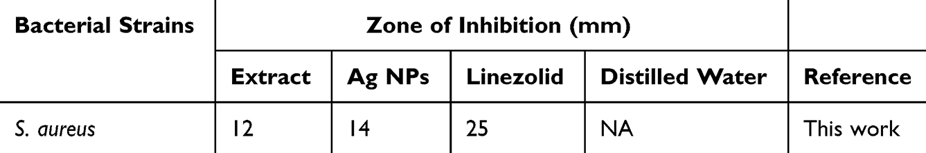 |
Table 1 Antibacterial Effect of Plant Ext., Ag NPs (Zone of Inhibition) |
 |
Figure 5 Antibacterial activities of MeOH plant extract and synthesized Ag NPs. |
Urease Inhibitory Activity
O. turpethum’s methanolic extract significantly reduced urease activity, with a percentage inhibition of 72.65%. The proportion of inhibition for the green synthesized silver nanoparticles, however, was 96.09%, indicating even inhibitory efficacy. The IC50 value for the nanoparticles was found to be 25.65±0.97 µg.mL−1, whereas the IC50 value for the methanolic extract was found to be 68.02±1.22 µg.mL−1 as shown in Table 2 According to these findings, the crude methanolic extract has a less inhibitory effect on urease activity than the green synthetic silver nanoparticles made from O. turpethum.
 |
Table 2 Enzyme Inhibitory Potential of Crude Extract Silver Nanoparticles Using Methanolic Extract of O. turpethum |
α-Glucosidase Activity
An enzyme involved in the breakdown of carbohydrates, -glucosidase, was inhibited by the MeOH extract and greenly synthesized AgNPs. Although the inhibitory effects were present in both samples, the nanoparticles showed a higher level of inhibition. These findings show the potential of NPs from O. turpethum as -glucosidase inhibitors, which may be investigated for treating disorders involving glucose metabolism. The nanoparticles were evaluated at a concentration of 0.25 µg.mL−1, whilst the methanolic extract was examined at a concentration of 0.2 µg.mL−1. The table displays the % inhibition. With a percentage inhibition of 30.76%, the methanolic extract showed a moderate inhibitory action against -glucosidase. The greenly synthesized AgNPs, on the other hand, had improved inhibitory activity, with a percentage inhibition of 48.09% as indicated in Table 2.
Carbonic Anhydrase II Enzyme Activity
The carbonic anhydrase II enzyme was moderately inhibited by the methanolic extract of O. turpethum, with a percentage inhibition of 45.09%. The proportion of inhibition for the green synthesized silver nanoparticles was 85.09%, in comparison, which showed much stronger inhibitory activity. The nanoparticles’ IC50 value was determined to be 0.66±0.80 µg.mL−1 (Table 2). These results indicate that as compared to the crude methanolic extract, the green synthesized silver nanoparticles had a more effective inhibitory impact on the carbonic anhydrase II enzyme. The information shows that, in comparison to the methanolic extract, the nanoparticles have a substantially stronger inhibitory impact. The nanoparticles’ lower IC50 values indicate that they are more effective in inhibiting carbonic anhydrase II enzyme activity.
Xanthine Oxidase Activity
The xanthine oxidase enzyme was significantly inhibited by the methanolic extract of O. turpethum, with a percentage inhibition of 66.98%. With a percentage inhibition of 90.97%, the green synthesized silver nanoparticles, however, demonstrated an even stronger inhibitory efficacy. The IC50 value for the nanoparticles was 14.23±1.01 µg.mL−1, whereas the IC50 value for the methanolic extract was determined to be 92.09±1.98 µg.mL−1. According to the data given in Table 2, when compared to the crude methanolic extract, green synthesized silver nanoparticles from O. turpethum have a stronger inhibitory effect on xanthine oxidase activity.
In vivo Biological Screening
Analgesic Effect
For the analgesic activity, the methanolic extract showed increased efficacy with higher doses, achieving percentage effects of 47.89% ± 2.50 at 25 mg/kg, 59.34% ± 2.26* at 50 mg/kg, and 65.87% ± 2.01** at 100 mg/kg. In comparison, the silver nanoparticles demonstrated a notably higher analgesic activity even at lower doses. A 2.5 mg/kg dose of nanoparticles yielded an effect of 61.45% ± 1.63**which was comparable to the 100 mg/kg dose of the methanolic extract. The analgesic effect increased further with the nanoparticles, reaching 75.09% ± 1.95*** at 5 mg/kg and 81.98% ± 1.85*** at 10 mg/kg. Diclofenac sodium at 1.5 mg/kg served as the standard analgesic and exhibited the highest effect at 86.02% ± 0.24*** as shown in the Table 3
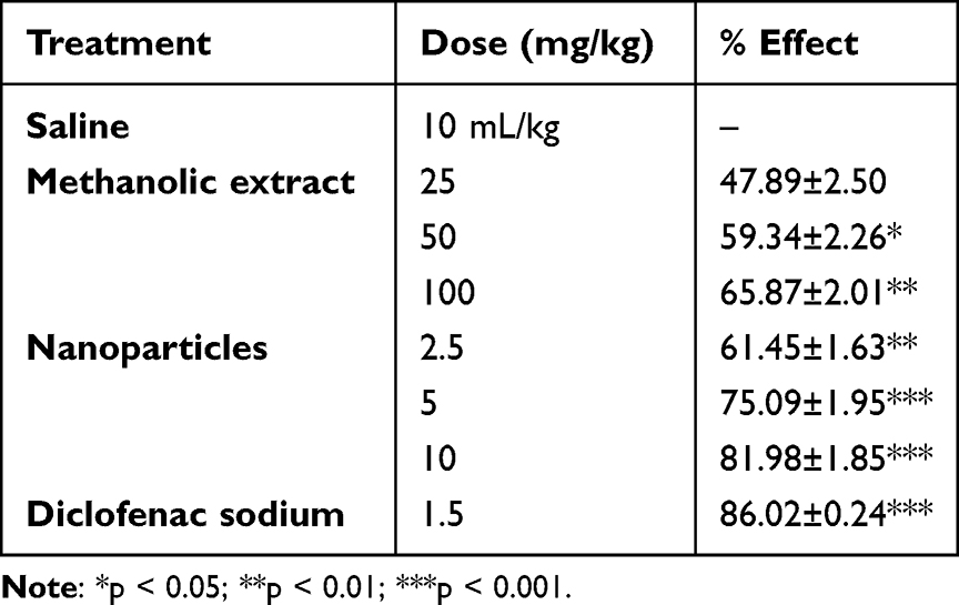 |
Table 3 Analgesic Activity of Crude Extract Ag NPs Using Methanolic Extract of O. turpethum |
Sedative Activities
The methanolic extract displayed a dose-dependent sedative effect, with 25, 50, and 100 mg/kg doses producing sedative effects of 30.98% ± 2.00, 39.12% ± 1.98, and 50.09% ± 1.87**respectively. While the silver nanoparticles also showed a same trend in their sedative activity, with higher doses resulting in higher effects. At a dose of 2.5 mg/kg, the nanoparticles produced a sedative effect of 14.98±1.20***which increased to 25.09% ± 1.23** at 5 mg/kg and further to 35.09±1.65** at 10 mg/kg. Diazepam at 0.25 mg/kg, used as the reference sedative, showed a minimal effect of 1.14% ± 1.00*** in this model as shown in the Table 4.
 |
Table 4 Sedative Activity of Crude Extract Silver Nanoparticles Using Methanolic Extract of O. turpethum |
Discussion
Plants serve as rich sources of phytochemicals with diverse therapeutic applications, and these compounds possess inherent capabilities for reducing and stabilizing processes conducive to nanoparticle formation.48,49 In this study, we utilized the methanolic extract of O. turpethum to synthesize silver nanoparticles (AgNPs) with the aim of enhancing their biological applications. The nanoparticle synthesis followed a standardized protocol documented in the literature.42 The successful synthesis of AgNPs was substantiated through UV spectroscopy, revealing an absorption peak around 400 nm in the spectrum a characteristic feature commonly associated with silver nanoparticles.50 The confluence of observed color changes and the specific absorption peak in the UV-Vis spectrum collectively provided robust confirmation of the effective synthesis of silver nanoparticles. Mechanistically, When an AgNO₃ solution is mixed with the plant extract, bioactive compounds such as flavonoids, terpenoids, and phenolics reduce Ag+ to Ago. This reduction is marked by a color change, typically to yellowish-brown, due to surface plasmon resonance. The reduced silver atoms aggregate to form nuclei, which grow into nanoparticles as more silver is reduced. Simultaneously, the phytochemicals stabilize the nanoparticles by capping their surfaces, preventing aggregation and ensuring uniform size. The synthesized nanoparticles underwent comprehensive characterization employing FTIR, SEM, and EDS. In the FTIR spectrum, alterations in the intensity of peaks in both the crude extract and AgNPs indicated the involvement of functional groups in the reduction process. Additionally, the disappearance of the sharp peak at 1425 cm−1 in the NPs suggested its role in the synthesis process SEM analysis demonstrated the uniform growth of the synthesized nanoparticles, providing insight into their structural characteristics. Furthermore, EDS analysis confirmed the presence of Ag ions in the reaction mixture, underscoring the purity of the synthesized nanoparticles. The biological activities of the synthesized NPs were also evaluated. The significant inhibitory activity of Ag NPs against S. aureus underscores their antimicrobial potential. These results are consistent with previous studies reported in the literature, reinforcing the reliable antimicrobial properties of Ag NPs,42,51 and validating the reliable antibacterial properties of silver nanoparticles. The antibacterial activity of AgNPs synthesized from O. turpethum was evaluated against Staphylococcus aureus, with the nanoparticles exhibiting a zone of inhibition of 14 mm. The antibacterial activities of the green synthesized NPs are due to the production of ROS and the interaction of the metal ions with the cell membrane, which disrupts the cell membrane.52–54 This result is comparable to the findings reported by Shahid et al (2024), where AgNPs synthesized from Callistemon viminalis also showed a similar zone of inhibition (14.5 mm) against S. aureus.55 Abdussalam-Mohammed et al, 2025 also reported the antibacterial activities of green synthesized Ag NPs, and comparable results have been observed.11 The inhibition of enzymes is also due to the various effective interactions including hydrogen bonding, dipole-dipole interaction and Van der Waals forces of the NPs functionalized with secondary metabolites with the active sites of the enzyme. The enzyme inhibition activity of the AgNPs in this study was promising. Specifically, the AgNPs exhibited 96.09% inhibition of urease at 0.25 µg/mL, with an IC50 value of 25.65±0.97 µg/mL. For xanthine oxidase, the AgNPs showed a 90.97% inhibition at the same concentration, with an IC50 value of 14.23±1.01 µg/mL. Gul et al (2021), reported the inhibition of enzymes where Ricinus communis-mediated AgNPs from roots (R-AgNPs) showed 94.2% inhibition against urease with an IC50 value of 36.81±0.05 µg/mL and 83.6% inhibition against xanthine oxidase (IC50 value 3.60±0.04 µg/mL). The R. communis leaf-mediated AgNPs (L-AgNPs) showed a similar trend in inhibition, with 92.1% urease inhibition and 83% inhibition against xanthine oxidase.56 The enzyme inhibition results from O. turpethum AgNPs in this study are in line with previous reports, highlighting the potential of plant-mediated AgNPs for therapeutic applications. The results indicate a promising avenue for the development of antibacterial agents derived from O. turpethum. Moreover, the significant inhibitory effects on various enzymes, evident in urease, α-glucosidase, carbonic anhydrase II, and xanthine oxidase assays were also observed. In the context of urease inhibitory activity, the superior efficacy of Ag NPs compared to the crude methanolic extract suggests enhanced potential for urease inhibition, offering a promising avenue for the development of therapeutic urease inhibitors This shows that the urease inhibitory potential of silver nanoparticles made using O. turpethum has increased, which may be related to the unique features and capabilities of the nanoparticles.57 Additionally, the heightened inhibitory activity of AgNPs against α-glucosidase, in comparison to the methanolic extract, suggests potential applications in managing disorders related to glucose metabolism. The lower IC50 values, indicative of a stronger inhibitory impact on carbonic anhydrase II enzyme activity, further underline the potential effectiveness of green-synthesized AgNPs as inhibitors, encouraging further exploration into their therapeutic implications in physiological processes. The substantial inhibitory effect on xanthine oxidase activity by these nanoparticles suggests their potential in therapeutic applications for disorders associated with purine metabolism. The enhanced physicochemical characteristics and improved interactions of the nanoparticles with the enzyme may be responsible for their increased inhibitory activity, which results in a more dramatic inhibition of xanthine oxidase. The in vivo results demonstrate that the silver nanoparticles of O. turpethum exhibit enhanced analgesic effects compared to the methanolic extract. The nanoparticles achieved significant analgesic activity at much lower doses, with a 2.5 mg/kg dose producing an effect like that of the highest dose of the methanolic extract (100 mg/kg). This suggests that the nanoparticle formulation increases the bioavailability and systemic absorption of active compounds, allowing for more effective pain relief at lower concentrations. Furthermore, the highest analgesic efficacy observed at 10 mg/kg of nanoparticles (81.98% ± 1.85***) was close to the standard diclofenac sodium (86.02% ± 0.24***), indicating the potential of these nanoparticles as a potent alternative analgesic. Conversely, the sedative effects of the silver nanoparticles showed a same trend with increasing doses. While the methanolic extract displayed a straightforward dose-dependent increase in sedative activity, the nanoparticles’ highest sedative effect was recorded at 10 mg/kg. The low sedative effect of diazepam in this particular study model, despite being a standard reference drug, suggests that the parameters used to assess sedation may differ from conventional models. These findings highlight the promising analgesic potential of silver nanoparticles derived from O. turpethum. The observed sedative effects suggest that these AgNPs could be further explored as therapeutic agents for managing conditions related to anxiety, stress, and sleep disorders. Similarly, the analgesic properties could provide a new avenue for pain management, offering a more eco-friendly alternative to conventional synthetic drugs. However, further research is needed to clarify the mechanisms behind the reduced sedative effects of the nanoparticles and to optimize their formulation for potential therapeutic use. More research is needed to delve deeper into the active ingredients, optimize nanoparticle production, and ensure their safety and efficacy in humans. But these initial findings offer a compelling case for further exploration, potentially leading to exciting new medicines rooted in this ancient medicinal plant.
While this work successfully demonstrates the green synthesis of AgNPs and evaluates their biological activities, there are a few limitations. First, the synthesis process, although effective on a small scale, may require optimization for large-scale production, particularly for industrial or clinical applications. Additionally, while the in vitro results are promising, further investigation into the in vivo toxicity, biocompatibility, and long-term stability of the synthesized AgNPs is necessary for validating their clinical potential. Lastly, the exact mechanisms underlying the biological activities of the AgNPs, including their interactions with cellular components, remain to be fully explored.
Conclusion
This study highlights the potential of green-synthesized AgNPs from O. turpethum for biomedical applications. The AgNPs demonstrated significant antibacterial and inhibition of enzymes, confirming their effectiveness as antimicrobial agents. Additionally, their dose-dependent sedative and analgesic effects suggest therapeutic potential for pain and neurological disorders. This eco-friendly and cost-effective synthesis method offers a sustainable approach to developing novel therapeutic agents, reducing reliance on traditional chemical methods. Future studies could further explore the clinical applications and long-term stability and in-depth toxicity studies of these AgNPs.
Ethical Approval
This study was conducted following ethical guidelines and regulations for research involving animals. Ethical approval was obtained from the Bio-Pharmacokinetics Centre, Makkah, Saudi Arabia, under committee approval number H-02–K-072-2024-047. The Bio-Pharmacokinetics Centre is an accredited ethical review body in Saudi Arabia. All experimental procedures adhered to the relevant institutional and national guidelines for treating and using laboratory animals.
Acknowledgment
The author greatly acknowledged the Department of Pharmaceutical Science, College of Pharmacy, Umm Al-Qura University, Makkah, Saudi Arabia for providing experimental facilities.
Author Contributions
Saud Bawazeer (SB): The author made a significant contribution to the work reported, whether that is in the conception, study design, execution, acquisition of data, analysis and interpretation, or in all these areas; took part in drafting, revising or critically reviewing the article; gave final approval of the version to be published; has agreed on the journal to which the article has been submitted; and agree to be accountable for all aspects of the work.
Disclosure
The author declares that they have no conflicts of interest.
References
1. Aljohny BO, Ahmad Z, Shah SA, Anwar Y, Khan SA. Cellulose acetate composite films fabricated with zero‐valent iron nanoparticles and its use in the degradation of persistent organic pollutants. Appl Organomet Chem. 2020;34(11):e5892. doi:10.1002/aoc.5892
2. Riaz M, Khan N, Khan SA, et al. Enhanced catalytic reduction/degradation of organic pollutants and antimicrobial activity with metallic nanoparticles immobilized on copolymer modified with NaY zeolite films. J Mol Liq. 2022;359:119246. doi:10.1016/j.molliq.2022.119246
3. Shah SA, Ahmad Z, Khan SA, et al. Biomass impregnated zero-valent Ag and Cu supported-catalyst: evaluation in the reduction of nitrophenol and discoloration of dyes in aqueous medium. J Organomet Chem. 2021;938:121756. doi:10.1016/j.jorganchem.2021.121756
4. Varadavenkatesan T, Nagendran V, Vinayagam R, Goveas LC, Selvaraj R. Effective degradation of dyes using silver nanoparticles synthesized from Thunbergia grandiflora leaf extract. Bioresour Technol Rep. 2024;27:101914. doi:10.1016/j.biteb.2024.101914
5. Ahmad Z, Shah SA, Khattak I, et al. Melia azedarach impregnated co and ni zero-valent metal nanoparticles for organic pollutants degradation: validation of experiments through statistical analysis. J Mater Sci-Mater El. 2020;31:16938–16950.
6. Selvaraj R, Nagendran V, Varadavenkatesan T, Goveas LC, Vinayagam R. Stable silver nanoparticles synthesis using Tabebuia aurea leaf extract for efficient water treatment: a sustainable approach to environmental remediation. Chem Eng Res Des. 2024;208:456–463. doi:10.1016/j.cherd.2024.07.012
7. Rukhsar M, Ahmad Z, Rauf A, Zeb H, Ur-Rehman M, Hemeg HA. An overview of iron oxide (Fe3O4) nanoparticles: from synthetic strategies, characterization to antibacterial and anticancer applications. Crystals. 2022;12(12):1809. doi:10.3390/cryst12121809
8. Rauf A, Rashid U, Atta A, et al. Antiproliferative activity of lignans from Olea ferruginea: in vitro evidence supported by docking studies. Front Biosci-Landmark. 2023;28(9):216. doi:10.31083/j.fbl2809216
9. Nakamura S, Sato M, Sato Y, et al. Synthesis and application of silver nanoparticles (Ag NPs) for the prevention of infection in healthcare workers. Int J mol Sci. 2019;20(15):3620. doi:10.3390/ijms20153620
10. Vadakkan K, Rumjit NP, Ngangbam AK, Vijayanand S, Nedumpillil NK. Novel advancements in the sustainable green synthesis approach of silver nanoparticles (AgNPs) for antibacterial therapeutic applications. Coord. Chem. Rev. 2024;499:215528.
11. Abdussalam-Mohammed W, Edbey K, Farhat HE, Shah P, Shamsi SS, Bhattarai A. Facile green synthesis of novel AgNPs using hyoscyamus leaf extract as capping agent: characterization and their potential antibacterial activities. Inorg Chem Commun. 2025;173:113893. doi:10.1016/j.inoche.2025.113893
12. Sharma NK, Vishwakarma J, Rai S, Alomar TS, AlMasoud N, Bhattarai A. Green route synthesis and characterization techniques of silver nanoparticles and their biological adeptness. ACS omega. 2022;7(31):27004–27020. doi:10.1021/acsomega.2c01400
13. Vadakkan K, Hemapriya J, Ngangbam AK, Sathishkumar K, Mapranathukaran VO. Biofilm inhibition of Staphylococcus aureus by silver nanoparticles derived from Hellenia speciosa rhizome extract. Microb Pathogenesis. 2024;196:106933. doi:10.1016/j.micpath.2024.106933
14. Parmar S, Kaur H, Singh J, Matharu AS, Ramakrishna S, Bechelany M. Recent advances in green synthesis of Ag NPs for extenuating antimicrobial resistance. Nanomaterials. 2022;12(7):1115. doi:10.3390/nano12071115
15. Hassanien AS, Khatoon UT. Synthesis and characterization of stable silver nanoparticles, Ag-NPs: discussion on the applications of Ag-NPs as antimicrobial agents. Phys B Condens Matter. 2019;554:21–30. doi:10.1016/j.physb.2018.11.004
16. Yin Z, J ZIS, Mei ML, Li Q, Chu CH, Chu CH. The antibacterial mechanism of silver nanoparticles and its application in dentistry. Int j Nanomed. 2020;Volume 15:2555–2562. doi:10.2147/IJN.S246764
17. Chandra A, Bhattarai A, Yadav AK, Adhikari J, Singh M, Giri B. Green synthesis of silver nanoparticles using tea leaves from three different elevations. ChemistrySelect. 2020;5(14):4239–4246. doi:10.1002/slct.201904826
18. Alomar TS, AlMasoud N, Awad MA, et al. Designing green synthesis-based silver nanoparticles for antimicrobial theranostics and cancer invasion Prevention. Int j Nanomed. 2024;Volume 19:4451–4464. doi:10.2147/IJN.S440847
19. Huang J, Li Q, Sun D, et al. Biosynthesis of silver and gold nanoparticles by novel sundried Cinnamomum camphora leaf. Nanotechnology. 2007;18(10):105104. doi:10.1088/0957-4484/18/10/105104
20. MubarakAli D, Thajuddin N, Jeganathan K, Gunasekaran M. Plant extract mediated synthesis of silver and gold nanoparticles and its antibacterial activity against clinically isolated pathogens. Colloids Surf B. 2011;85(2):360–365. doi:10.1016/j.colsurfb.2011.03.009
21. Ahmad T, Husain MK, Tariq M, et al. A review on Operculina turpethum: a potent herb of Unani system of medicine. J Pharmacogn Phytochem. 2017;6(1):23–26.
22. Kohli K, Nipanikar S, Kadbhane K. A comprehensive review on trivrit [Operculina turpethum syn. ipomoea turpethum]. Int J Pharm Bio Sci. 2010;1(4).
23. Choudhary N, Prasad SB, Singh A, et al. Phytochemistry and pharmacological potential of Operculina turpethum. Plant Arch. 2020;20:683–692.
24. Bhande RM, Kumar P, Mahurkar NK, Setty SR. Pharmacological screening of root of Operculina turpethum and its formulations. Acta Pharma Sci. 2006;48(1).
25. Gupta S, Ved A. Operculina turpethum (Linn.) Silva Manso as a medicinal plant species: a review on bioactive components and pharmacological properties. Pharmacogn revi. 2017;11(22):158. doi:10.4103/phrev.phrev_6_17
26. Wall M, Wani M, Brown D, et al. Effect of tannins on screening of plant extracts for enzyme inhibitory activity and techniques for their removal. Phytomedicine. 1996;3(3):281–285. doi:10.1016/S0944-7113(96)80067-5
27. Kumar S, Basumatary IB, Sudhani HP, et al. Plant extract mediated silver nanoparticles and their applications as antimicrobials and in sustainable food packaging: a state-of-the-art review. Trends Food Sci Technol. 2021;112:651–666. doi:10.1016/j.tifs.2021.04.031
28. Shaik MR, Khan M, Kuniyil M, et al. Plant-extract-assisted green synthesis of silver nanoparticles using Origanum vulgare L. extract and their microbicidal activities. Sustainability. 2018;10(4):913. doi:10.3390/su10040913
29. Alshameri AW, Owais M. Antibacterial and cytotoxic potency of the plant-mediated synthesis of metallic nanoparticles Ag NPs and ZnO NPs: a review. OpenNano. 2022;8:100077. doi:10.1016/j.onano.2022.100077
30. Borase HP, Salunke BK, Salunkhe RB, et al. Plant extract: a promising biomatrix for ecofriendly, controlled synthesis of silver nanoparticles. Appl. Biochem. Biotechnol. 2014;173:1–29. doi:10.1007/s12010-014-0831-4
31. Ghaffari-Moghaddam M, Hadi-Dabanlou R, Khajeh M, Rakhshanipour M, Shameli K. Green synthesis of silver nanoparticles using plant extracts. Korean J Chem Eng. 2014;31:548–557. doi:10.1007/s11814-014-0014-6
32. Kumar PV, Pammi S, Kollu P, Satyanarayana K, Shameem U. Green synthesis and characterization of silver nanoparticles using Boerhaavia diffusa plant extract and their anti bacterial activity. Ind Crops Prod. 2014;52:562–566. doi:10.1016/j.indcrop.2013.10.050
33. Azizi M, Sedaghat S, Tahvildari K, Derakhshi P, Ghaemi A. Synthesis of silver nanoparticles using Peganum harmala extract as a green route. Green Chem Lett Rev. 2017;10(4):420–427. doi:10.1080/17518253.2017.1395081
34. Alam M, Uddin G, Rashid U, et al. In vitro and in silico xanthine oxidase inhibitory potential of benzofuran isolated from viburnum grandiflorum wall. Ex DC. S Afr J Bot. 2021;143:359–362. doi:10.1016/j.sajb.2021.01.010
35. Rauf A, Khan IA, Muhammad N, et al. Phytochemical composition, in vitro urease, α-glucosidase and phosphodiesterase inhibatroy potency of Syzygium cumini (Jamun) fruits. S Afr J Bot. 2021;143:418–421. doi:10.1016/j.sajb.2021.04.006
36. Rauf A, Raza M, Saleem M, et al. Carbonic anhydrase and urease inhibitory potential of various plant phenolics using in vitro and in silico methods. Chem Biodivers. 2017;14(6):e1700024. doi:10.1002/cbdv.201700024
37. Asimuddin M, Shaik MR, Adil SF, et al. Azadirachta indica based biosynthesis of silver nanoparticles and evaluation of their antibacterial and cytotoxic effects. J King Saud Univ Sci. 2020;32(1):648–656. doi:10.1016/j.jksus.2018.09.014
38. Chi NTL, Narayanan M, Chinnathambi A, et al. Fabrication, characterization, anti-inflammatory, and anti-diabetic activity of silver nanoparticles synthesized from Azadirachta indica kernel aqueous extract. Environ Res. 2022;208:112684. doi:10.1016/j.envres.2022.112684
39. Younas M, Rasool MH, Khurshid M, et al. Moringa oleifera leaf extract mediated green synthesis of silver nanoparticles and their antibacterial effect against selected gram-negative strains. Biochem. Syst. Ecol. 2023;107:104605. doi:10.1016/j.bse.2023.104605
40. Anju T, Parvathy S, Veettil MV, et al. Green synthesis of silver nanoparticles from Aloe vera leaf extract and its antimicrobial activity. Materials Today: Proceedings. 2021;43:3956–3960.
41. Khan M, Tarek F, Nuzat M, Momin M, Hasan M. Rapid biological synthesis of silver nanoparticles from Ocimum sanctum and their characterization. J Nanosci. 2017;2017(1):1693416. doi:10.1155/2017/1693416
42. Rahman H, Rauf A, Khan SA, et al. Green synthesis of silver nanoparticles using rhazya stricta decne extracts and their anti-microbial and anti-oxidant activities. Crystals. 2023;13(3):398. doi:10.3390/cryst13030398
43. Parekh J, Jadeja D, Chanda S. Efficacy of aqueous and methanol extracts of some medicinal plants for potential antibacterial activity. Turk J Biol. 2005;29(4):203–210.
44. Muhammad N, Saeed M, Khan H. Antipyretic, analgesic and anti-inflammatory activity of Viola betonicifolia whole plant. BMC Complementary Alternative Med. 2012;12:1–8. doi:10.1186/1472-6882-12-59
45. Rauf A, Al-Awthan YS, Khan IA, et al. In vivo anti-inflammatory, analgesic, muscle relaxant, and sedative activities of extracts from Syzygium cumini (L.) Skeels in mice. Evid Based Complt Alternat Med. 2022;2022:1–7. doi:10.1155/2022/6307529
46. Balachandar R, Gurumoorthy P, Karmegam N, et al. Plant-mediated synthesis, characterization and bactericidal potential of emerging silver nanoparticles using stem extract of Phyllanthus pinnatus: a recent advance in phytonanotechnology. J Cluster Sci. 2019;30:1481–1488. doi:10.1007/s10876-019-01591-y
47. Ebrahimzadeh MA, Naghizadeh A, Amiri O, Shirzadi-Ahodashti M, Mortazavi-Derazkola S. Green and facile synthesis of Ag nanoparticles using Crataegus pentagyna fruit extract (CP-AgNPs) for organic pollution dyes degradation and antibacterial application. Bioorg. Chem. 2020;94:103425. doi:10.1016/j.bioorg.2019.103425
48. Dzobo K. The role of natural products as sources of therapeutic agents for innovative drug discovery. Comprehensive Pharmacology. 2022;408.
49. Ovais M, Khalil AT, Islam NU, et al. Role of plant phytochemicals and microbial enzymes in biosynthesis of metallic nanoparticles. Appl Microbiol Biotechnol. 2018;102:6799–6814. doi:10.1007/s00253-018-9146-7
50. Bryaskova R, Pencheva D, Nikolov S, Kantardjiev T. Synthesis and comparative study on the antimicrobial activity of hybrid materials based on silver nanoparticles (AgNps) stabilized by polyvinylpyrrolidone (PVP). J Chem Bio. 2011;4:185–191. doi:10.1007/s12154-011-0063-9
51. Mohanta YK, Biswas K, Panda SK, et al. Phyto‐assisted synthesis of bio‐functionalised silver nanoparticles and their potential anti‐oxidant, anti‐microbial and wound healing activities. IET Nanobiotechnol. 2017;11(8):1027–1034. doi:10.1049/iet-nbt.2017.0017
52. Chan YB, Aminuzzaman M, Rahman MK, et al. Green synthesis of ZnO nanoparticles using the mangosteen (Garcinia mangostana L.) leaf extract: comparative preliminary in vitro antibacterial study. Green Processing and Synthesis. 2024;13(1):20230251. doi:10.1515/gps-2023-0251
53. Selvanathan V, Aminuzzaman M, Tan LX, et al. Synthesis, characterization, and preliminary in vitro antibacterial evaluation of ZnO nanoparticles derived from soursop (Annona muricata L.) leaf extract as a green reducing agent. J Mater Res Technol. 2022;20:2931–2941. doi:10.1016/j.jmrt.2022.08.028
54. Heng MH, Win YF, Cheah ESG, et al. Microwave-assisted green synthesis, characterization, and in vitro antibacterial activity of NiO nanoparticles obtained from lemon peel extract. Green Processing and Synthesis. 2024;13(1):20240071. doi:10.1515/gps-2024-0071
55. Khan S, Rauf A, Aljohani AS, et al. Green synthesis of silver and gold nanoparticles in Callistemon viminalis extracts and their antimicrobial activities. Bioprocess Biosyst Eng. 2024;1–15.
56. Gul A, Fozia S, Shaheen A, et al. Green synthesis, characterization, enzyme inhibition, antimicrobial potential, and cytotoxic activity of plant mediated silver nanoparticle using Ricinus communis leaf and root extracts. Biomolecules. 2021;11(2):206. doi:10.3390/biom11020206
57. Ahmad K, Asif HM, Afzal T, et al. Green synthesis and characterization of silver nanoparticles through the Piper cubeba ethanolic extract and their enzyme inhibitory activities. Front Chem. 2023;11:1065986. doi:10.3389/fchem.2023.1065986
 © 2025 The Author(s). This work is published and licensed by Dove Medical Press Limited. The
full terms of this license are available at https://www.dovepress.com/terms.php
and incorporate the Creative Commons Attribution
- Non Commercial (unported, 3.0) License.
By accessing the work you hereby accept the Terms. Non-commercial uses of the work are permitted
without any further permission from Dove Medical Press Limited, provided the work is properly
attributed. For permission for commercial use of this work, please see paragraphs 4.2 and 5 of our Terms.
© 2025 The Author(s). This work is published and licensed by Dove Medical Press Limited. The
full terms of this license are available at https://www.dovepress.com/terms.php
and incorporate the Creative Commons Attribution
- Non Commercial (unported, 3.0) License.
By accessing the work you hereby accept the Terms. Non-commercial uses of the work are permitted
without any further permission from Dove Medical Press Limited, provided the work is properly
attributed. For permission for commercial use of this work, please see paragraphs 4.2 and 5 of our Terms.