")
Back to Journals » International Journal of Nanomedicine » Volume 19
How Advanced are Nanocarriers for Effective Subretinal Injection?
Authors Guan JX, Wang YL, Wang JL
Received 21 May 2024
Accepted for publication 28 August 2024
Published 10 September 2024 Volume 2024:19 Pages 9273—9289
DOI https://doi.org/10.2147/IJN.S479327
Checked for plagiarism Yes
Review by Single anonymous peer review
Peer reviewer comments 4
Editor who approved publication: Professor Farooq A. Shiekh
Jia-Xin Guan,1,2 Yan-Ling Wang,1,2,* Jia-Lin Wang1,2,*
1Department of Ophthalmology, Beijing Friendship Hospital, Capital Medical University, Beijing, People’s Republic of China; 2Institute of Ophthalmology, Capital Medical University, Beijing, People’s Republic of China
*These authors contributed equally to this work
Correspondence: Jia-Lin Wang; Yan-Ling Wang, Department of Ophthalmology, Beijing Friendship Hospital, Capital Medical University, Beijing, 100050, People’s Republic of China, Email [email protected]; [email protected]
Abstract: Subretinal injection (SR injection) is a commonly used method of ocular drug delivery and has been mainly applied for the treatment of neovascular age-associated macular degeneration (nAMD) and sub-macular hemorrhage (SMH) caused by nAMD, as well as various types of hereditary retinopathies (IRD) such as Stargardt’s disease (STGD), retinitis pigmentosa (RP), and a series of fundus diseases such as Leber’s congenital dark haze (LCA), choroidal defects, etc. The commonly used carriers of SR injection are mainly divided into viral and non-viral vectors. Leber’s congenital amaurosis (LCA), choroidal agenesis, and a series of other fundus diseases are also commonly treated using SR injection. The commonly used vectors for SR injection are divided into two categories: viral vectors and non-viral vectors. Viral vectors are a traditional class of SR injection drug carriers that have been extensively studied in clinical treatment, but they still have many limitations that cannot be ignored, such as poor reproduction efficiency, small loading genes, and triggering of immune reactions. With the rapid development of nanotechnology in the treatment of ocular diseases, nanovectors have become a research hotspot in the field of non-viral vectors. Nanocarriers have numerous attractive properties such as low immunogenicity, robust loading capacity, stable structure, and easy modification. These valuable features imply greater safety, improved therapeutic efficacy, longer duration, and more flexible indications. In recent years, there has been a growing interest in nanocarriers, which has led to significant advancements in the treatment of ocular diseases. Nanocarriers have not only successfully addressed clinical problems that viral vectors have failed to overcome but have also introduced new therapeutic possibilities for certain classical disease types. Nanocarriers offer undeniable advantages over viral vectors. This review discusses the advantages of subretinal (SR) injection, the current status of research, and the research hotspots of gene therapy with viral vectors. It focuses on the latest progress of nanocarriers in SR injection and enumerates the limitations and future perspectives of nanocarriers in the treatment of fundus lesions. Furthermore, this review also covers the research progress of nanocarriers in the field of subretinal injection and highlights the value of nanocarrier-mediated SR injection in the treatment of fundus disorders. Overall, it provides a theoretical basis for the application of nanocarriers in SR injection.
Keywords: nanoparticles, subretinal injection, drug delivery, non-viral vectors, nanomedicine, gene therapy
Introduction
SR injection is a commonly used method for intraocular drug delivery. It is widely utilized in the treatment of nAMD1 and SMH due to nAMD,2 as well as different types of IRDs (eg RPE65-associated LCA,3 CHM-associated choroidal agenesis,4 MERTK-associated RP,5 and ABCA4-associated STGD6). SR injection has become the preferred therapy for treating IRD, and various vectors have been developed and tailored for different types of IRD.7 In the case of SMH, the preferred method is subretinal injections of tissue plasminogen activator (tPA), which is considered the gold standard.8 Moreover, for treating nAMD, which involves neovascularization, a promising approach involves combining different drugs that inhibit neovascularization with carriers and injecting them into the subretinal space (SRS). This method has shown a positive impact on disease control.9
A series of therapeutic methods, such as laser therapy, vitrectomy surgery, and gene therapy through intravitreal drug delivery routes, have been developed over the years. However, these methods still have shortcomings, including poor therapeutic effect and short duration. These shortcomings can be perfectly remedied by SR injection. SR injection can deliver drugs directly to the SRS, which is the potential space between photoreceptors and retinal pigment epithelial (RPE) cells in the outer layer of the retina. This avoids the need for long-distance delivery through the vitreous body and the barrier effect of the inner limiting membrane (ILM). As a result, the drugs can directly contact the target cells.10 Furthermore, drugs delivered by SR injection do not elicit an immune reaction with the body due to the special immune privilege of SRS.11 Therefore, SR injection is generally considered to have many advantages, including good transduction efficiency, delivery efficiency, and safety.
Viruses are traditionally used as vectors for subretinal injection, with adeno-associated virus (AAV) being the most widely used. Recombinant adeno-associated virus (rAAV)-mediated SR injection provides a more effective and safer way to treat fundus disorders. It has demonstrated favorable efficacy in in vivo tests in both small animals (eg, mice) and large animals (eg, dogs).12–14 Furthermore, in 2018, the first gene therapy drugs using rAAV vectors for the treatment of RPE65-deficient IRD were introduced.15 However, viral vectors also have significant drawbacks. Studies have shown that subretinal injection of AAV can activate the immune response, leading to intraocular inflammation and the production of neutralizing antibodies that bind to AAV. This weakens the efficacy of the treatment.16 In recent years, non-viral vectors, particularly nanocarriers, have been found to offer the advantages of good safety and high therapeutic efficiency. As a result, they have attracted research interest in the field of subretinal injection.
Nanocarriers tend to be around 100 nm in size.17 The most commonly used materials include liposomes, inorganic nanocarriers and polymer nanocarriers.18,19 So far, nanotechnology has been useful in almost all fields of medicine, including imaging, sensors, and drug delivery. With the recent development of nanotechnology, nanocarriers have been well-developed and applied in the field of SR injection for the treatment of diseases such as macular degeneration and hereditary retinopathy. Table 1 lists the key research conducted in the field of nanocarrier-mediated subretinal injection in recent years. These studies will be described in detail in the section titled ‘Recent Progress in Subretinal Injection of Nanocarriers’. Nanocarriers have emerged as a promising class of injection carriers for sustained-release (SR) delivery due to their numerous advantages. These include extended duration of action, enhanced safety, and targeted and precise delivery.17 By surpassing the limitations of conventional viral carriers, they offer a novel approach for the treatment of fundus diseases through subretinal injection.
The aim of this paper is to present the research progress of nanocarriers in SR injection. Firstly, we outline the application scope, advantages, and current research status of SR injection. Then, we discuss the characteristics and research hotspots of viral vectors and non-viral vectors commonly used in SR injection. Additionally, we focus on the recent research focus of non-viral vectors, specifically the latest research progress of nanocarriers in SR injection. We also evaluate the value of nanocarriers in SR injection for the treatment of fundopathy and provide a future outlook. This can serve as a reference for subsequent clinical research.
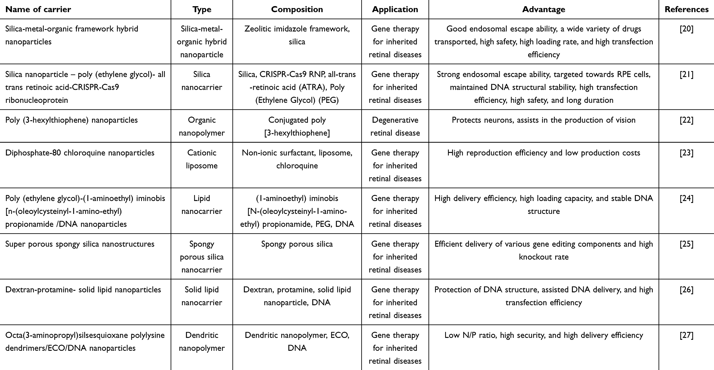 |
Table 1 Key Studies on Nanocarriers for Subretinal Injection |
How Important the Subretinal Injection is?
SR injection is a widely used method of delivering drugs into the eye, specifically targeting the SRS, located between the RPE cells and the photoreceptors.28 It has been demonstrated that SR injection is the most efficient route for delivering genetic material to both photoreceptor and RPE cells, with the outer retinal layer exhibiting the highest level of transgene expression.29 In recent years, SR injection has gained considerable attention in both preclinical and clinical investigations of fundus diseases.
In the disease areas where SR injection is most widely used, namely SMH due to nAMD and the treatment of various types of IRD, many studies have demonstrated that SR injection outperforms conventional therapies. SMH is a vision-threatening complication that occurs secondary to nAMD. It involves the formation of clots due to coagulation and contraction of blood in the SRS, which pulls on the retina. Additionally, iron in the blood is toxic to the photoreceptors. Conventional treatments for SMH include laser therapy, vitrectomy, and intravitreal injection (IVT) of anti-VEGF drugs. However, these conventional therapies have been shown to cause significant damage to the photoreceptors and retina.30 Recently, it has been suggested that SR injection of tissue plasminogen activator (tPA) can be a viable solution to this problem. tPA can be directly and evenly delivered to the SRS through SR injection, where it can interact with the clot and create a more favorable environment for subsequent blood replacement. This method effectively reduces the incidence of medically induced injury.6
Gene therapy is a viable and effective option for a variety of different types of IRDs.31 The traditional method of administration for gene therapy in IRDs is via IVT. However, as research has advanced, it has become evident that subretinal injection appears better results compared to IVT. The success of SR injection is well supported by theoretical evidence.32 To provide a comprehensive overview, we have compiled a list of approved clinical trials focusing on subretinal injection in the past five years, which is presented in Table 2.
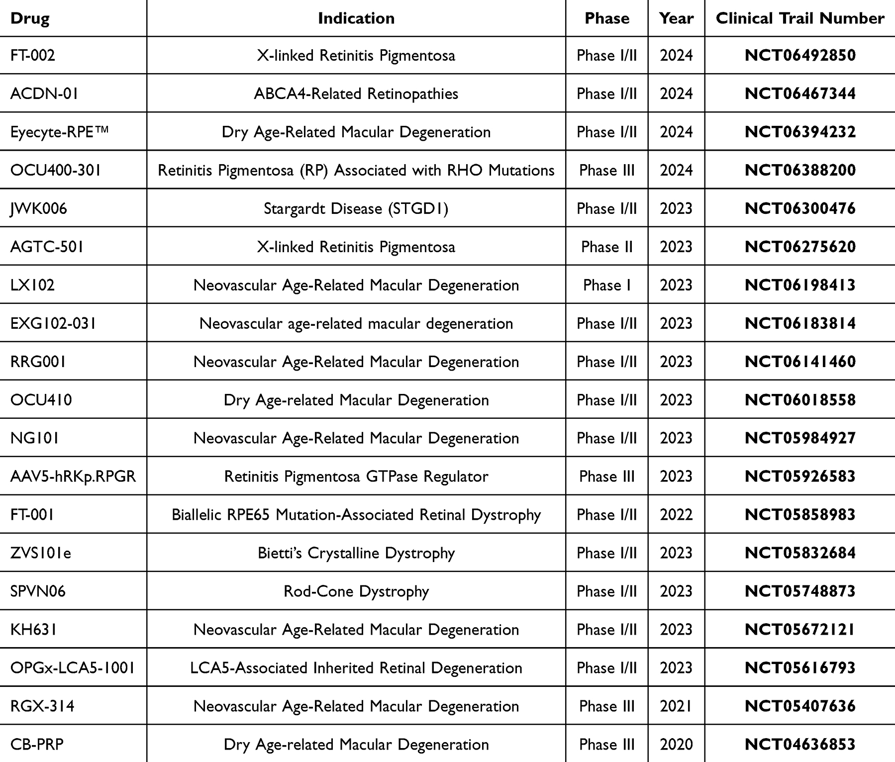 |
Table 2 Clinical Trials Related to Subretinal Injection |
Firstly, when treating eye-related diseases via the IVT route, drugs based on viral vectors are immediately diluted in the vitreous, and only a few reach the target cells. In order to achieve the desired efficacy, the concentration of the administered drug must be increased, and a high concentration of the virus often implies the possibility of eliciting a stronger immune response.33 SR injection allows the vector to penetrate the dense vitreous body without being diluted. Therefore, the effective dose required for SR injection treatment is much smaller than that for IVT, and a smaller therapeutic dose represents a higher safety profile.
Secondly, SR injection has the capability to traverse the dense ILM and directly deliver therapeutic vectors to RPE cells and photoreceptors. This effectively overcomes the barrier posed by the ILM to drug delivery and enhances transduction efficiency. In contrast, gene therapy drugs administered IVT also need to cross the ILM, which serves as both a physical and biological barrier between the vitreous and the retina.34 A study conducted by Woodard et al demonstrated that acetyl heparin sulfate proteoglycans (HSPGs) are abundant in the ILM and play a crucial role in facilitating key processes for postretinal transduction of AAV.35 Only AAV serotypes 2 and 3 are capable of postretinal transduction, therefore, utilizing the IVT route for drug delivery not only significantly limits the applicability of AAV carriers in treating fundus disorders but also introduces additional obstacles in the delivery process, consequently reducing therapeutic efficiency.
Thirdly, SR injection delivers the vector into the SRS, which not only avoids exposing the viral vector to the bloodstream, where it could be neutralized by antibodies, but also does not trigger an adaptive immune response. This not only enhances therapeutic efficacy and safety, but also enables repeat treatment with SR injection. The closely connected RPE cells on one side of the subretinal space, along with the retinal vascular endothelial cells on the other side, contribute to the restricted anatomy of the SRS as well as its immune privilege.36 This immune privilege helps maintain the stability of the retina and photoreceptors by producing its own immunomodulatory factors, unlike the vitreous body, which is connected to the body’s circulatory system.37 Epidemiological data have shown that a certain percentage of individuals in many regions of the world have anti-AAV neutralizing antibodies, ranging from 30% to 60%.38 The use of IVT for drug delivery would result in the viral vectors binding to the neutralizing antibodies, thereby reducing the delivery efficiency.
In addition, SR injection inevitably leads to small-scale retinal detachments, which can cause damage to the delicate retina. This disadvantage, which causes medical harm, has significantly hindered the advancement of SR injection in clinical practice for treating ocular diseases. However, recent evidence suggests that patients’ retinal structure and function can be restored within one month after the occurrence of medical damage.39 Moreover, in the majority of patients, the improvement in ocular function resulting from SR injection has outweighed the damage caused by the procedure.40,41
What are the Commonly Used Carriers for Subretinal Injection?
What Role Does the Viral Vector Play in Subretinal Injection?
Viral vectors are the traditional vectors for subretinal injection. The original genome of the virus has been replaced with the desired therapeutic genes for different diseases and delivered to the SRS.42 The virally delivered genes integrate with the genome of the target cells through endocytosis to repair genetic defects.43 Many species of viruses have shown effectiveness in ocular gene delivery.44 Commonly used viral vectors include lentivirus (LVV), adenovirus (AdV), and adeno-associated virus (AAV), among others. Each type of virus has its unique advantages.
Lentivirus
Lentiviruses (LVVs) are single-stranded RNA retroviruses with a loading capacity of about 8–9 kb.45 A significant advantage of LVs over other viral vectors is their robust gene loading capacity. This is expected to further address IRD caused by larger gene mutations. It has been shown that the non-pathogenic equine infectious anemia virus (EIAV) can be used as a gene therapy for STGD disease, which is currently undergoing clinical trials.46 However, the gene integration ability, as well as the transduction ability, of lentiviruses is not satisfactory and has the disadvantage of low therapeutic efficiency.
Adenovirus
Adenovirus (AdV) is a double-stranded DNA virus with a larger gene loading capacity of 30–40 kb compared to other viruses. The transduction pathway of AdV is capable of activating the immune pathway and stimulating the body to produce adaptive immunity. Therefore, it has a significant advantage in the production of vaccine vectors.47 Additionally, AdV has the ability to selectively infect cancer cells and induce an immune response to kill cancer cells, providing a promising approach in gene therapy for cancer.48 However, it is important to note that AdV carries the risk of inducing a severe immune response. When neutralizing antibodies are present in the organism, the transduction efficiency is greatly reduced, posing challenges for long-term repeated gene therapy.
Adeno-Associated Virus
Adeno-associated virus (AAV) is a small, single-stranded DNA virus that is named because it can only replicate in the presence of Ad. AAV is widely considered the preferred vector for retinal gene therapy due to several advantages, including greater safety, lower inflammatory response, improved therapeutic efficacy, and longer duration of treatment.49 AAV has multiple serotypes, with AAV type 2 (AAV2) currently being the most commonly used viral vector for ocular drug delivery. This is because AAV2 has the ability to bind to HSPG on ILM and undergo post-retinal transduction.35 Recently, Wiley et al50 observed that AAV type 1 (AAV1) and type 4 (AAV4) have the highest affinity for the photoreceptor cell layer. Therefore, they could also be used as therapeutic alternatives to IRD vectors.
Recent Advances in Viral Vectors
Despite the proven advantages of viral vectors such as long duration, rapid diffusion, and high therapeutic efficiency, there are still unresolved issues. One problem is the limited loading capacity of AAV, which can only accommodate around 4.7 kb.34 This poses restrictions on delivering large target genes and prevents the use of gene editing with CRISPR-Cas9 combinations.51 To address this, a novel dual AAV system has been developed. The mechanism involves loading the target gene into two independent AAV vectors, which then exert gene therapy effects upon recombination in target cells.52 An example of this approach is demonstrated by Li et al,6 who designed a dual AAV8-ABCA4 viral vector to deliver the ABCA4 gene for the treatment of STGD disease. Another example is the treatment of Usher syndrome by Riaz et al53 who used a double AAV vector to deliver the PCDH15 gene into the retina of Usher mice. This resulted in sustained recovery of visual function in the mice. Therefore, the dual AAV system retains the advantages of AAV vectors while overcoming the limitations of small gene load, thus bridging the gap in gene therapy using viral vectors.
In addition, although efforts have been made to select viruses with low immunogenicity as therapeutic vectors, it is still difficult to avoid binding to neutralizing antibodies in body fluids. This not only reduces the efficiency of transduction but also decreases safety. For this reason, studies have been devoted to isolating AAV from other animals as vectors to evade binding to human antiviral antibodies. Bello et al54 extracted porcine-derived AAV and found that it did not neutralize human immunoglobulin G (IgG). Moreover, the authors also noted that porcine-derived AAV could cross the biological barrier of the eye and produce transduction efficiencies comparable to those of conventional AAV vectors. This is a meaningful new attempt to enhance the efficacy and safety of viral vectors.
How Critical is Nanotechnology in Retinal Diseases Treating?
In order to overcome the limitations of current viral vector-mediated subretinal injections, there has been an effort to investigate novel nanotechnology-based vectors. Nanocarriers are a new class of carrier systems that are independent of viral carriers and have sizes ranging from 10–1000 nm.55 The mechanism of nanocarriers for the treatment of eye-related diseases is to combine a component or drug with a specific function with the structure of the nanocarrier and deliver the drug to the target cells or tissues as efficiently as possible through different routes of administration. In this paper, we focus on the subretinal injection route.56 Once the nanopreparations are injected into the subretinal space, various types of modified nanocarriers can perform specific functions (eg, evading cell phagocytosis, targeting drug delivery, stimulating specific cells, and gene knockdown) to achieve therapeutic goals.
There have been numerous studies confirming the superior role of nanocarriers compared to traditional viral carriers in the field of SR injection. Nanotechnology not only addresses the limitations of viral vectors such as immune responses, production of antiviral neutralizing antibodies, low loading capacity, and high therapeutic cost, but also offers new advantages including reduced drug side effects, prolonged drug release, and improved transfection efficiency.57 For instance, nanocarriers used for subretinal injections have smaller sizes than viral particles, allowing them to better penetrate the mucous membranes or biological barriers of the eye. This not only provides better stability but also reduces eye irritation.58 The hydrophobicity and hydrophilicity of certain nanocarriers, particularly lipid-based ones, also play a crucial role in drug distribution.59 With the flexible chemical structure of nanocarriers, their surface properties can be tailored to suit various therapeutic scenarios. For example, lipophilic nanocarriers can be used for higher penetration efficiency when targeting RPE.60 Another advantage of nanocarriers is their high loading capacity, particularly in subretinal injection-mediated gene therapy. While traditional vectors for gene therapy are limited by size, nanocarriers overcome this disadvantage.61 Moreover, nanocarriers can be easily modified to include targeted transport, which can increase the drug concentration in target cells or tissues, minimize unnecessary dilution and wastage, and reduce drug side effects.62 However, viral particles in the field of subretinal injections lack these qualities.
Subretinal injections are commonly used in the treatment of retina-related diseases as an invasive method of drug delivery. These injections have several advantages, including fast onset of action and small dosage requirements due to their ability to cross the blood-retinal barrier.63 Moreover, nanocarriers offer a significantly higher safety profile compared to viral carriers. In cases where subretinal injection or other invasive drug delivery methods are necessary, nanocarriers have shown superior therapeutic effects with fewer injections. This is due to their enhanced stability, greater loading capacity, prolonged duration in the subretinal space, and higher drug concentration. Furthermore, nanocarriers are less irritating and lack viral immunogenicity due to their smaller size. Consequently, they provide a safer treatment option overall.64 In summary, nanocarriers are invaluable and surpass viral carriers in the widely used route of drug delivery, particularly in subretinal injections.
To date, various nanocarriers, such as liposomes, nanoparticles, nanomicelles, and nanogels, have been developed to varying degrees.65 In the last five years, an increasing number of nanocarriers have been utilized in subretinal injections for various retina-related diseases, with some proving to be effective in clinical trials. For instance, Wang et al improved nanocarrier-mediated gene therapy by enhancing the nanocarrier’s loading capacity, escape ability, and transfection efficiency.20 Giovanni et al developed a nanocarrier capable of transmitting biosignals to aid in vision generation under light-stimulated conditions for treating retinal degenerative diseases.22 Nanocarrier-mediated subretinal injection therapies have been employed for a wide range of retina-related diseases, including STGD, RP, LCA, choroidal agenesis, and others. Compared to viral vectors, nanocarriers significantly expand the potential treatment options for various diseases and offer novel therapeutic approaches for conventional disease therapies.
In conclusion, nanocarriers enable various levels of modification of nanocarriers based on the requirements of the therapeutic regimen and also offer the opportunity to flexibly formulate the physical and chemical characteristics of nanocarriers with great versatility.65 Considered a safer alternative to viral vectors, nanocarriers have shown advantages that go beyond those of traditional viral vectors and have already made significant progress in the field of fundus disease therapy.66
Liposome
Liposomes are spherical nanovesicles formed by phospholipid bilayers.67 They are primarily composed of phospholipids and cholesterol. Liposomal vesicles are amphiphilic, which means they can carry both hydrophilic drugs in the core region and lipophilic drugs in the lipid bilayer.68 This carrier has no limitations on loading different types of drug molecules and possesses excellent biocompatibility and a high safety profile. Currently, liposomes are mainly used in the treatment of retinal diseases. For example, Abrishami et al69 loaded anti-VEGF drugs into liposomes to treat choroidal neovascularization (CNV). In conclusion, liposomes have become a common carrier for ocular drug delivery systems due to their excellent biocompatibility and corneal permeability.
Polymer Nanocarriers
Common types of polymer nanocarriers, such as dendritic polymer macromolecules, nanospheres, solid lipid nanoparticles, nanomicelles, and nanocapsules, are widely used in various applications.70 However, there is still ample room for the development of polymer nanocarriers in the field of subretinal injection. This chapter primarily outlines the related research on nanopolymers in the field of subretinal injection.
Sánchez-López et al71 investigated novel poly(propylglycolide) glycoside (PLGA) nanoparticles for drug delivery. These nanoparticles exhibited a loading efficiency of 85% and the ability to release the drug in a sustained manner. In a separate study, Ye et al72 prepared PLGA nanoparticles loaded with bevacizumab. They observed a significant increase in the half-life of the drug and the mean concentration of the drug after delivery into the eye. This finding is particularly encouraging because PLGA nanoparticles offer the advantage of prolonged drug delivery, which helps overcome the limitations of ocular drug delivery, such as repetitive dosing and frequency of dosing.73
Solid lipid nanoparticles (SLNPs) are a type of nanocarrier that is gaining recognition. SLNPs consist of a solid lipid core and are considered to be small drug delivery systems that combine lipids with nanopolymers. One of the key advantages of SLNPs is their ability to target drug delivery without causing toxicity. In a study conducted by Delgado et al, SLNPs were utilized as carriers for DNA delivery in the treatment of hereditary retinal diseases. The findings from the experiment demonstrated that this carrier exhibited exceptionally high safety and delivery efficiency, both in mice and at the cellular level.26
Dendritic nanopolymers are nanomacromolecules with a highly branched structure, where each branch end can serve a distinct function. This property makes dendrimer nanopolymers suitable for delivering various types of drugs, including hydrophilic and lipophilic ones. In a study by Sun et al27 a dendrimer nanocarrier was developed for the treatment of hereditary retinal diseases. The dendrimer’s structure not only helps maintain the stability of DNA but also collaborates with other lipid nanostructures to improve gene delivery efficiency and extend the duration of drug action. Moreover, the safety of this approach is well assured.
In general, there are many types of polymer nanocarriers, which provide strong support for the clinical treatment of retina-related diseases.74
Inorganic Nanoparticles
Commonly used materials for the production of inorganic nanoparticles (NPs) include gold, silica, and cerium oxide. Inorganic NPs encompass a wide range of nanocarriers such as gold nanoparticles (AuNPs), silver nanoparticles (AgNPs), cerium oxide nanoparticles (Nanoceria-CeO2-NPs), mesoporous silica nanoparticles (MSNs) and others. Numerous studies have demonstrated that AuNPs possess excellent antioxidant and antiangiogenic properties. Specifically, they inhibit angiogenesis by down-regulating the expression of VEGFR-2.75 Kim et al76 found that AuNPs can suppress cell proliferation and migration in a retinal neovascularization model. Apaolaza et al77 developed a type of AuNPs that not only exhibit good stability but also have the ability to inhibit RPE cell death, reduce the production of VEGF, interleukins, and other substances, thereby achieving the inhibition of vascular proliferation. Similar findings were observed in the case of cerium oxide nanoparticles, as experiments conducted by Guo et al78 demonstrated that these nanoparticles inhibited cell proliferation and migration and reduced the production of transforming growth factor (TGF-β).
Anbukkarasi et al found that AgNPs have the ability to scavenge oxidative substances, such as DPPH, from tissues and demonstrated a delaying effect on cataract formation in rats.79 Gurunathan et al also found that AgNPs can inhibit angiogenesis by inhibiting the PI3K/Akt signaling pathway.80 MSNs are one of the most well-studied inorganic nanoparticles. Among the subtypes, MSNs have the advantage of greatly prolonging the duration of drug action due to the presence of mesopores. Kim et al found that MSNs loaded with brimonidine had a drug action time of up to 8 hours.81 Unfortunately, inorganic nanocarriers have not yet been widely used in the field of subretinal injections. With the continuous development of nanotechnology, inorganic NPs are expected to be a new approach to combat retinal neovascularization diseases.
Recent Progress in Subretinal Injection of Nanocarriers
Silica-Metal-Organic Framework Hybrid Nanoparticles
Among inorganic NPs, silica NPs are a class of commonly used nanocarriers, which have been widely used in the medical field because of their unique stability, low toxicity and ease of modification,82 for example, in the direction of tumour diagnosis and treatment related to cancer. Recently Miriam et al83 doped silica NPs with dyes to make the first investigational drug for cancer-targeted molecular imaging. Lu et al84 found a micro MSNs with good biocompatibility at appropriate drug concentrations and preferentially accumulated in tumour tissues, thus MSNs loaded with relevant drugs can be be used to inhibit tumour growth. Not only that, silica NPs have also contributed equally well in the treatment of ophthalmic diseases. Liao et al85 used MSNs loaded with pilocarpine for the treatment of glaucoma in rabbits, and it was shown that the MSNs possessed the ability to release the drug persistently and had pH-responsive properties, which successfully acted to lower the IOP.
In the field of silica NPs mediating SR injection, Wang et al20 combined metal-organic frameworks (MOFs) with Zn coordination to form ions. They then used 2-methylimidazole (2-MIM) to form a zeolite imidazolate backbone (ZIF). Silica was combined with ZIF to form a pH-responsive silica-metal-organic framework hybridized NP (SMOF NP). Among other things, ZIF enables the superior pH responsiveness of SMOF NPs and enhances the ability of the nanocarriers to escape endocytosis.86 This has been demonstrated in in vitro assays. For different loading cargoes, SMOF NPs showed superb loading efficiency as well as loading content. In different cell lines, the transfection efficiency of SMOF NPs ranged from 1.2- to 1.9-fold higher than that of the commercially available transfection agent Lipofectamine (Lipo). The gene knockdown efficiency mediated by SMOF NPs was 1.3-fold higher than that of the Lipo group, with a gene correction efficiency of 1.4-fold. Additionally, even high concentrations of SMOF NPs did not show significant cytotoxicity.
Conventional pH-responsive silica NPs have great difficulties in designing their structures,87 and GSH-responsive silica NPs tend to be unstable and difficult to synthesise,88 whereas SMOF NPs, as a new type of structurally stable and more efficient silica NPs, can be better adapted to the needs of retinal gene therapy. Besides, SMOF NPs can efficiently transport various types of hydrophilic cargoes, such as hydrophilic drugs, nucleic acids, and CRISPR-Cas9 gene editors, which greatly expands the scope of application of SMOF NPs in ophthalmic diseases. Moreover, the authors showed in their conclusion that ATRA-coupled SMOF NPs successfully induced efficient genome editing in mouse RPE cells via SR. In summary, SMOF NPs are expected to play a greater role in the field of IRD gene therapy due to their excellent delivery efficiency, escape ability, and high safety.
Silica Nanoparticle – Poly (Ethylene Glycol)- All Trans Retinoic Acid -CRISPR-Cas9 Ribonucleoprotein
Wang et al21 reported the development of glutathione (GSH)-responsive silica nanoparticles (SNPs) with imidazole-containing components integrated into their structure to enhance endosomal escape capabilities. When conjugated with all-trans retinoic acid (ATRA) and CRISPR-Cas9 ribonucleoproteins (RNPs), these polymeric nanoparticles demonstrated targeted delivery to retinal pigment epithelial (RPE) cells, GSH-triggered responsiveness, and effective gene editing. In cellular assays, SNP-PEG exhibited a 1.3-fold increase in nucleic acid delivery efficiency, a 1.3-fold higher gene knockdown rate, and a 1.1-fold improvement in gene correction compared to the commercially available transfection agent Lipofectamine (Lipo). Notably, modifying the surface of negatively charged SNPs with positive or neutral charges further enhanced DNA transfection efficiency by 1.6-fold. Additionally, SNP-PEG achieved a loading efficiency of up to 90% with minimal cytotoxicity, while the Lipo-treated cells exhibited a reduced survival rate of 77%.
It has also been found that nanocarriers treated with PEG can mask the charge carried on the carrier surface and achieve improved printing efficiency by preventing the aggregation and binding of proteins.89 Koirala et al combined nanocarriers containing fluorescent protein genes with PEG and found that the structure of DNA was much more stable, and that a large number of fluorescent signals could still be detected after 30 days of subretinal injection. All of these studies proved that nanocarriers containing PEG can act as a protective agent for DNA, which is undoubtedly a valuable finding.90
CRISPR-Cas9 RNP is a highly efficient and specific gene editor, but it is highly susceptible to degradation and has low transfection efficiency.91,92 SNP-PEG vectors have solved the shortcomings of the instability of CRISPR-Cas9 editors and broken through many obstacles to CRISPR-Cas9-mediated gene therapy; the development of SNP-PEG has led to a more sensitive, efficient and safe gene editing vector, providing a new idea for gene therapy for retinal diseases.
Poly (3-Hexylthiophene) Nanoparticles
The conjugated polymer poly[3-hexylthiophene] (P3HT) is an organic photovoltaic polymer that depolarizes neurons and generates action potentials when stimulated with visible light, thereby delivering signals to neuronal cells. P3HT NPs have great potential for the treatment of neurological disorders due to their ability to provide electrical signals to neuronal cells and because of their unique conductivity and biocompatibility. P3HT NPs are used as carriers in drug delivery systems to enhance nerve regeneration and repair for the treatment of spinal cord and peripheral neuropathy.93 Among ophthalmic diseases, retinal degenerative diseases are the most common cause of severe visual impairment in the aging population, the most prevalent of which are AMD and RP.94 Because AMD and RP often involve degeneration of photoreceptors, P3HT NPs have been proposed to treat these diseases by restoring the activity of retinal neurons.95 The study of Arijit et al96 has demonstrated that P3HT can be used in the treatment of spinal and peripheral neuropathy. The study has demonstrated that P3HT has promising results in the treatment of retinal degenerative diseases.
Giovanni et al22 made a new attempt in conjunction with the SR injection technique to fabricate a retinal nanoprosthesis as an interface with retinal neurons for the treatment of retinal degenerative diseases. The device consisted of a layer of P3HT and a layer of conductive material, which was uniformly distributed within the SRS of the diseased rats by SR injection. Upon encountering a light stimulus, the P3HT NPs stimulated the primary visual cortex through a change in electrical charge for the purpose of assisting in the production of vision. The experimental results showed that P3HT NPs were able to improve light sensitivity and spatial discrimination in the lesioned rats until the ability was similar to that of healthy rats, and uniformly covered more than 80% of the retina without pro-inflammatory effects on the retina. At this stage, although methods such as gene therapy or stem cell therapy have been investigated for retinal degenerative diseases, they have not yet reached the stage of clinical treatment.97,98 Therefore, the research related to P3HT NPs opens up a new line of thought for the treatment of this type of disease.
Diphosphate-80 Chloroquine Nanoparticles
It has been shown that chloroquine (CQ) can achieve anti-inflammatory and immunosuppressive effects by inhibiting endosomal toll-like receptor signaling and reducing cytokine production.99–101 In the treatment of ophthalmic diseases, CQ is often used for the treatment of dry eye because it can alleviate the drying stress and inflammatory state of the cornea. Julia et al102 conducted a meta-analysis of clinical studies on the treatment of dry eye with CQ and indicated that CQ can effectively alleviate the symptoms of dry eye.
In addition to this, CQ is a known endosome-disrupting molecule that can be used as a cell pre-treatment drug to enhance gene delivery.103 Mashal et al23 made chloroquine diphosphate-containing nano-vesicles (DPP80-CQ) by combining a non-ionic surfactant with cationic liposomes and doped with chloroquine diphosphate. Doping chloroquine into the nanovesicles can assist the nanovesicles in evading phagocytosis by endosomes and increase the delivery efficiency of the carrier. The results showed that DPP80-CQ was able to better evade endosomal phagocytosis and transfected RPE cells with excellent delivery efficiency compared to DPP80, a nanovesicle without chloroquine doping.
Nanoformulated vectors for gene delivery systems, in contrast to traditional viral vectors, include the advantages of low production costs and easy modification of vectors on demand, in addition to higher loading.104 On the basis of which DPP80-CQ optimises the capacity of the vectors and overcomes the challenge of enhancing the reproduction efficiency, a problem that must be solved for the development of nanoformulated vectors. Therefore, the use of DPP80-CQ nanovesicle-mediated SR injection can reduce the number of injections for patients, thereby reducing the incidence of adverse reactions.
Poly (Ethylene Glycol)-(1-Aminoethyl) Iminobis [N-(Oleoylcysteinyl-1-Amino-Ethyl) Propionamide /DNA Nanoparticles
(1-Aminoethyl) iminobis [N-(oleoylcysteinyl-1-aminoethyl) propionamide] is a multifunctional lipid particle (ECO) that has been experimentally demonstrated to have pH sensitivity, excellent endosomal escape, and stable nucleic acid delivery, and is being used as a simple and smart class of gene delivery vehicles.105 PEG-ECO/DNA NPs loaded with relevant DNA were developed for the treatment of LCA by researchers as early as 2017 and were tested in both cellular and animal assays. The results showed that both PEG-ECO/DNA NPs had good transduction efficiency and could treat mouse models suffering from human LCA.106
STGD is an inherited retinal disease due to mutations in the ABCA4 gene,107 which does not have the ability to remove toxic substances such as N-methyldihydroxyretinyl-N-retinoid ethanolamine (A2E) from RPE cells, thus causing irreversible damage to the retina.108 Sun et al24 developed a novel nanoliposome using pH sensitive ECO with a plasmid capable of stably expressing the ABCA4 gene to form a stable structure and then delivered to the SRS. In fact, PEG-ECO/DNA NPs were able to be uniformly expressed in the retina, resulting in a significant decrease in the rate of progression of STDG, with a single injection being effective for up to 1 year and multiple injections being more effective. Besides, the retinal inflammation induced by PEG-ECO/DNA NPs was much less than that of AAV vectors, and the safety was also strongly proved in the experiment.
The traditional therapy for STGD is the application of AAV-mediated gene therapy, however, the insufficient loading capacity of AAV leads to many limitations in the delivery targeting the ABCA4 gene.109 With greater loading capacity as well as more stable drug release, PEG-ECO/DNA NPs not only fill the gap in the treatment of gene therapy for STDG diseases, but also make up for the traditional AAV vector therapies. Therefore, SR injection with PEG-ECO/DNA NPs is a promising approach for gene therapy of STDG.
Super Porous Spongy Silica Nanostructures
Spongy porous silica nanostructures (SN) are highly efficient nanocarriers that are widely used in various drug delivery scenarios. Marzieh et al110 designed a super porous spongy SN and compared it with two other different morphologies of SN in order to study the delivery advantages of super porous spongy SN. It was shown that the super porous spongy SN has the best ability to load and adsorb drugs and is a potential nanocarrier for drug delivery. Marzieh et al also showed in their study that the application of SN loaded with adriamycin can bring better anticancer effect than the group without SN, so SN is also expected to play a greater ability in antitumour therapy.
Kim et al25 developed an SN as a carrier as a nanoplatform for delivering Cas9-RNP or base editor RNP (BE-RNP) for gene therapy of various IRDs. The results showed that the loading efficiency of SN for Cas9-RNP was 25%, which was higher than several nanomaterials reported in the market.86 And the release rate of RNP could reach 70% within 12 h of the SN carrier being ingested into cells. In addition to this, the gene editing efficiency of SN-based delivery vector was about 10 times higher than that of the commercially available transfection agent Lipo, and more than 90% cell survival was maintained under the condition of high concentration of SN. The authors also mentioned that the SN-based delivery platform can deliver a variety of RNPs, including Cas9-RNP and BE-RNP, and has shown good efficacy in both human and mouse cells.
Successful in vivo gene editing by delivering Cas9 nuclease RNP or base editor RNP (BE-RNP) via non-viral vectors has been previously reported, but ensuring the stability and homogeneity of RNP and the vector is difficult. This often leads to unsatisfactory results of vector-mediated gene editing.111,112 In contrast, SN overcame the deficiencies of the original vector and promoted the development of SR injection in vivo gene editing. This was achieved through its biocompatibility with various RNPs, high loading rate, high knockout rate, high release rate, and good safety.
Dextran-Protamine-Solid Lipid Nanoparticles
Delgado et al26 developed a SLNs based vector for gene therapy of retinal diseases represented by X-linked juvenile retinoschisis (XLRS). The nanovectors consist of SLNs, dextran, Fischer protein and a plasmid containing the target gene. Ichthyosin has a nuclear localisation signal and protects DNA from DNA degrading enzymes in vivo. Ichthyosin assists DNA to integrate better with RPE cells. Rajala et al exploited this property of Ichthyosin-assisted DNA delivery and developed Ichthyosin-liposome nanocomplexes for delivery of DNA. They delivered the nanocarriers directly to RPE cells of RPE65-deficient mice by subretinal injection and found persistent and efficient gene expression in the mice that promoted vision recovery in mice.113 Dextran was able to promote endocytosis mediated by lattice proteins, which improved the cellular uptake and translocation of nanocarriers.
In in vitro experiments, it was shown that dextran as well as fisetin in this nanocarrier could enhance the transfection and expression of target genes. Solid lipid nanocarriers loaded with green fluorescent protein (EGFP) were delivered into the retinal space of rats by subretinal injection, and good expression of EGFP was detected in the rat retina. In addition, it has also been shown that dextran-fisetin-SLNs can reduce photoreceptor loss and repair the retina, and maintain gene expression for 2 months after subretinal injection, which is not only long-lasting but also has a powerful effect on restoring vision.114 Studies have shown that the powerful ability of subretinal injection of dextran-fisetin-SLNs to deliver nucleic acids offers a better option for gene therapy for retinal diseases, with a potential that cannot be ignored.
Octa(3-Aminopropyl) Silsesquioxane Polylysine Dendrimers/ECO/DNA Nanoparticles
Gene therapies have been widely used in the treatment of retinal diseases. However, the negative charge of nucleic acids makes it difficult for DNA-loaded nanocarriers to cross cell membranes that are also negatively charged. To solve this problem, some researchers have doped a large number of cations into the nanocarriers for improving the reproduction efficiency.115 However, the large amount of positive charge also has greater cytotoxicity and poses a safety issue.116 Therefore, we need a nanocarrier that is both safe and efficient for optimising subretinal injection-mediated gene therapy.117
Sun et al27 developed an octa(3-aminopropyl) silsesquioxane polylysine dendrimers called G4 nanoglobule. G4 not only has a low positive to negative charge ratio (N/P ratio), but also mimics the function of histones to achieve the purpose of encapsulating and concentrating DNA.118 They also developed a pH-sensitive multifunctional lipid (ECO) that can form a stable structure with nucleic acids to facilitate cellular uptake and gene delivery.119 Sun et al combined G4 with ECO to design a novel nanocarrier system for gene delivery.
In vitro experiments conducted by Sun et al assessed the efficacy of the G4/ECO/DNA nanocarrier system in ARPE-19 cells, focusing on cell viability, endosomal escape, and transfection efficiency. The results revealed that cell viability following transfection with G4/ECO/DNA in serum-free medium was approximately 60%, compared to only 40% with the commercial transfection agent Lipo2000. Notably, in the presence of 10% serum, cell viability increased to about 90% for G4/ECO/DNA transfected cells. The G4/ECO/DNA nanocarrier system also exhibited excellent endosomal escape properties when loaded with fluorescent genes. Under 10% serum conditions, the transfection rate of G4/ECO/DNA reached an impressive 95%, significantly surpassing that of Lipo2000. In vivo, Sun et al administered GFP-loaded G4/ECO/DNA via subretinal injection in mice, resulting in strong GFP signals within the retinal layer, indicating successful transfection of RPE cells and sustained gene expression.
G4 is responsible for safely condensing DNA in environments with a low positive-to-negative charge ratio, while ECO enhances the transfection efficiency of target genes. The G4/ECO/DNA nanocarrier has demonstrated robust functionality in both in vitro and in vivo models, suggesting its potential as a safe and effective platform for gene delivery.
The Future Prospective
This review discusses research advancements related to SR injection and the progress of nanocarriers in this field. We begin by highlighting SR injection as a prominent drug delivery method for ocular diseases. SR injection offers several advantages: (1) it bypasses the internal limiting membrane (ILM), enhancing drug delivery efficiency; (2) it traverses the vitreous body without dilution, allowing for direct contact with target cells, thereby significantly reducing the effective dose required per treatment and improving tolerability and safety; (3) it delivers the drug carrier directly to the immune-privileged subretinal space (SRS), avoiding neutralization by antibodies and minimizing immune reactions, which supports the feasibility of long-term and multiple injections and broadens the application scope. SR injection has demonstrated efficacy in treating various IRDs, nAMD, and SMH resulting from nAMD, addressing a critical gap in the treatment of retinal diseases and proving to be a clinically significant technique.
Traditional vectors for SR injection are predominantly viral, with AAV vectors being the most commonly used due to their long-term transduction, rapid diffusion, high therapeutic efficiency, low inflammatory response, and ease of vector production. AAV vectors are frequently employed in gene therapy for IRDs, integrating therapeutic genes into the patient’s genome to repair defective genes. However, viral vectors are limited by their small gene loading capacity and potential for antigen-antibody reactions.
With the development of nanotechnology, novel non-viral vectors have gradually become a hot topic in SR injection in recent years, and this paper highlights the latest findings of nanocarriers in SR injection. Nanocarriers are designed to address the unavoidable drawbacks of viral carriers and offer surprising advantages such as reduced side effects, prolonged drug release, and easy modification of the carriers. However, along with the advantages come challenges that need to be addressed.
Firstly, while recent advances in nanocarrier-mediated subretinal injection have shown promise in cellular and animal studies, these results may not fully translate to clinical trials, potentially leading to divergent outcomes.63 Additionally, ethical considerations must be carefully addressed when transitioning from in vitro to in vivo experiments.120
Secondly, nanocarriers offer significant advantages due to their versatility, allowing for modifications tailored to specific therapeutic needs. This flexibility presents extensive opportunities for developing optimized carrier structures. Moreover, nanocarriers can be engineered to respond to various stimuli—such as light, pH, ultrasound, and enzymes—enabling more precise drug delivery and controlled release.121
Thirdly, assessing the toxicity of nanocarriers and the impact of their degradation products on human health is crucial. Various metrics are used to evaluate cellular health, including inflammatory response markers and lactate dehydrogenase levels, which indicate cell integrity.122 However, discrepancies in safety data between animal and clinical trials necessitate meticulous attention when advancing to human studies.123
In conclusion, nanocarrier-mediated subretinal injection has significantly advanced the treatment of retinal diseases and represents a promising class of non-viral carriers. However, a thorough evaluation of the clinical therapeutic effects of these nanocarriers is essential. Further detailed studies are needed to optimize their safety and therapeutic efficacy, which will be crucial for advancing the field of nanocarrier research and enhancing clinical outcomes.
Acknowledgments
Supported by the National Natural Science Foundation of China (NO.82271124), and the training Fund for Open Projects at Clinical Institutes and Departments of Capital Medical University (CCMU2022ZKYXZ001). The funding organizations had no role in the design or conduct of this research.
Disclosure
The authors report no conflicts of interest in this work.
References
1. Rakoczy EP, Lai C-M, Magno AL, et al. Gene therapy with recombinant adeno-associated vectors for neovascular age-related macular degeneration: 1 year follow-up of a Phase 1 randomised clinical trial. Lancet. 2015;386(10011):2395–2403. doi:10.1016/S0140-6736(15)00345-1
2. Ogata M, Oh H, Nakata A, et al. Displacement of submacular hemorrhage secondary to age-related macular degeneration with subretinal injection of air and tissue plasminogen activator. Sci Rep. 2022;12(1):22139. doi:10.1038/s41598-022-26289-6
3. Maguire AM, Russell S, Chung DC, et al. Durability of voretigene neparvovec for biallelic RPE65-mediated inherited retinal disease: phase 3 results at 3 and 4 years. Ophthalmology. 2021;128(10):1460–1468. doi:10.1016/j.ophtha.2021.03.031
4. Lam BL, Davis JL, Gregori NZ, et al. Choroideremia gene therapy phase 2 clinical trial: 24-month results. Am J Ophthalmol. 2019;197:65–73. doi:10.1016/j.ajo.2018.09.012
5. Ghazi NG, Abboud EB, Nowilaty SR, et al. Treatment of retinitis pigmentosa due to MERTK mutations by ocular subretinal injection of adeno-associated virus gene vector: results of a Phase I trial. Hum Genet. 2016;135(3):327–343. doi:10.1007/s00439-016-1637-y
6. Li R, Jing Q, She K, et al. Split AAV8 mediated ABCA4 expression for gene therapy of mouse stargardt disease (STGD1). Hum Gene Ther. 2023;34(13–14):616–628. doi:10.1089/hum.2023.017
7. Maguire AM, Russell S, Wellman JA, et al. Efficacy, safety, and durability of voretigene neparvovec-rzyl in RPE65 mutation-associated inherited retinal dystrophy: results of phase 1 and 3 trials. Ophthalmology. 2019;126(9):1273–1285. doi:10.1016/j.ophtha.2019.06.017
8. Stanescu-Segall D, Balta F, Jackson TL. Submacular hemorrhage in neovascular age-related macular degeneration: a synthesis of the literature. Surv Ophthalmol. 2016;61(1):18–32. doi:10.1016/j.survophthal.2015.04.004
9. Holmgaard AB, Askou AL, Jensen EG, et al. Targeted knockout of the vegfa gene in the retina by subretinal injection of RNP complexes containing cas9 protein and modified sgRNAs. Mol Ther. 2021;29(1):191–207. doi:10.1016/j.ymthe.2020.09.032
10. Dalkara D, Byrne LC, Klimczak RR, et al. In vivo-directed evolution of a new adeno-associated virus for therapeutic outer retinal gene delivery from the vitreous. Sci Transl Med. 2013;5(189):189ra76. doi:10.1126/scitranslmed.3005708
11. Dalkara D, Sahel JA. Gene therapy for inherited retinal degenerations. C R Biol. 2014;337(3):185–192. doi:10.1016/j.crvi.2014.01.002
12. Buck TM, Wijnholds J. recombinant adeno-associated viral vectors (rAAV)-vector elements in ocular gene therapy clinical trials and transgene expression and bioactivity assays. Int J Mol Sci. 2020;21(12):4197.
13. Acland GM, Aguirre GD, Ray J, et al. Gene therapy restores vision in a canine model of childhood blindness. Nat Genet. 2001;28(1):92–95. doi:10.1038/ng0501-92
14. Beltran WA, Cideciyan AV, Lewin AS, et al. Gene therapy rescues photoreceptor blindness in dogs and paves the way for treating human X-linked retinitis pigmentosa. Proc Natl Acad Sci U S A. 2012;109(6):2132–2137. doi:10.1073/pnas.1118847109
15. Russell S, Bennett J, Wellman JA, et al. Efficacy and safety of voretigene neparvovec (AAV2-hRPE65v2) in patients with RPE65-mediated inherited retinal dystrophy: a randomised, controlled, open-label, phase 3 trial. Lancet. 2017;390(10097):849–860. doi:10.1016/S0140-6736(17)31868-8
16. Wu Z, Yang H, Colosi P. Effect of genome size on AAV vector packaging. Mol Ther. 2010;18(1):80–86. doi:10.1038/mt.2009.255
17. Kamaleddin MA. Nano-ophthalmology: applications and considerations. Nanomedicine. 2017;13(4):1459–1472. doi:10.1016/j.nano.2017.02.007
18. Singh AP, Biswas A, Shukla A, et al. Targeted therapy in chronic diseases using nanomaterial-based drug delivery vehicles. Signal Transduct Target Ther. 2019;4:33. doi:10.1038/s41392-019-0068-3
19. Xu Q, Kambhampati SP, Kannan RM. Nanotechnology approaches for ocular drug delivery. Middle East Afr J Ophthalmol. 2013;20(1):26–37. doi:10.4103/0974-9233.106384
20. Wang Y, Shahi PK, Xie R, et al. A pH-responsive silica-metal-organic framework hybrid nanoparticle for the delivery of hydrophilic drugs, nucleic acids, and CRISPR-Cas9 genome-editing machineries. J Control Release. 2020;324:194–203. doi:10.1016/j.jconrel.2020.04.052
21. Wang Y, Shahi PK, Wang X, et al. In vivo targeted delivery of nucleic acids and CRISPR genome editors enabled by GSH-responsive silica nanoparticles. J Control Release. 2021;336:296–309. doi:10.1016/j.jconrel.2021.06.030
22. Maya-Vetencourt JF, Manfredi G, Mete M, et al. Subretinally injected semiconducting polymer nanoparticles rescue vision in a rat model of retinal dystrophy. Nat Nanotechnol. 2020;15(8):698–708. doi:10.1038/s41565-020-0696-3
23. Mashal M, Attia N, Martínez-Navarrete G, et al. Gene delivery to the rat retina by non-viral vectors based on chloroquine-containing cationic niosomes. J Control Release. 2019;304:181–190. doi:10.1016/j.jconrel.2019.05.010
24. Sun D, Sun W, Gao S-Q, et al. Effective gene therapy of Stargardt disease with PEG-ECO/pGRK1-ABCA4-S/MAR nanoparticles. Mol Ther Nucleic Acids. 2022;29:823–835. doi:10.1016/j.omtn.2022.08.026
25. Kim S, Jeong YK, Cho CS, et al. Enhancement of gene editing and base editing with therapeutic ribonucleoproteins through in vivo delivery based on absorptive silica nanoconstruct. Adv Healthc Mater. 2023;12(4):e2201825. doi:10.1002/adhm.202201825
26. Delgado D, Del Pozo-Rodríguez A, Solinís MÁ, et al. Dextran and protamine-based solid lipid nanoparticles as potential vectors for the treatment of X-linked juvenile retinoschisis. Hum Gene Ther. 2012;23(4):345–355. doi:10.1089/hum.2011.115
27. Sun D, Maeno H, Gujrati M, et al. Self-assembly of a multifunctional lipid with core-shell dendrimer DNA nanoparticles enhanced efficient gene delivery at low charge ratios into RPE cells. Macromol Biosci. 2015;15(12):1663–1672. doi:10.1002/mabi.201500192
28. Peng Y, Tang L, Zhou Y. Subretinal injection: a review on the novel route of therapeutic delivery for vitreoretinal diseases. Ophthalmic Res. 2017;58(4):217–226. doi:10.1159/000479157
29. Cai X, Conley SM, Nash Z, et al. Gene delivery to mitotic and postmitotic photoreceptors Via compacted DNA nanoparticles results in improved phenotype in a mouse model of retinitis pigmentosa. FASEB J. 2010;24(4):1178–1191. doi:10.1096/fj.09-139147
30. Olivier S. Subretinal recombinant tissue plasminogen activator injection and pneumatic displacement of thick submacular hemorrhage in Age-Related macular degeneration. Ophthalmology. 2004;111(6):1201–1208. doi:10.1016/j.ophtha.2003.10.020
31. Dhurandhar D, Sahoo NK, Mariappan I, et al. Gene therapy in retinal diseases: a review. Indian J Ophthalmol. 2021;69(9):2257–2265. doi:10.4103/ijo.IJO_3117_20
32. Irigoyen C, Amenabar Alonso A, Sanchez-Molina J, et al. Subretinal injection techniques for retinal disease: a review. J Clin Med. 2022;11(16):4717. doi:10.3390/jcm11164717
33. Reichel FF, Peters T, Wilhelm B, et al. Humoral immune response after intravitreal but not after subretinal AAV8 in primates and patients. Invest Ophthalmol Vis Sci. 2018;59(5):1910–1915. doi:10.1167/iovs.17-22494
34. Ross M, Ofri R. The future of retinal gene therapy: evolving from subretinal to intravitreal vector delivery. Neural Regen Res. 2021;16(9):1751–1759. doi:10.4103/1673-5374.306063
35. Woodard KT, Liang KJ, Bennett WC, et al. Heparan sulfate binding promotes accumulation of intravitreally delivered adeno-associated viral vectors at the retina for enhanced transduction but weakly influences tropism. J Virol. 2016;90(21):9878–9888. doi:10.1128/JVI.01568-16
36. Dosmar E, Walsh J, Doyel M, et al. Targeting ocular drug delivery: an examination of local anatomy and current approaches. Bioengineering. 2022;9(1):41. doi:10.3390/bioengineering9010041
37. Strauss O. The retinal pigment epithelium in visual function. Physiol Rev. 2005;85(3):845–881. doi:10.1152/physrev.00021.2004
38. Calcedo R, Vandenberghe L, Gao G, et al. Worldwide epidemiology of neutralizing antibodies to adeno-associated viruses. J Infect Dis. 2009;199(3):381–390. doi:10.1086/595830
39. Simunovic MP, Xue K, Jolly JK, et al. Structural and functional recovery following limited iatrogenic macular detachment for retinal gene therapy. JAMA Ophthalmol. 2017;135(3):234–241. doi:10.1001/jamaophthalmol.2016.5630
40. Jacobson SG. Gene therapy for Leber congenital amaurosis caused by RPE65 mutations: safety and efficacy in 15 children and adults followed up to 3 years. Arch Ophthalmol. 2012;130(1):9–24. doi:10.1001/archophthalmol.2011.298
41. MacLaren RE, Groppe M, Barnard AR, et al. Retinal gene therapy in patients with choroideremia: initial findings from a phase 1/2 clinical trial. Lancet. 2014;383(9923):1129–1137. doi:10.1016/S0140-6736(13)62117-0
42. Trapani I, Puppo A, Auricchio A. Vector platforms for gene therapy of inherited retinopathies. Prog Retin Eye Res. 2014;43:108–128. doi:10.1016/j.preteyeres.2014.08.001
43. Nonnenmacher M, Weber T. Intracellular transport of recombinant adeno-associated virus vectors. Gene Ther. 2012;19(6):649–658. doi:10.1038/gt.2012.6
44. Moore NA, Morral N, Ciulla TA, et al. Gene therapy for inherited retinal and optic nerve degenerations. Expert Opin Biol Ther. 2018;18(1):37–49. doi:10.1080/14712598.2018.1389886
45. Sahu B, Chug I, Khanna H. The ocular gene delivery landscape. Biomolecules. 2021;11(8):1135. doi:10.3390/biom11081135
46. Binley K, Widdowson P, Loader J, et al. Transduction of photoreceptors with equine infectious anemia virus lentiviral vectors: safety and biodistribution of StarGen for Stargardt disease. Invest Ophthalmol Vis Sci. 2013;54(6):4061–4071. doi:10.1167/iovs.13-11871
47. Wold WS, Toth K. Adenovirus vectors for gene therapy, vaccination and cancer gene therapy. Curr Gene Ther. 2013;13(6):421–433. doi:10.2174/1566523213666131125095046
48. Ginn SL, Amaya AK, Alexander IE, et al. Gene therapy clinical trials worldwide to 2017: an update. J Gene Med. 2018;20(5):e3015. doi:10.1002/jgm.3015
49. Hauswirth WW. Retinal gene therapy using adeno-associated viral vectors: multiple applications for a small virus. Hum Gene Ther. 2014;25(8):671–678. doi:10.1089/hum.2014.2530
50. Wiley LA, Burnight ER, Kaalberg EE, et al. Assessment of adeno-associated virus serotype tropism in human retinal explants. Hum Gene Ther. 2018;29(4):424–436. doi:10.1089/hum.2017.179
51. Wang D, Zhang F, Gao G. CRISPR-Based Therapeutic Genome Editing: strategies and In Vivo Delivery by AAV Vectors. Cell. 2020;181(1):136–150. doi:10.1016/j.cell.2020.03.023
52. Trapani I, Tornabene P, Auricchio A. Large gene delivery to the retina with AAV vectors: are we there yet? Gene Ther. 2021;28(5):220–222. doi:10.1038/s41434-020-0174-4
53. Riaz S, Sethna S, Duncan T, et al. Dual AAV-based PCDH15 gene therapy achieves sustained rescue of visual function in a mouse model of Usher syndrome 1F. Mol Ther. 2023;31(12):3490–3501. doi:10.1016/j.ymthe.2023.10.017
54. Bello A, Chand A, Aviles J, et al. Novel adeno-associated viruses derived from pig tissues transduce most major organs in mice. Sci Rep. 2014;4:6644. doi:10.1038/srep06644
55. Amrutkar CS, Patil SB. Nanocarriers for ocular drug delivery: recent advances and future opportunities. Indian J Ophthalmol. 2023;71(6):2355–2366. doi:10.4103/ijo.IJO_1893_22
56. Patra JK, Das G, Fraceto LF, et al. Nano based drug delivery systems: recent developments and future prospects. J Nanobiotechnology. 2018;16(1):71. doi:10.1186/s12951-018-0392-8
57. Srinivasarao DA, Lohiya G, Katti DS. Fundamentals, challenges, and nanomedicine-based solutions for ocular diseases. Wiley Interdiscip Rev Nanomed Nanobiotechnol. 2019;11(4):e1548. doi:10.1002/wnan.1548
58. Burton MJ, Ramke J, Marques AP, et al. The lancet global health commission on global eye health: vision beyond 2020. Lancet Glob Health. 2021;9(4):e489–e551. doi:10.1016/S2214-109X(20)30488-5
59. Reimondez-Troitiño S, Csaba N, Alonso MJ, et al. Nanotherapies for the treatment of ocular diseases. Eur J Pharm Biopharm. 2015;95(Pt B):279–293. doi:10.1016/j.ejpb.2015.02.019
60. Ranta VP, Mannermaa E, Lummepuro K, et al. Barrier analysis of periocular drug delivery to the posterior segment. J Control Release. 2010;148(1):42–48. doi:10.1016/j.jconrel.2010.08.028
61. Shen J, Kim J, Tzeng SY, et al. Suprachoroidal gene transfer with nonviral nanoparticles. Sci Adv. 2020;6(27):eaba1606. doi:10.1126/sciadv.aba1606
62. Su Z, Dong S, Zhao S-C, et al. Novel nanomedicines to overcome cancer multidrug resistance. Drug Resist Updat. 2021;58:100777. doi:10.1016/j.drup.2021.100777
63. Han H, Li S, Xu M, et al. Polymer- and lipid-based nanocarriers for ocular drug delivery: current status and future perspectives. Adv Drug Deliv Rev. 2023;196:114770. doi:10.1016/j.addr.2023.114770
64. Kotterman MA, Yin L, Strazzeri JM, et al. Antibody neutralization poses a barrier to intravitreal adeno-associated viral vector gene delivery to non-human primates. Gene Ther. 2015;22(2):116–126. doi:10.1038/gt.2014.115
65. Kim HM, Woo SJ. Ocular drug delivery to the retina: current innovations and future perspectives. Pharmaceutics. 2021;13(1):108. doi:10.3390/pharmaceutics13010108
66. Bordet T, Behar-Cohen F. Ocular gene therapies in clinical practice: viral vectors and nonviral alternatives. Drug Discov Today. 2019;24(8):1685–1693. doi:10.1016/j.drudis.2019.05.038
67. Guimarães D, Cavaco-Paulo A, Nogueira E. Design of liposomes as drug delivery system for therapeutic applications. Int J Pharm. 2021;601:120571. doi:10.1016/j.ijpharm.2021.120571
68. Lalu L, Tambe V, Pradhan D, et al. Novel nanosystems for the treatment of ocular inflammation: current paradigms and future research directions. J Control Release. 2017;268:19–39. doi:10.1016/j.jconrel.2017.07.035
69. Abrishami M, Ganavati SZ, Soroush D, et al. Preparation, characterization, and in vivo evaluation of nanoliposomes-encapsulated bevacizumab (avastin) for intravitreal administration. Retina. 2009;29(5):699–703. doi:10.1097/IAE.0b013e3181a2f42a
70. Akhter MH, Ahmad I, Alshahrani MY, et al. Drug delivery challenges and current progress in nanocarrier-based ocular therapeutic system. Gels. 2022;8(2):82. doi:10.3390/gels8020082
71. Sánchez-López E, Esteruelas G, Ortiz A, et al. Dexibuprofen biodegradable nanoparticles: one step closer towards a better ocular interaction study. Nanomaterials. 2020;10(4):720. doi:10.3390/nano10040720
72. Ye Z, Ji Y-L, Ma X, et al. Pharmacokinetics and distributions of bevacizumab by intravitreal injection of bevacizumab-PLGA microspheres in rabbits. Int J Ophthalmol. 2015;8(4):653–658. doi:10.3980/j.issn.2222-3959.2015.04.02
73. Tsai CH, Wang P-Y, Lin I-C, et al. Ocular drug delivery: role of degradable polymeric nanocarriers for ophthalmic application. Int J Mol Sci. 2018;19(9):2830. doi:10.3390/ijms19092830
74. Zhang C, Yin Y, Zhao J, et al. An update on novel ocular nanosystems with possible benefits in the treatment of corneal neovascularization. Int J Nanomed. 2022;17:4911–4931. doi:10.2147/IJN.S375570
75. Darweesh RS, Ayoub NM, Nazzal S. Gold nanoparticles and angiogenesis: molecular mechanisms and biomedical applications. Int J Nanomed. 2019;14:7643–7663. doi:10.2147/IJN.S223941
76. Kim JH, Kim MH, Jo DH, et al. The inhibition of retinal neovascularization by gold nanoparticles via suppression of VEGFR-2 activation. Biomaterials. 2011;32(7):1865–1871. doi:10.1016/j.biomaterials.2010.11.030
77. Apaolaza PS, Busch M, Asin-Prieto E, et al. Hyaluronic acid coating of gold nanoparticles for intraocular drug delivery: evaluation of the surface properties and effect on their distribution. Exp Eye Res. 2020;198:108151. doi:10.1016/j.exer.2020.108151
78. Guo DD, Li QN, Li CM, et al. Zinc oxide nanoparticles inhibit murine photoreceptor-derived cell proliferation and migration via reducing TGF-β and MMP-9 expression in vitro. Cell Prolif. 2015;48(2):198–208. doi:10.1111/cpr.12163
79. Anbukkarasi M, Thomas PA, Sheu J-R, et al. In vitro antioxidant and anticataractogenic potential of silver nanoparticles biosynthesized using an ethanolic extract of tabernaemontana divaricata leaves. Biomed Pharmacother. 2017;91:467–475. doi:10.1016/j.biopha.2017.04.079
80. Gurunathan S, Lee K-J, Kalishwaralal K, et al. Antiangiogenic properties of silver nanoparticles. Biomaterials. 2009;30(31):6341–6350. doi:10.1016/j.biomaterials.2009.08.008
81. Kim SN, Ko SA, Park CG, et al. Amino-functionalized mesoporous silica particles for ocular delivery of brimonidine. Mol Pharm. 2018;15(8):3143–3152. doi:10.1021/acs.molpharmaceut.8b00215
82. Lee JE, Lee N, Kim T, et al. Multifunctional mesoporous silica nanocomposite nanoparticles for theranostic applications. Acc Chem Res. 2011;44(10):893–902. doi:10.1021/ar2000259
83. Benezra M, Penate-Medina O, Zanzonico PB, et al. Multimodal silica nanoparticles are effective cancer-targeted probes in a model of human melanoma. J Clin Invest. 2011;121(7):2768–2780. doi:10.1172/JCI45600
84. Lu J, Liong M, Li Z, et al. Biocompatibility, biodistribution, and drug-delivery efficiency of mesoporous silica nanoparticles for cancer therapy in animals. Small. 2010;6(16):1794–1805. doi:10.1002/smll.201000538
85. Liao YT, Lee C-H, Chen S-T, et al. Gelatin-functionalized mesoporous silica nanoparticles with sustained release properties for intracameral pharmacotherapy of glaucoma. J Mater Chem B. 2017;5(34):7008–7013. doi:10.1039/C7TB01217A
86. Alsaiari SK, Patil S, Alyami M, et al. Endosomal escape and delivery of CRISPR/cas9 genome editing machinery enabled by nanoscale zeolitic imidazolate framework. J Am Chem Soc. 2018;140(1):143–146. doi:10.1021/jacs.7b11754
87. Yuan P, Zhang H, Qian L, et al. Intracellular delivery of functional native antibodies under hypoxic conditions by using a biodegradable silica nanoquencher. Angew Chem Int Ed Engl. 2017;56(41):12481–12485. doi:10.1002/anie.201705578
88. Prasetyanto EA, Bertucci A, Septiadi D, et al. Breakable hybrid organosilica nanocapsules for protein delivery. Angew Chem Int Ed Engl. 2016;55(10):3323–3327. doi:10.1002/anie.201508288
89. Khar RK, Warsi MH, Akhter S, et al. Nano-vectors for the ocular delivery of nucleic acid-based therapeutics. Indian J Pharm Sci. 2010;72(6):675–688. doi:10.4103/0250-474X.84575
90. Koirala A, Makkia RS, Cooper MJ, et al. Nanoparticle-mediated gene transfer specific to retinal pigment epithelial cells. Biomaterials. 2011;32(35):9483–9493. doi:10.1016/j.biomaterials.2011.08.062
91. Glass Z, Li Y, Xu Q. Nanoparticles for CRISPR-Cas9 delivery. Nat Biomed Eng. 2017;1(11):854–855. doi:10.1038/s41551-017-0158-x
92. Song H, Yu M, Lu Y, et al. Plasmid DNA delivery: nanotopography matters. J Am Chem Soc. 2017;139(50):18247–18254. doi:10.1021/jacs.7b08974
93. Yi H, Patel R, Patel KD, et al. Conducting polymer-based scaffolds for neuronal tissue engineering. J Mater Chem B. 2023;11(46):11006–11023. doi:10.1039/D3TB01838E
94. Pascolini D, Mariotti SP. Global estimates of visual impairment: 2010. Br J Ophthalmol. 2012;96(5):614–618. doi:10.1136/bjophthalmol-2011-300539
95. Manfredi G, Colombo E, Barsotti J, et al. Photochemistry of organic retinal prostheses. Annu Rev Phys Chem. 2019;70(1):99–121. doi:10.1146/annurev-physchem-042018-052445
96. Maity A, Perotto S, Moschetta M, et al. Resonant enhancement of polymer-cell optostimulation by a plasmonic metasurface. ACS Omega. 2022;7(47):42674–42680. doi:10.1021/acsomega.2c04812
97. Ziccardi L, Cordeddu V, Gaddini L, et al. Gene therapy in retinal dystrophies. Int J Mol Sci. 2019;20(22):5722. doi:10.3390/ijms20225722
98. Maeda A, Mandai M, Takahashi M. Gene and induced pluripotent stem cell therapy for retinal diseases. Annu Rev Genomics Hum Genet. 2019;20:201–216. doi:10.1146/annurev-genom-083118-015043
99. Mariette X, Gottenberg JE. Pathogenesis of Sjögren’s syndrome and therapeutic consequences. Curr Opin Rheumatol. 2010;22(5):471–477. doi:10.1097/BOR.0b013e32833c36c5
100. Kuznik A, Benčina M, Švajger U, et al. Mechanism of endosomal TLR inhibition by antimalarial drugs and imidazoquinolines. J Immunol. 2011;186(8):4794–4804. doi:10.4049/jimmunol.1000702
101. Goodman SM. Rheumatoid arthritis: perioperative management of biologics and DMARDs. Semin Arthritis Rheum. 2015;44(6):627–632. doi:10.1016/j.semarthrit.2015.01.008
102. Prinz J, Maffulli N, Fuest M, et al. Chloroquine and hydroxychloroquine in the treatment of dry eye disease. Diseases. 2023;11(2):85. doi:10.3390/diseases11020085
103. Mahato RI. Water insoluble and soluble lipids for gene delivery. Adv Drug Deliv Rev. 2005;57(5):699–712. doi:10.1016/j.addr.2004.12.005
104. Morille M, Passirani C, Vonarbourg A, et al. Progress in developing cationic vectors for non-viral systemic gene therapy against cancer. Biomaterials. 2008;29(24–25):3477–3496. doi:10.1016/j.biomaterials.2008.04.036
105. Wang XL, Ramusovic S, Nguyen T, et al. Novel polymerizable surfactants with pH-sensitive amphiphilicity and cell membrane disruption for efficient siRNA delivery. Bioconjug Chem. 2007;18(6):2169–2177. doi:10.1021/bc700285q
106. Sun D, Sahu B, Gao S, et al. Targeted multifunctional lipid ECO Plasmid DNA nanoparticles as efficient non-viral gene therapy for Leber’s congenital amaurosis. Mol Ther Nucleic Acids. 2017;7:42–52. doi:10.1016/j.omtn.2017.02.005
107. Tsybovsky Y, Molday RS, Palczewski K. The ATP-binding cassette transporter ABCA4: structural and functional properties and role in retinal disease. Adv Exp Med Biol. 2010;703:105–125.
108. Molday RS, Garces FA, Scortecci JF, et al. Structure and function of ABCA4 and its role in the visual cycle and Stargardt macular degeneration. Prog Retin Eye Res. 2022;89:101036. doi:10.1016/j.preteyeres.2021.101036
109. Lipinski DM, Thake M, MacLaren RE. Clinical applications of retinal gene therapy. Prog Retin Eye Res. 2013;32:22–47. doi:10.1016/j.preteyeres.2012.09.001
110. Heidari NM, Koshani R, Munguia-Lopez JG, et al. Biotemplated hollow mesoporous silica particles as efficient carriers for drug delivery. ACS Appl Bio Mater. 2021;4(5):4201–4214. doi:10.1021/acsabm.0c01671
111. Alghuthaymi MA, Ahmad A, Khan Z, et al. Exosome/liposome-like nanoparticles: new carriers for CRISPR genome editing in plants. Int J Mol Sci. 2021;22(14):7456. doi:10.3390/ijms22147456
112. Chen G, Abdeen AA, Wang Y, et al. A biodegradable nanocapsule delivers a Cas9 ribonucleoprotein complex for in vivo genome editing. Nat Nanotechnol. 2019;14(10):974–980. doi:10.1038/s41565-019-0539-2
113. Rajala A, Wang Y, Zhu Y, et al. Nanoparticle-assisted targeted delivery of eye-specific genes to eyes significantly improves the vision of blind mice in vivo. Nano Lett. 2014;14(9):5257–5263. doi:10.1021/nl502275s
114. Apaolaza PS, Del Pozo-Rodríguez A, Torrecilla J, et al. Solid lipid nanoparticle-based vectors intended for the treatment of X-linked juvenile retinoschisis by gene therapy: in vivo approaches in Rs1h-deficient mouse model. J Control Release. 2015;217:273–283. doi:10.1016/j.jconrel.2015.09.033
115. Kolli S, Wong S-P, Harbottle R, et al. pH-triggered nanoparticle mediated delivery of siRNA to liver cells in vitro and in vivo. Bioconjug Chem. 2013;24(3):314–332. doi:10.1021/bc3004099
116. Kim ST, Saha K, Kim C, et al. The role of surface functionality in determining nanoparticle cytotoxicity. Acc Chem Res. 2013;46(3):681–691. doi:10.1021/ar3000647
117. Neu M, Fischer D, Kissel T. Recent advances in rational gene transfer vector design based on poly(ethylene imine) and its derivatives. J Gene Med. 2005;7(8):992–1009. doi:10.1002/jgm.773
118. Kaneshiro TL, Wang X, Lu ZR. Synthesis, characterization, and gene delivery of poly-L-lysine octa(3-aminopropyl) silsesquioxane dendrimers: nanoglobular drug carriers with precisely defined molecular architectures. Mol Pharm. 2007;4(5):759–768. doi:10.1021/mp070036z
119. Xu R, Wang XL, Lu ZR. New amphiphilic carriers forming pH-sensitive nanoparticles for nucleic acid delivery. Langmuir. 2010;26(17):13874–13882. doi:10.1021/la1024185
120. Baltimore D, Berg P, Botchan M, et al. Biotechnology. A prudent path forward for genomic engineering and germline gene modification. Science. 2015;348(6230):36–38. doi:10.1126/science.aab1028
121. Karimi M, Ghasemi A, Sahandi Zangabad P, et al. Smart micro/nanoparticles in stimulus-responsive drug/gene delivery systems. Chem Soc Rev. 2016;45(5):1457–1501. doi:10.1039/c5cs00798d
122. Liu LC, Chen YH, Lu DW. Overview of recent advances in nano-based ocular drug delivery. Int J Mol Sci. 2023;24(20):15352. doi:10.3390/ijms242015352
123. Huang YW, Cambre M, Lee HJ. The toxicity of nanoparticles depends on multiple molecular and physicochemical mechanisms. Int J Mol Sci. 2017;18(12):2702. doi:10.3390/ijms18122702
 © 2024 The Author(s). This work is published and licensed by Dove Medical Press Limited. The
full terms of this license are available at https://www.dovepress.com/terms.php
and incorporate the Creative Commons Attribution
- Non Commercial (unported, 3.0) License.
By accessing the work you hereby accept the Terms. Non-commercial uses of the work are permitted
without any further permission from Dove Medical Press Limited, provided the work is properly
attributed. For permission for commercial use of this work, please see paragraphs 4.2 and 5 of our Terms.
© 2024 The Author(s). This work is published and licensed by Dove Medical Press Limited. The
full terms of this license are available at https://www.dovepress.com/terms.php
and incorporate the Creative Commons Attribution
- Non Commercial (unported, 3.0) License.
By accessing the work you hereby accept the Terms. Non-commercial uses of the work are permitted
without any further permission from Dove Medical Press Limited, provided the work is properly
attributed. For permission for commercial use of this work, please see paragraphs 4.2 and 5 of our Terms.