Back to Journals » ImmunoTargets and Therapy » Volume 14
Increased Risk of Dermatomyositis in Patients with Psoriasis: A Retrospective Cohort Study
Authors Chen M, Tian N, Cui R, Zhang H, Wang Q, Tong Q, Chen Z, Wang YH, Wei JC, Dai SM
Received 22 October 2024
Accepted for publication 20 February 2025
Published 1 March 2025 Volume 2025:14 Pages 139—149
DOI https://doi.org/10.2147/ITT.S500811
Checked for plagiarism Yes
Review by Single anonymous peer review
Peer reviewer comments 2
Editor who approved publication: Professor Michael Shurin
Miao Chen,1,* Na Tian,1,* Ran Cui,1 Hua Zhang,1 Qian Wang,1 Qiang Tong,1 Zhiyong Chen,1 Yu-Hsun Wang,2 James Cheng-Chung Wei,3– 6,* Sheng-Ming Dai1,*
1Department of Rheumatology and Immunology, Shanghai Sixth People’s Hospital Affiliated to Shanghai Jiao Tong University School of Medicine, Shanghai, People’s Republic of China; 2Department of Medical Research, Chung Shan Medical University Hospital, Taichung, Taiwan; 3Shanxi Bethune Hospital, Shanxi Academy of Medical Sciences, Third Hospital of Shanxi Medical University, Tongji Shanxi Hospital, Taiyuan, People’s Republic of China; 4Department of Allergy, Immunology & Rheumatology, Chung Shan Medical University Hospital, Taichung, Taiwan; 5Graduate Institute of Integrated Medicine, China Medical University, Taichung, Taiwan; 6Institute of Medicine/Department of Nursing, Chung Shan Medical University, Taichung, Taiwan
*These authors contributed equally to this work
Correspondence: Sheng-Ming Dai, Department of Rheumatology and Immunology, Shanghai Sixth People’s Hospital Affiliated to Shanghai Jiao Tong University School of Medicine, Shanghai, People’s Republic of China, Email [email protected] James Cheng-Chung Wei, Institute of Medicine, Chung Shan Medical University, Taichung, Taiwan, Email [email protected]
Purpose: This study aimed to investigate the risk of dermatomyositis among patients with psoriasis in a large population.
Patients and Methods: Individuals aged ≥ 20 years with records in the TriNetX database from January 1, 2002 to December 31, 2022 were included. Diagnoses of psoriasis, non-psoriasis, dermatomyositis, and associated comorbidities were established using the International Classification of Diseases, 10th Revision, Clinical Modification (ICD-10-CM) code. Patients who were diagnosed with dermatomyositis before the index date were excluded. Propensity score matching (PSM) was performed in a 1:1 ratio between the psoriasis group and non-psoriasis group. Kaplan–Meier curves were used to determine the cumulative incidence of dermatomyositis, and the Cox proportional hazard model was used to estimate the hazard ratio between the two groups.
Results: After PSM, 301018 individuals were included in the psoriasis and non-psoriasis groups, respectively. A higher risk of dermatomyositis was identified in patients with psoriasis than in those without (HR: 2.41, 95% CI: 2.01– 2.89). This elevated risk was further confirmed in various subgroup analyses. Specifically, patients with PsA exhibited a higher incidence of dermatomyositis than those without PsA (HR, 1.73; 95% CI, 1.32– 2.28). Patients treated with interleukin-17 inhibitors (IL-17i) showed a significantly higher risk of developing dermatomyositis compared to those naïve to biological agents (HR, 5.79; 95% CI, 1.57– 21.31). In the European, Middle East, and Africa network and Asia-Pacific network, the risk of dermatomyositis in patients with psoriasis was higher than that in patients without psoriasis (HR (95% CI): 4.77 (1.40– 16.10) and 2.50 (1.33– 4.66), respectively).
Conclusion: This study revealed a higher risk of dermatomyositis in patients with psoriasis than in those without. The psoriatic patients with PsA or those who had received IL-17i treatment demonstrated a significantly higher risk of developing dermatomyositis.
Keywords: dermatomyositis, psoriasis, psoriatic arthritis, biological agents, incidence
Introduction
Idiopathic inflammatory myopathies (IIM) encompass a collection of autoimmune inflammatory muscle diseases involving the skin and visceral organs. Dermatomyositis is one of the most common phenotypes of IIM, accounting for 35–50% of IIM cases.1 Dermatomyositis is characterized by proximal muscle weakness, a typical rash, systemic manifestations, and the presence of myositis-specific antibodies (MSAs). Typical rashes of dermatomyositis consist of a periorbital heliotrope rash with edema, Gottron papules over extensor surfaces, and photosensitive erythema on the face, neck, and anterior chest (V sign).2 The prevalence of dermatomyositis is estimated to be 1–6 per 100,000 adults in the United States.3 Dermatomyositis affects both sexes, but it is more common in females, with a 2:1 ratio of females to males.4 The pathogenesis of dermatomyositis is complex and involves genetic, environmental, and immune mechanisms.1 The multifactorial genetic background, particularly human leukocyte antigen (HLA) class; and environmental stimuli as a trigger result in the inflammatory cascade in dermatomyositis, including type 1 interferon (IFN) response, activation of T and B cells, and overexpression of major histocompatibility complex (MHC) class 1.5
Psoriasis is a chronic immune-mediated inflammatory skin disease that affects over 60 million individuals worldwide.6 Plaque psoriasis is the most common form of psoriasis, characterized by symmetric, well-demarcated, pink plaques covered with silvery scales. Psoriasis can manifest on any skin surface; however, the knee and elbow extensors, lumbosacral area, and scalp are the most commonly affected sites. Psoriasis impacts not just the skin, but also internal organs like the digestive, metabolic, and cardiovascular systems.7 Consequently, the notion of a psoriatic syndrome has been introduced in recent years. The pathogenesis of psoriasis is multifactorial, and includes genetic, environmental, and immune factors. According to genetic and immunological studies, the T helper 17 (Th17) cells and interleukin-23 (IL-23) pathways are the main drivers of psoriasis pathogenesis, and immunological studies.8 As an autoimmune disease itself, psoriasis has been reported to coexist with other autoimmune disorders. Various autoimmune diseases have been reported in psoriatic patients, like Hashimoto thyroiditis, Crohn’s disease, diabetes mellitus, vitiligo, and connective tissue diseases (CTD) including systemic lupus erythematosus (SLE), systemic sclerosis (SSc), Sjögren syndrome, and dermatomyositis.9
Psoriasis and dermatomyositis are both autoimmune diseases that can affect the skin. Keratotic erythema on the extensor surfaces of joints, such as the elbows and knees, is a common feature observed in both diseases. Common underlying immune mechanisms, including plasmacytoid dendritic cell (pDC) activation, type 1 IFN response, and the IL-17-IL-23 pathway, may be involved in both psoriasis and dermatomyositis.5,7 There were isolated case-reports on the concurrence of psoriasis and dermatomyositis.10 The risk of dermatomyositis in the psoriasis patient population remains to be thoroughly investigated on a larger scale. Biological agents are commonly used medications for psoriasis therapy, including tumor necrosis factor inhibitors (TNFi), interleukin 17 inhibitors (IL-17i) and interleukin 12/23 inhibitors (IL-12/23i). Individual cases have been reported regarding the onset of dermatomyositis following biological therapy in patients with psoriasis.11,12 Given the role of these cytokines in both conditions, these findings appear contradictory, and it is crucial to determine whether this paradoxical phenomenon is prevalent in the general population. To address these issues, we conducted this study in a large population based on the TriNetX database to investigate the association between psoriasis and the incidence of dermatomyositis. The association of biological therapy for psoriasis and the risk of dermatomyositis has also been investigated.
Materials and Methods
Data Sources
This retrospective study was conducted using the TriNetX network database ((https://trinetx.com/). It is a large global federated health research network that contains over 250 million patient records from over 120 health-care organizations (HCOs). The database is updated in real-time, meaning that data is continuously and instantaneously refreshed as new information becomes available. This real-time database is built upon real-world data (RWD) periodically synchronized from the servers of participating HCOs. It incorporates updates on demographics, diagnoses, laboratory values, medications, and vital status, ensuring a comprehensive and current resource for research and clinical analysis. No protected health information or personal privacy is available on the TriNetX platform. Informed consent was not required in the present study because only de-identified information and statistical summaries were obtained from the database. The use of the TriNetX platform in this study was approved by the Institutional Review Board of Chung Shan Medical University Hospital (No. CS2-21176).
Subjects and Study Design
This retrospective cohort study utilized de-identified electronic health records from the TriNetX platform, a US-based collaborative network that includes approximately 118 million patients.13 The exposed group consisted of patients aged 20 years and older with psoriasis, identified between January 1, 2002, and December 31, 2022. Psoriasis was defined using the International Classification of Diseases, 10th Revision, Clinical Modification (ICD-10-CM) code L40, with at least two recorded diagnoses during the study period. The comparison group was defined as individuals aged 20 and older who underwent a general examination without complaint, suspected, or reported diagnosis (ICD-10-CM = Z00) and who have never been diagnosed with psoriasis. The index date for the psoriasis group was defined as the date of the initial diagnosis of psoriasis. For the non-psoriasis group, the index date was defined as the date of the first general examination. Additionally, individuals who had been diagnosed with dermatomyositis prior to the index date were excluded from both groups (Figure 1). The primary outcome was the initial diagnosis of dermatomyositis with ICD-10-CM code M33, as well as the related diagnoses, including M33 for dermatopolymyositis, M33.0 for juvenile dermatomyositis, M33.1 for other dermatomyositis, and M33.9 for unspecified dermatopolymyositis. M33.2 for polymyositis without skin eruptions was excluded. The amyopathic dermatomyositis (ADM) was included under M33.1. Individuals were followed until the onset of dermatomyositis or the last recorded event in the US collaborative network.
 |
Figure 1 Flowchart of study design. Abbreviations: US, United States; BMI, body mass index. |
Covariates Definitions
The following comorbidities were identified using the ICD-10-CM code within one year before the index date: hypertensive disease (I10-I16), ischemic heart disease (I20-I25), cerebrovascular diseases (I60-I69), diabetes mellitus (E08-E13), disorders of lipoprotein metabolism (E78), liver disease (K70-K77), depressive disease (F32), and history of nicotine dependence (Z87.891). Medication within one year prior to the index date was stratified into hormones/synthetics/modifiers and antirheumatic drugs. The biological therapies included etanercept, adalimumab, ustekinumab, secukinumab, ixekizumab and infliximab (Supplementary Table 1). Socioeconomic status was identified using problems related to education and literacy (ICD-10-CM = Z55), problems related to employment and unemployment (ICD-10-CM = Z56), occupational exposure to risk factors (ICD-10-CM = Z57), and problems related to housing and economic circumstances (ICD-10-CM = Z59).
Statistical Analysis
Statistical analyses were conducted using the built-in tools of the TriNetX platform. To reduce the impact of confounding factors, propensity score matching (PSM) was performed at a one-to-one ratio between the psoriasis and non-psoriasis groups by demographic parameters, including age, sex, race, socioeconomic status, BMI, medical utilization, comorbidities, and medication. In the match, a greedy nearest neighbor matching with a caliper of 0.1 pooled standard deviations of the two groups were used. The standardized mean difference (SMD) was used to compare the baseline characteristics of the two cohorts after PSM. A SMD <0.1 is considered well matched. Kaplan–Meier curves were used to determine the cumulative incidence of dermatomyositis, and the log rank test was used to test significance. The Cox proportional hazards model was used to estimate hazard ratios between the two groups. In addition, to further investigate the association between different psoriasis subgroups and dermatomyositis, this study conducted the following subgroup analyses: the presence of psoriatic arthritis (ICD-10-CM = L40.5), the use of biological agents categorized into TNFi, IL-17i and IL-12/23i. To examine the effects of biologics on the occurrence of dermatomyositis in psoriasis patients, we included psoriasis patients on the index date and excluded individuals who were diagnosed with dermatomyositis either prior to or within six months of the psoriasis index date. The biologics-treated group was set as psoriasis patients with treatment history of biologics on the index date. The control group was set as psoriasis patients without biologics treatment history on the index date as well as during the follow-up period. All statistical analyses were performed on the TriNetX platform, which utilizes R 4.0.2 as its underlying statistical software.
Results
Demographic Characteristics of the Subjects
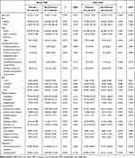
A total of 312176 psoriatic patients aged 20 years or older with records of at least two instances’ records from January 1, 2002 to December 31, 2022 were identified in the TriNetX database. Individuals in the same age range, without a diagnosis of psoriasis counted for 12104309 were identified as the control group. After excluding individuals diagnosed with dermatomyositis before the index date, PSM was performed for the psoriasis and non-psoriasis groups. Finally, 301018 individuals were selected from each group (Figure 1). After PSM, the demographic characteristics of patients with and without psoriasis were comparable. All SMDs between the two groups were <0.1 (Table 1).
 |
Table 1 Demographic Characteristics of Psoriasis Patients and Non-Psoriasis Patients |
Risk of Dermatomyositis in Psoriasis and Non-Psoriasis Groups and Subgroup Analyses
Four hundred and thirty-six patients with psoriasis developed dermatomyositis during the follow-up period. The risk of dermatomyositis in the psoriasis and non-psoriasis groups was compared using three models adjusted for different variables in the PSM. There were respectively 143, 141, and 159 individuals who developed dermatomyositis in the non-psoriasis group in Models 1–3 (Table 2). After PSM based on variables such as age, sex, race, socioeconomic status, BMI, medical utilization, comorbidities, and medication use, the risk of dermatomyositis in the psoriasis group remained significantly higher than that in the non-psoriasis group (HR: 2.41, 95% CI: 2.01–2.89) (Table 2). Kaplan–Meier curves also showed a significantly higher incidence of dermatomyositis in the psoriasis group than that in the non-psoriasis group (P<0.001 in the Log rank test (Figure 2). The higher risk of dermatomyositis among patients with psoriasis was further confirmed by subgroup analysis (Figure 3 and Supplementary Table 2).
 |
Table 2 Risk of Dermatomyositis in Psoriasis Patients Compared to Non-Psoriasis Patients |
 |
Figure 2 Kaplan–Meier analysis for risk of dermatomyositis among patients with psoriasis compared with the non-psoriasis group. |
 |
Figure 3 Forest plots for subgroup analysis of risk of dermatomyositis exposed to psoriasis compared to the non-psoriasis group. Abbreviation: BMI, body mass index. |
Influence of PsA and Biological Agents Use on Dermatomyositis Risk
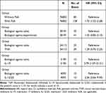
Patients with PsA exhibited a significantly increased risk of developing dermatomyositis compared to those without PsA (HR, 1.73; 95% CI: 1.32–2.28). There was no significant difference in the risk of developing dermatomyositis between psoriatic patients who had been treated with biological agents, including TNFi, IL-17i, and IL-12/23i, and those who were naïve to such treatments (HR, 1.35; 95% CI, 0.85–2.13). Despite this, Kaplan-Meier survival analysis revealed a significantly higher incidence of dermatomyositis among psoriatic patients who had undergone biological agent therapy (P < 0.001 in the Log rank test) compared to those who were biological agent-naïve (Figure 4). Among the different types of biological agents, psoriatic patients treated with IL-17i showed a significantly elevated risk of developing dermatomyositis (HR, 5.79; 95% CI, 1.57–21.31) (Table 3).
 |
Table 3 Risk of Dermatomyositis in Different Groups of Psoriasis Patients |
Risk of Dermatomyositis in Different TriNetX Network Database
The risk of dermatomyositis in patients with psoriasis compared with that in patients without psoriasis was further explored in different racial population databases. In the European, Middle East, and Africa (EMEA) network, including Bulgaria, Germany, Italy, Lithuania, Malaysia, Poland, Spain, and the UK, as well as the APAC (Asia-Pacific) network, including Australia, India, Malaysia, Singapore, and Taiwan, patients with psoriasis showed a significantly higher risk of developing dermatomyositis than those without psoriasis (HR (95% CI): 4.77 (1.40–16.10) and 2.50 (1.33–4.66), respectively) (Table 4).
 |
Table 4 Risk of Dermatomyositis in Psoriasis Patients Compared to Non-Psoriasis Patients in Different TriNetX Network Database |
Discussion
In this retrospective study, the effect of psoriasis on the incidence of dermatomyositis was investigated using a propensity score-matched cohort from the TriNetX network. The risk of dermatomyositis in patients with psoriasis was significantly higher than that in patients without psoriasis, after adjusting for various potential confounding variables. The psoriatic patients with PsA or those experienced treatment of IL-17i exhibited a higher risk of developing dermatomyositis compared to those who did not use biologics.
The occurrence of dermatomyositis in patients with psoriasis has been previously reported in separated cases.10 This is the first study to provide evidence that patients with psoriasis have an elevated risk of dermatomyositis based on a large population cohort. Both psoriasis and dermatomyositis are systemic autoimmune inflammatory diseases, with cutaneous manifestations serving as primary indicators of both disorders. Clinically, both psoriasis and dermatomyositis can present with a rash accompanied by cutaneous keratosis on the extended sides of the knee and elbow. The pathogenesis of psoriasis and dermatomyositis remains incompletely understood, but genetic background, environment stimuli and immune imbalance were thought to play important roles in it.1,6 Genome-wide association studies have identified numerous susceptible loci for psoriasis and dermatomyositis.14,15 Although there is no reported overlap in susceptible loci for these two diseases, the MHC was found to be the major genetic region associated with both psoriasis and dermatomyositis.14,15 In terms of external triggers for induction of skin lesions, Koebner phenomenon which describes the onset of skin lesions resulting from physical stress, injury or trauma would be stimuli for onset both of the two diseases.9 In addition, drug and viral infection have been identified as established environmental trigger factors to both diseases.2,6 For dermatomyositis, ultraviolet (UV) radiation is proposed as a trigger for the occurrence of skin lesions.1 UV exposure or sunlight exposure was found to be associated with dermatomyositis flares.16 However, the UV-based phototherapy, including UVB or psoralen UVA (PUVA) is effective for psoriasis treatment and widely applied clinically.17 Consequently, it is proposed that the UV exposure may be imputed to the occurrence of dermatomyositis among patients with psoriasis under UV phototherapy.
Aberrant immune response plays a crucial role in the pathogenesis of dermatomyositis, psoriasis, and autoimmune inflammatory diseases. Common cellular and cytokine imbalances, such as dendritic cells and IFN, are shared by these two diseases. In psoriasis, dendritic cells play a key role in both the onset and maintenance stages of psoriasis.18 Increased infiltration of pDC has been identified in the skin lesions of psoriasis patients compared to the normal skin of healthy controls.19 In the early phase of psoriasis, pDCs produce IFN and TNF-a, which causes the maturation of resident dermal DC and the differentiation of monocytes into inflammatory DC (iDC). Increased iDCs produce cytokines, such as IL-23, IL-12, and TNF-α, which activate the differentiation of naive T cells into Th17 and Th22 cells. The secretion of IL-17 and IL-22 from Th-17 and Th-22 can induce the proliferation and abnormal differentiation of keratinocytes, which act as the main immune cells in psoriasis.18 Similar to psoriasis, IFNs and DCs play a pivotal role in triggering and perpetuating dermatomyositis.20 Various subsets of dendritic cells have been recently identified in skeletal muscle and skin lesions in dermatomyositis. IFN-inducible proteins, such as CXC chemokines (CXCL9, CXCL10, and CXCL11), are commonly overexpressed in dermatomyositis skin and muscle.21 In addition, these two diseases share signaling pathways, such as the IL-17-IL-23 axis. The central role of the IL-17-IL-23 axis in the pathogenesis of psoriasis has been established in many previous studies. Biological agents targeting inflammatory cytokines, including IL-17 and IL-23, have been widely used in psoriasis therapy.8,22 Recent studies have reported IL-17-IL-23 axis also participates in the onset of dermatomyositis. The infiltration of Th17 cells in cutaneous lesions of dermatomyositis implies an activating role of IL-17.23 Increased serum IL-17 levels have been identified in dermatomyositis and are associated with disease activity.24 In a murine model of myositis, IL-23 was found to mediate the muscle damage in myositis.25 The IL-17-IL-23 axis is also recognized for its significant role in the pathogenesis of PsA. Targeting this axis has been shown to ameliorate both musculoskeletal and cutaneous symptoms in patients with PsA. Prior research indicates that severe psoriasis is associated with a higher prevalence of PsA.26 The heightened activity of the IL-17-IL-23 axis in psoriatic patients with PsA, as compared to those without PsA, potentially confers an increased risk of developing dermatomyositis, which was observed in the present study.
The psoriatic patients who had been treated with biological agents, especially IL-17i, showed a higher risk of developing dermatomyositis than those who did not. As mentioned above, psoriasis and dermatomyositis share common inflammatory cytokines such as IL-17 and TNF-α. Biologics targeting these cytokines have been used to treat both diseases. Paradoxically, the application of biologics in patients with psoriasis increases the risk of dermatomyositis. There have been several previous reports on the association of biologic treatments, such as IL-17i or TNFi, with the occurrence or exacerbation of dermatomyositis in patients with psoriasis.11,12 Similar contradictory reactions have also been observed with the use of IL-17 inhibitors to induce systemic lupus erythematosus. The mechanism by which biological use increases the risk of dermatomyositis remains elusive. Biologics targeting one cytokine can alter the balance of the inflammatory circuit, leading to the accumulation of other inflammatory cytokines. This is so-called “cytokine shift” or “paradoxical reaction” hypothesis. For example, the TNFi or IL-17i targeting TNF or IL-17 may indirectly influence other cytokines that interact with these targets, including type I IFN. This interaction could potentially elevate the risk of developing dermatomyositis. On the other hand, the psoriasis patients experience biological agent therapy may have sever disease condition and have higher activities of IL-17-IL-23 axis, and thus have higher risk to develop dermatomyositis. However, since the dosage and duration of biological agent use could not be adjusted, the results based on whether biological agents experience may not be reliable. Furthermore, the relatively small sample sizes within different categories of biological agents can also affect the reliability of the results.
The present study has some limitations. First, psoriasis and dermatomyositis were diagnosed according to the International Classification of Diseases codes in the TriNetX network. Miscoding of these two diseases is occasionally observed in the clinical setting. Some dermatomyositis patients present without muscle involvement and only show cutaneous manifestations, which are called clinically amyopathic dermatomyositis (CADM). Under these circumstances, the cutaneous symptoms of dermatomyositis, especially those affecting the extensor surface of the joint (Gottron’s sign), may be misdiagnosed as psoriasis. Second, the severity of psoriasis was not adjusted for in the subgroup analysis, which may have affected the risk of dermatomyositis.
Conclusion
In conclusion, the current study is the first to revealed that psoriasis increased the risk of dermatomyositis development based on a large population. The psoriatic patients with PsA or those who had received IL-17i treatment demonstrated a significantly higher risk of developing dermatomyositis. These findings suggest the need for clinicians to screen patients with psoriasis for dermatomyositis, especially when they present with atypical rashes. The mechanism underlying the association between these two diseases needs to be explored in the future.
Funding
This study was supported by Shanghai Sixth People’s Hospital Fund (ynts202307) and Bethune Charity Foundation (J202301E036).
Disclosure
The authors report no conflicts of interest in this work.
References
1. DeWane ME, Waldman R, Lu J. Dermatomyositis: clinical features and pathogenesis. J Am Acad Dermatol. 2020;82(2):267–281. doi:10.1016/j.jaad.2019.06.1309
2. Robinson AB, Reed AM. Clinical features, pathogenesis and treatment of juvenile and adult dermatomyositis. Nat Rev Rheumatol. 2011;7(11):664–675. doi:10.1038/nrrheum.2011.139
3. Furst DE, Amato AA, Iorga ŞR, Gajria K, Fernandes AW. Epidemiology of adult idiopathic inflammatory myopathies in a U.S. managed care plan. Muscle Nerve. 2012;45(5):676–683. doi:10.1002/mus.23302
4. Aussy A, Boyer O, Cordel N. Dermatomyositis and immune-mediated necrotizing myopathies: a window on autoimmunity and cancer. Front Immunol. 2017;8:992. doi:10.3389/fimmu.2017.00992
5. Kao L, Chung L, Fiorentino DF. Pathogenesis of dermatomyositis: role of cytokines and interferon. Curr Rheumatol Rep. 2011;13(3):225–232. doi:10.1007/s11926-011-0166-x
6. Griffiths CEM, Armstrong AW, Gudjonsson JE, Barker J. Psoriasis. Lancet. 2021;397(10281):1301–1315. doi:10.1016/s0140-6736(20)32549-6
7. Armstrong AW, Read C. Pathophysiology, clinical presentation, and treatment of psoriasis: a review. JAMA. 2020;323(19):1945–1960. doi:10.1001/jama.2020.4006
8. Blauvelt A, Chiricozzi A. The immunologic role of IL-17 in psoriasis and psoriatic arthritis pathogenesis. Clin Rev Allergy Immunol. 2018;55(3):379–390. doi:10.1007/s12016-018-8702-3
9. Yamamoto T. Psoriasis and connective tissue diseases. Int J mol Sci. 2020;21(16):5803. doi:10.3390/ijms21165803
10. Chu D, Yang W, Niu J. Concurrence of dermatomyositis and psoriasis: a case report and literature review. Front Immunol. 2024;15:1345646. doi:10.3389/fimmu.2024.1345646
11. Dicaro D, Bowen C, Dalton SR. Dermatomyositis associated with anti-tumor necrosis factor therapy in a patient with psoriasis. J Am Acad Dermatol. 2014;70(3):e64–5. doi:10.1016/j.jaad.2013.11.012
12. Perna DL, Callen JP, Schadt CR. Association of treatment with secukinumab with exacerbation of dermatomyositis in a patient with psoriasis. JAMA Dermatol. 2022;158(4):454–456. doi:10.1001/jamadermatol.2021.6011
13. Palchuk MB, London JW, Perez-Rey D, et al. A global federated real-world data and analytics platform for research. JAMIA Open. 2023;6(2):ooad035. doi:10.1093/jamiaopen/ooad035
14. Dand N, Stuart PE, Bowes J, et al. GWAS meta-analysis of psoriasis identifies new susceptibility alleles impacting disease mechanisms and therapeutic targets. medRxiv. 2023. doi:10.1101/2023.10.04.23296543
15. Miller FW, Cooper RG, Vencovský J, et al. Genome-wide association study of dermatomyositis reveals genetic overlap with other autoimmune disorders. Arthritis Rheum. 2013;65(12):3239–3247. doi:10.1002/art.38137
16. Mamyrova G, Rider LG, Ehrlich A, et al. Environmental factors associated with disease flare in juvenile and adult dermatomyositis. Rheumatology. 2017;56(8):1342–1347. doi:10.1093/rheumatology/kex162
17. Li Y, Cao Z, Guo J, et al. Assessment of efficacy and safety of UV-based therapy for psoriasis: a network meta-analysis of randomized controlled trials. Ann Med. 2022;54(1):159–169. doi:10.1080/07853890.2021.2022187
18. Kamata M, Tada Y. Dendritic cells and macrophages in the pathogenesis of psoriasis. Front Immunol. 2022;13:941071. doi:10.3389/fimmu.2022.941071
19. Wollenberg A, Wagner M, Günther S, et al. Plasmacytoid dendritic cells: a new cutaneous dendritic cell subset with distinct role in inflammatory skin diseases. J Invest Dermatol. 2002;119(5):1096–1102. doi:10.1046/j.1523-1747.2002.19515.x
20. de Padilla CM, Reed AM. Dendritic cells and the immunopathogenesis of idiopathic inflammatory myopathies. Curr Opin Rheumatol. 2008;20(6):669–674. doi:10.1097/BOR.0b013e3283157538
21. Jiang J, Zhao M, Chang C, Wu H, Lu Q. Type I interferons in the pathogenesis and treatment of autoimmune diseases. Clin Rev Allergy Immunol. 2020;59(2):248–272. doi:10.1007/s12016-020-08798-2
22. Miossec P, Kolls JK. Targeting IL-17 and TH17 cells in chronic inflammation. Nat Rev Drug Discov. 2012;11(10):763–776. doi:10.1038/nrd3794
23. Tournadre A, Porcherot M, Chérin P, Marie I, Hachulla E, Miossec P. Th1 and Th17 balance in inflammatory myopathies: interaction with dendritic cells and possible link with response to high-dose immunoglobulins. Cytokine. 2009;46(3):297–301. doi:10.1016/j.cyto.2009.02.013
24. Silva MG, Oba-Shinjo SM, Marie SKN, Shinjo SK. Serum interleukin-17A level is associated with disease activity of adult patients with dermatomyositis and polymyositis. Clin Exp Rheumatol. 2019;37(4):656–662.
25. Umezawa N, Kawahata K, Mizoguchi F, et al. Interleukin-23 as a therapeutic target for inflammatory myopathy. Sci Rep. 2018;8(1):5498. doi:10.1038/s41598-018-23539-4
26. Scher JU, Ogdie A, Merola JF, Ritchlin C. Preventing psoriatic arthritis: focusing on patients with psoriasis at increased risk of transition. Nat Rev Rheumatol. 2019;15(3):153–166. doi:10.1038/s41584-019-0175-0
 © 2025 The Author(s). This work is published and licensed by Dove Medical Press Limited. The
full terms of this license are available at https://www.dovepress.com/terms.php
and incorporate the Creative Commons Attribution
- Non Commercial (unported, 3.0) License.
By accessing the work you hereby accept the Terms. Non-commercial uses of the work are permitted
without any further permission from Dove Medical Press Limited, provided the work is properly
attributed. For permission for commercial use of this work, please see paragraphs 4.2 and 5 of our Terms.
Recommended articles
Comorbid Psoriasis and Metabolic Syndrome: Clinical Implications and Optimal Management
De Brandt E, Hillary T
Psoriasis: Targets and Therapy 2022, 12:113-126
Published Date: 25 May 2022
Reducing the Risk of Developing Psoriatic Arthritis in Patients with Psoriasis
Gisondi P, Bellinato F, Maurelli M, Geat D, Zabotti A, McGonagle D, Girolomoni G
Psoriasis: Targets and Therapy 2022, 12:213-220
Published Date: 10 August 2022
JAK Inhibitors as Potential Therapeutic Strategy for the Dilemma of Psoriasis Concurrent with Dermatomyositis in the SARS-CoV-2 Era
Xu Q, Xu N
Clinical, Cosmetic and Investigational Dermatology 2023, 16:1059-1062
Published Date: 20 April 2023
Cutaneous Adverse Events After COVID-19 Vaccination
Weschawalit S, Pongcharoen P, Suthiwartnarueput W, Srivilaithon W, Daorattanachai K, Jongrak P, Chakkavittumrong P
Clinical, Cosmetic and Investigational Dermatology 2023, 16:1473-1484
Published Date: 8 June 2023
The Combination of IL-6, PLR and Nail Psoriasis: Screen for the Early Diagnosis of Psoriatic Arthritis
Liu X, Zhao Y, Mu Z, Jia Y, Liu C, Zhang J, Cai L
Clinical, Cosmetic and Investigational Dermatology 2023, 16:1703-1713
Published Date: 28 June 2023


