")
Back to Journals » International Journal of Nanomedicine » Volume 20
Medicinal Plant-Derived Exosome-Like Nanovesicles as Regulatory Mediators in Microenvironment for Disease Treatment
Authors Feng Z , Huang J, Fu J, Li L, Yu R , Li L
Received 20 March 2025
Accepted for publication 7 June 2025
Published 30 June 2025 Volume 2025:20 Pages 8451—8479
DOI https://doi.org/10.2147/IJN.S526287
Checked for plagiarism Yes
Review by Single anonymous peer review
Peer reviewer comments 2
Editor who approved publication: Prof. Dr. Anderson Oliveira Lobo
Zhiying Feng,1,* Jiawang Huang,1,* Jingmin Fu,1 Lixin Li,2 Rong Yu,1 Ling Li3
1College of Traditional Chinese Medicine of Hunan University of Chinese Medicine, Changsha, Hunan, 410208, People’s Republic of China; 2College of Medicine of Hunan University of Chinese Medicine, Changsha, Hunan, 410208, People’s Republic of China; 3College of Integrated Traditional Chinese and Western Medicine of Hunan University of Chinese Medicine, Hunan, Changsha, 410208, People’s Republic of China
*These authors contributed equally to this work
Correspondence: Ling Li, Email [email protected] Rong Yu, Email [email protected]
Abstract: Medicinal plant-derived exosome-like nanovesicles is a kind of vesicles with a diameter of 30– 300 nm extracted from medicinal plants, which can regulate the microenvironment of various systems, diseases and cells through multiple drug delivery routes, so as to achieve the purpose of alleviating and treating diseases. In this paper, the extraction and identification methods of nanovesicles derived from medicinal plants were summarized. Then, according to the systematic classification of diseases, the effects and mechanisms of the treatment of diseases and the regulation of the microenvironment by the nanovesicles derived from medicinal plants were summarized and analyzed. This paper finds Medicinal plant-derived exosome-like nanovesicles, as plant-derived nanoscale vesicles, can act more efficiently on various microenvironments of the body such as inflammation, tumors, immunity and the intestine through various administration methods such as oral administration and intravenous injection according to the characteristics of the disease, achieving the effect of treating diseases and alleviating symptoms. Medicinal plant-derived exosome-like nanovesicles have initiated a new model of transboundary communication regulation involving plants in the human microenvironment, using their own advantages to regulate the human microenvironment. Meanwhile, it can also be used as a drug carrier to achieve a dual communication regulation effect. It provides ideas for the follow-up research and clinical use of nanovesicles derived from medicinal plants.
Keywords: medicinal plants, nanovesicles, microenvironment, systemic disease mechanism
Introduction
The microenvironment is the interstitial, tissue, organ, or humoral component of the cell, which is involved in constituting the environment of survival. The stability of microenvironment is an important condition for maintaining normal physiological activities. Abnormal changes of microenvironment components can cause pathological changes in cells and tissues.1 Under normal physiological function, the microenvironment of organ tissues affects the normal physiological activities of organ tissues. The thymus microenvironment regulates the phenotype and number of hematopoietic cells by regulating chemokines,2 bone marrow microenvironment has an important influence on the immune, hematopoietic and motor systems of the body.3 In addition, microenvironments exist in the lungs, pancreas, spleen and other organs, and play an important role in their physiology and pathology.4–6 In pathological conditions, the regulation of microenvironment also plays an important role in the occurrence and development of diseases, including tumor microenvironment, immune microenvironment, inflammatory microenvironment, and tissue damage microenvironment. In particular, the tumor microenvironment has become one of the targets of drug therapy in recent years.7,8 The regulation of microenvironment such as immunity, inflammation and tissue damage is also important for the occurrence and development of diseases, which directly or indirectly affects the disease process and the effect of drug therapy.
Medicinal plant is a plant used in medicine to prevent and treat diseases. The whole or part of Medicinal plant is used as Medicinal raw material, which is widely used in ethnomedicine. A lot of scientific research has been carried out on the monomer extracted from Medicinal plant. And widely used in clinical, such as paclitaxel, berberine and so on.9 In 1983, Pan BTd et al extracted exosomes for the first time from sheep reticulocytes in vitro.10 In 2009, Canal’s research group found that the intercellular fluid in the leaves of sunflower seedlings contained plant-derived exosome-like nanovesicles, it was first demonstrated that plants can secrete plant-derived exosome-like nanovesicles (PLDENs) into cells.11 Medicinal plant-derived exosome-like nanovesicles (MPLDENs) are extracellular vesicles secreted by cells of medicinal plants and are rich in proteins, nucleic acids, lipids and other bioactive substances,12 it has anti-inflammatory, anti-tumor, immunomodulatory and anti-oxidative stress effects.13,14 In the past 15 years, research on the application and mechanism of MPLDENs in the improvement and treatment of various diseases has become a research hotspot. From the different biological characteristics of MPLDENs, it is discussed that MPLDENs, as a new type of natural nanomaterial, have high biocompatibility and a series of functions such as drug delivery.15,16 In the past 15 years, research on the application and mechanism of MPLDENs in the improvement and treatment of various diseases has become a research hotspot. From the different biological characteristics of MPLDENs, it is discussed that MPLDENs, as a new type of natural nanomaterial, have high biocompatibility and a series of functions such as drug delivery.13,17 Compared with other extracts or monomers of medicinal plants, MPLDENs, as natural biological nanovesicles, have rich endogenous substances and active ingredients, and possess multiple therapeutic effects. Due to the biological characteristics of MPLDENs, MPLDENs have high lipid solubility. Therefore, MPLDENs significantly increase drug absorption and drug membrane permeability. Meanwhile, many related studies on MPLDENs have confirmed that MPLDENs are targeted to organs. The concentration of MPLDENs in the target organs is much higher than that in other organs, which is conducive to increasing the drug concentration in the target organs and can significantly enhance the therapeutic effect.
Due to the large number of research articles on MPLDENs, the extraction methods and administration methods of MPLDENs vary to some extent in different articles. Not only that, in the articles related to MPLDENs studying the same disease, the microenvironment and regulatory mechanisms involved in different articles also have certain differences. Therefore, this review comprehensively outlines the basic characteristics and extraction and identification methods of MPLDENs, summarizes the extraction methods of MPLDENs from different medicinal plant sources, and visually expounds the three main extraction methods. Meanwhile, the therapeutic effects of MPLDENs in diseases of various systems are classified. Meanwhile, the therapeutic effects of MPLDENs in diseases of various systems are classified, We mainly emphasize the impact of MPLDENs on cells, tissues and the microenvironment where diseases are located, and further summarize the specific mechanisms of MPLDENs regulation on the microenvironment. In addition, our goal is to explore how to apply the biological and therapeutic characteristics of MPLDENs and provide perspectives and ideas for further research on MPLDENs and the expansion of clinical application of MPLDENs.
Medicinal Plant-Derived Exosome-Like Nanovesicles
Biological Characters and Characteristics of MPLDENs
MPLDENs are around 30–300 nm in size,18 The particle size and morphology of different MPLDENs have certain differences.19 The size of MPLDENs extracted from most of the Mosaic organs, such as tea leaves, honeysuckle flowers and aloe leaves, is small, with the particle size range below 150nm.20–22 The grain size of MPLDENs extracted from most of the root organs, such as polygala and ginseng, is larger, and the particle size ranges above 250nm.23–25 This is due to the subtle differences caused by the different compositions and structures of various organs in medicinal plants. The form of MPLDENs is small round or oval vesicles, and most MPLDENs have a typical saucer-like structure.26 The composition of MPLDENs mainly includes proteins, nucleic acids, lipids and other secondary metabolites, etc. Due to the differences in the growth environment of medicinal plants and autologous genes, the types and contents of proteins, nucleic acids and lipids in different MPLDENs are quite different, and the biological role played by MPLDENs is closely related to its composition. The researchers analyzed the proteins in MPLDENs and classified them into functional proteins, structural proteins and secreted proteins according to their functions.27,28 Functional proteins are involved in many biological processes such as cell expansion, protein folding, and heat stress.29,30 Wheat -derived PLDENs contain heat shock proteins and chaperone proteins that prevent heat stress in the rice during bad weather and help with protein folding.31 Nucleic acid is one of the main components of MPLDENs and plays an important role in the cell biology process. Due to the advantages of wide application and long-term effectiveness of miRNAs in treatment, miRNAs from MPLDENs have been widely extracted and identified and applied in the treatment and research of diseases.13 At the same time, MPLDENs can deliver siRNA by wrapping siRNA and thus play a role in disease treatment.32 Studies have shown that lipids, as the main components of MPLDENs, play a role in maintaining structural stability and participating in biological functions in vesicles. MPLDENs are phospholipid bilayer membrane structures, similar to the membrane structure of cell membrane, and contain a variety of lipid components, such as triglycerides, diacylglycerol, phosphatidylcholine and phosphatidylinositol.27,33,34 It has been reported that diacylglycerol and phosphatidic acid have a significant effect on membrane fusion and are involved in biological functions such as vesicle uptake, which is one of the advantages of exosome-like nanovesicles of medicinal plant origin into the human body35 (Figure 1).
 |
Figure 1 Medicinal plant-derived exosome-like nanovesicles diagram.13,18,27,32–34 |
Methods for Extraction, Purification and Identification of MPLDENs
MPLDENs can be extracted and purified by a variety of methods, the main extraction and purification methods of MPLDENs include differential ultra-high speed centrifugation, sucrose gradient density centrifugation, immunoaffinity capture, ultrafiltration, size exclusion chromatography, PEG co-precipitation and so on,27 the extraction and purification methods of exosome-like nanovesicles from medicinal plants were summarized (Table 1). Among the MPLDENs extraction methods, differential ultra-high speed centrifugation and sucrose gradient density centrifugation are the most widely accepted.18 It is also the most used extraction and purification method. Three extraction methods steps (Figure 2). According to the biological properties and characteristics of MPLDENs, the morphology, particle size, protein concentration, nucleic acid content, protein type, nucleic acid type and lipid analysis of plant vesicles were evaluated.36–38 The identification methods of plant vesicles were summarized, and the advantages of different methods were compared (Table 2).39–46
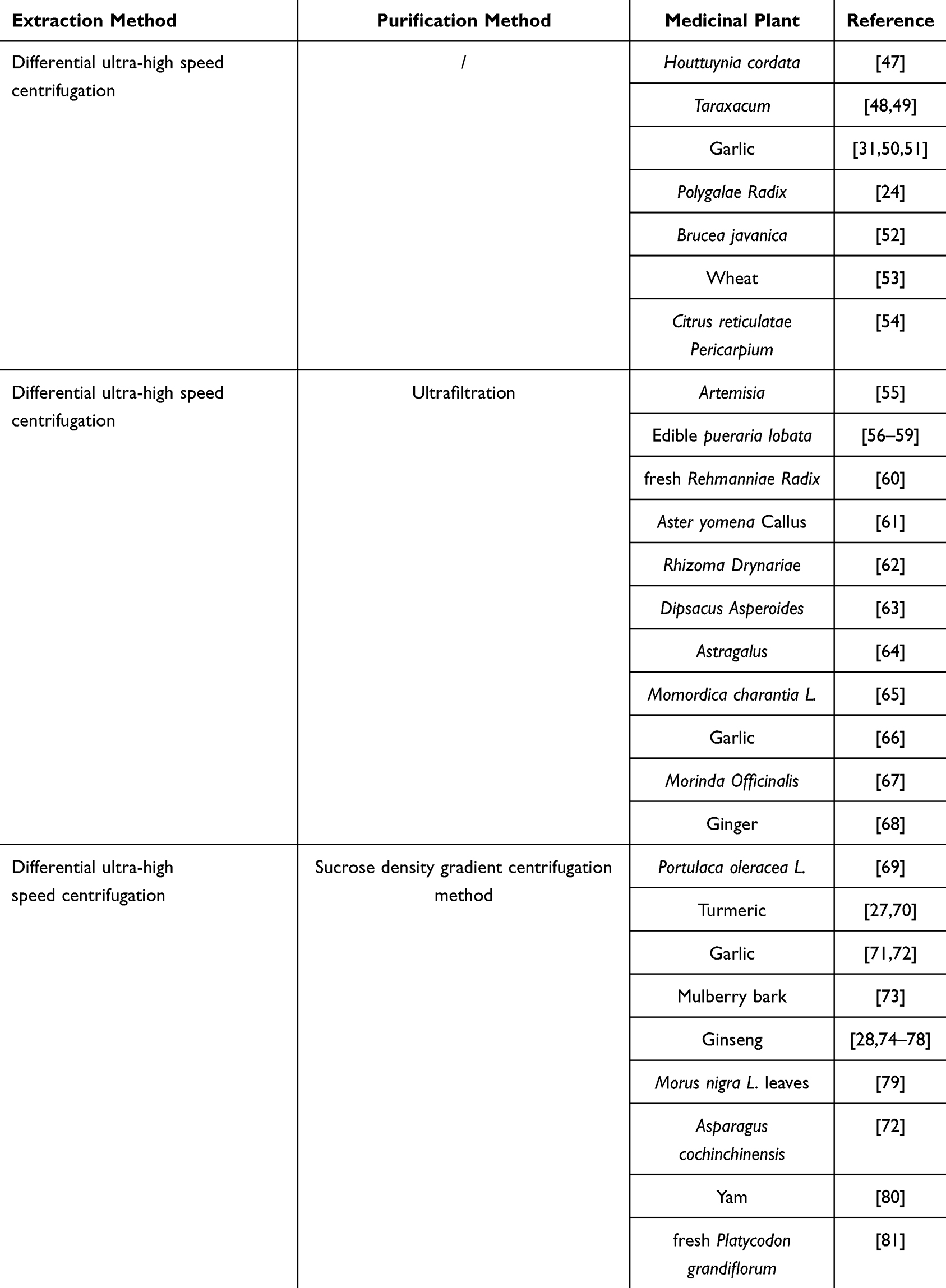 |
Table 1 Extraction methods of MPLDENs from medicinal plants |
 |
Table 2 Comparison of plant vesicles identification methods |
 |
Figure 2 Methods for extraction and purification of MDPLNs (1) Method A: Differential ultra-high speed centrifugation; (2) Method B: Method A: Differential ultra-high speed centrifugation unite ultrafiltration; (3) Method C: Differential ultra-high speed centrifugation unite sucrose density gradient centrifugation method. |
The Role and Mechanism of MPLDENs in Disease Treatment and Microenvironment Regulation
As a new type of therapeutic drug, MPLDENs enter the body through different modes of administration, so as treat the diseases occurring in each system in a targeted way, treating diseases by directly targeting disease tissues and cells and regulating the disease microenvironment. This section summarizes and discusses the therapeutic effect of MPLDENs on diseases of respiratory, digestive, motor, endocrine, circulatory, nervous and reproductive systems and skin diseases verified by in vivo and in vitro experiments and elucidates its mechanism of action on diseases and microenvironment regulation.
Respiratory System
MPLDENs are used in the treatment of respiratory diseases through different administration routes. The following diseases have been used in existing research Viral Pneumonia, Acute lung injury, Pulmonary fibrosis, Bronchial asthma (Figure 3 and Table 3).
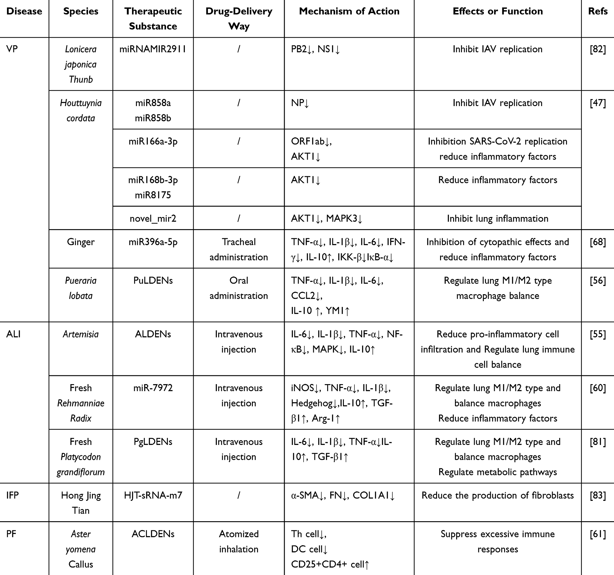 |
Table 3 MPLDENs for the treatment of respiratory diseases |
 |
Figure 3 Mechanism of MPLDENs in the treatment of respiratory diseases. |
Viral Pneumonia (VP)
VP is a kind of pulmonary inflammation caused by virus immersed in lung epithelial cells through respiratory tract. The main clinical manifestations are asthma, dry cough, fever and headache.84 Influenza A virus (IAV) and 2019 novel coronavirus (SARS-CoV-2) are the main types of viruses that cause VP in recent years.85,86
After the virus infects the body, it invades the lung and activates the immune system, recruits immune cells, transmits a large number of pro-inflammatory and anti-inflammatory cytokines, and shapes the pulmonary inflammatory microenvironment.87 In terms of improving the inflammatory microenvironment in the lungs, Houttuynia cordata-derived exosome-like nanovesicle miR168a-3p, miR168b-3p, novel_mir2 and miR8175 regulate the expression of PI3K-AKT pathway. novel_mir2 also regulates the expression of MAPK3.47 It can inhibit inflammation and improve the pulmonary inflammatory microenvironment. miR396a-5p from Ginger-derived exosome-like nanovesicles alleviates lung microenvironment inflammation by targeting the inhibition of NF-κb pathway mediated inflammation.68 Taraxacum-derived exosome-like nanovesicles encapsulated PGY-sRNA-6 targeting the RELA gene in the NF-kB complex, reducing p65 protein expression, reducing oxidative stress, and inhibiting lung inflammation.47 Pueraria lobata-derived exosome-like nanovesicles (PuLDENs) can regulate the balance of lung M1/M2 macrophages, increase the expression of M2 macrophages, improve the pulmonary inflammatory microenvironment, and reduce lung inflammation.48,56
In addition to improving the pulmonary inflammatory microenvironment, the proteins transcribed by the viral genome are closely related to the formation of the pulmonary microenvironment, and inhibiting the transcription of the viral genome is conducive to improving the pulmonary microenvironment.88 miRNAMIR2911 in Lonicera japonica Thunb-derived exosome-like nanovesicles targets multiple subtypes of IAV, including H1N1, H5N1, and H7N9. The expression of PB2 and NS1 proteins encoded by H1N1 was significantly reduced, thus significantly inhibiting the replication of H1N1 virus.82 In 2023, Zhu et al showed that miR858a and miR858b in Houttuynia cordata-derived exosome-like nanovesicles inhibited the expression of NP genes in IAV. miR166a-3p targets the inhibition of ORF1ab gene in SARS-CoV-2, reducing viral replication in vivo.47 At the same time, Ginger derived exosome-like miR396a-5p from nanovesicles inhibited the expression of viral S proteins and Nsp12 and Nsp13 proteins. The Cytopathic Effect (CPE) of SRAS-CoV-2 was inhibited in Vero E6 cells.68 This reduces the virus’s damage to cells and indirectly reduces the amount of virus in the body. In 2025,
The miRNAs in MPLDENs plays a major role in inhibiting virus replication, and miRNAs plays an important regulatory role in VP. The miRNAs produced in the body can inhibit virus infection symptoms and virus replication. In addition, viral infection can regulate the expression of miRNA in the body, regulate the pulmonary inflammatory microenvironment, and carry out immune escape through self-produced miRNA.89 Therefore, miRNAs in MPLDENs may not only inhibit viruses but also serve as endogenous miRNAs in the body to improve the pulmonary inflammatory microenvironment.
In conclusion, MPLDENs and their miRNAs can treat viral pneumonia by regulating the pulmonary inflammatory microenvironment and reducing the virus content in the body, preventing immune overactivation and increasing endogenous healing.90 Effective prevention of secondary pneumonia and acute respiratory distress syndrome.91,92 The two mechanisms are synergistic. Clinically, anti-inflammatory therapy and antiviral therapy are played by different drugs, and MPLDENs have a dual role, which can play a dual role and have a synergistic therapeutic effect. In addition to antiviral therapy, it can alleviate respiratory tract and fever symptoms such as asthma and dry cough in patients.
Acute Lung Injury (ALI)
ALI is the injury of alveolar epithelial cells and capillary endothelial cells caused by direct or indirect injury factors, which can lead to diffuse pulmonary interstitial and alveolar edema and acute hypoxic respiratory insufficiency, with high morbidity and high mortality.93 The damaged microenvironment formed by acute lung injury plays an important role in the development of ALI. By regulating the inflammatory microenvironment of ALI, M1 macrophages are reduced, M2 macrophages are increased, neutrophil infiltration is reduced, the secretion of proteins and inflammatory factors in lung tissue is inhibited, and the inflammatory microenvironment of ALI is improved, which can effectively alleviate ALI symptoms.94 In 2024, Ye et al found that Artemisia derived exosome-like nanovesicles (ALDENs) regulated the production of inflammatory factors and reduced inflammatory infiltration by inhibiting NF-κB and MAPK signaling pathways, and at the same time improved the mitochondrial function of macrophages by interacting with GABA receptors.55 Thus, it plays an anti-inflammatory role and prevents the progressive development of ALI. In 2023, Qiu et al showed that miR-7972 in fresh Rehmanniae Radix derived exosome-like nanovesicles extreme macrophages from M1 phenotype to M2 phenotype, reducing the production of inflammatory factors. miR-7972 inhibits Hedgehog pathway by targeting GPR161 protein and upstream and downstream GPR161 protein, thereby improving the inflammatory symptoms of ALI.60 By regulating the inflammatory microenvironment and immune microenvironment of ALI, MPLDENs and their miRNAs can alleviate the damage of alveolar epithelial cells and capillaries, control diffuse inflammation, and reduce pulmonary interstitial and alveolar edema. Fu et al showed that Platycodon grandiflorum-derived exosome-like nanovesicles can alleviate acute lung injury caused by IAV infection by regulating the mechanisms of immune and metabolic anti-inflammatory damage.81
Pulmonary Fibrosis (PF)
PF is a chronic interstitial lung disease characterized by abnormal activation of fibroblasts, excessive accumulation of extracellular matrix, inflammatory damage and abnormal lung remodeling.95 Hong Jing Tian-derived exosome-like nanovesicles and Taraxacum-derived exosome-like HJT-sRNA-m7 in nanovesicles (taraxion) reduce collagen and fibrin production by fibroblasts by inhibiting gene expression of α-SMA, Fibronectin (FN), and collagen type I α1 (COL1A1)48,96 and control the progressive development of pulmonary fibrosis. HJT-sRNA-m7 maintains extracellular matrix microenvironmental homeostasis and reduces epithelial mesenchymal transformation by reducing fibrin accumulation, thereby inhibiting myofibrocyte formation in lung mesenchyma,94,95 preventing fibroblasts from being over-activated and delaying pulmonary fibrosis.
Bronchial Asthma (BA)
BA is a chronic inflammatory disease of the airway, which is accompanied by damage and detachment of the airway epithelia, airway hyperresponsiveness and increased mucosal secretion, leading to symptoms such as cough, wheezing, shortness of breath, and eventually airway remodeling.96 Aster yomena Callus-derived exosome-like nanovesicles (ACLDENs) have a protective effect on allergic airway diseases by inhibiting the T cell response associated with asthma and increasing the level of regulatory T cells (Foxp3+CD25+CD4+). Inhibition of airway overimmune response, thereby relieving airway smooth muscle tension and spasm, reduce airway hyperreactivity. Aster yomena callus-derived exosome-like nanovesicles inhibit Th2 and mixed Th2/Th17 responses associated with asthma, reduce helper T cell infiltration, regulate the immune microenvironment of asthmatic97 and reduce mucosal secretion. Improve symptoms such as cough, wheezing, and shortness of breath.61
Digestive System
MPLDENs are used in the treatment of digestive system through different administration routes. The following diseases have been used in existing research: ulcerative colitis, digestive system tumors, acute liver failure (Figure 4 and Table 4).
 |
Table 4 MPLDENs for the treatment of digestive system |
 |
Figure 4 Mechanism of MPLDENs in digestive system. |
Ulcerative Colitis (UC)
UC is a chronic inflammatory disease that affects the function of the colon, with typical symptoms including diarrhea, rectal bleeding, and abdominal pain. MPLDENs treat ulcerative colitis by improving intestinal inflammation with immune microenvironment and regulating intestinal microenvironment.
In terms of MPLDENs improving intestinal inflammation and immune microenvironment, in 2019, Zhu et al‘s research shows that Portulaca oleracea-derived exosome-like nanovesicles (PoLDENs) down-regulated the expression of Zbtb7b in conventional CD4+ T cells through the Lactobacillus-Enterobacteriaceae-indole derivative axis.97 Zbtb7b plays a role in peripheral CD4+ T cells, inhibiting the expression of CD8 lineage genes, including the expression of CD8 and cytotoxic effector genes perforin and granzyme B, and inhibiting the expression of IFN-γ during effector differentiation.97 The conventional CD4+ T cells are reprogrammed into DP CD4+CD8+ T cells (DP cells), thereby regulating the immune microenvironment of ulcerative colitis, inhibiting the production of TNF-α, IL-1β, IL-6, IL-12 and other inflammatory factors, increasing the production of anti-inflammatory factors, and significantly alleviating the symptoms of colitis and has specific targeted therapeutic action.97 Edible pueraria lobata-derived exosome-like nanovesicles (PuLDENs) reduce the formation of M1-type macrophages and regulate the immune microenvironment by polarizing macrophages into M2 macrophages. Reduce the production of TNF-α, IL-1β, IL-6 and other inflammatory factors, reduce intestinal epithelial damage, promote the proliferation of intestinal epithelial cells and restore barrier function,56 thereby reducing the inflammatory response of UC and repairing tissue damage caused by UC. In 2022, Liu et al showed that Turmeric-derived exosome-like nanovesicles (TLDENs) improved mouse colitis and accelerated the resolution of colitis by regulating inflammatory factors and antioxidant gene HO-1, and the potential mechanism of its treatment may be related to the NF-κB pathway.70
TLDENs can not only alleviate the symptoms of ulcerative colitis by improving the intestinal inflammatory microenvironment, but Gao et al found that TLDENs can also promote the transformation of M1 macrophages into M2 macrophages, up-regulate the expression of tight junction protein and regulate the intestinal microbiota to restore the damaged intestinal epithelial barrier and protect the intestinal barrier function.27 The effects of TLDENs inhibited intestinal inflammation and reduced intestinal damage. Sophora japonica exosome-like nanovesicles reduce iron death in intestinal epithelial cells and regulate metabolic disorders by regulating HIF pathway, thereby repairing intestinal mucosa, and have potential therapeutic effects on ulcerative colitis.75 Ginseng-derived exosome-like nanovesicles (GinLDENs) improve the immune microenvironment of ulcerative colitis by regulating the polarization of macrophages, inhibiting the anti-inflammatory effect of NF-κB pathway.74 Zhao et al found that Garlic derived exosome-like nanovesicles (GaLDENs) treated ulcerative colitis by inhibiting the TLR4/MyD88/NF-κB signaling pathway, thereby inhibiting downstream cascade reaction and the release of inflammatory factors. Inhibition of TLR4 expression may act through Han-miR3630-5p in GaLDENs.50 In 2022, Sriwastva et al found that Mulberry bark-derived exosome-like nanovesicles (MBLDENs) activate the anti-inflammatory pathway mediated by AhR-COPS8, AhR signal transduction through the combination of MBLDENs derived heat shock protein HSPA8 (MBELN-HSPA8) and AhR to activate and improve the inflammatory microenvironment of colitis and alleviate colitis symptoms.73
In the regulation of intestinal microenvironment by MPLDENs, garlic-derived exosome-like nanovesicles (GaLDENs) can restore the relative abundance of Lachnospiraceae beneficial to intestinal microenvironment. Decreased the abundance of Helicobacter, which leads to chronic intestinal inflammation97 peu MIR2916-p3 in GaLDENs specifically promoted the growth of Bacteroides thetaiotaomicron, which is beneficial to the intestinal microenvironment.71 To change the intestinal microbiota spectrum of colitis mice, which has a preventive effect on the occurrence of ulcerative colitis. Ginseng-derived exosome-like nanovesicles (GinLDENs) reduce the ratio of Firmicutes/Bacteroides, regulate intestinal flora, and slow the progression of ulcerative colitis.74 Fresh Rehmannia exosome-like nanovesicles can reduce the abundance of Escherichia coli and regulate the microenvironment of intestinal flora by inhibiting the formation of Escherichia coli biofilms.60
In conclusion, MPLDENs can regulate the balance of M1 and M2 macrophages and CD4+ T cell genes to regulate the immune microenvironment, reduce the production of inflammatory factors and increase the expression of pro-inflammatory factors, thus inhibiting inflammatory response, improving the inflammatory microenvironment, regulating the type and abundance of intestinal flora, and optimizing the intestinal microenvironment for the treatment of ulcerative colitis. Relieve symptoms such as diarrhea, abdominal pain and blood in the stool, and repair the intestinal barrier effect, reducing the damage of ulcerative colitis to the colon. Intestinal microflora microenvironment, immune microenvironment and inflammatory microenvironment are closely related to the progression and treatment of ulcerative colitis. The immune microenvironment improves the intestinal microflora microenvironment and inflammatory microenvironment, and the intestinal microflora and inflammatory microenvironment affect the expression and function of macrophages and dendritic cells in the immune microenvironment.99
Digestive System Tumors
Colorectal cancer is a malignant tumor occurring in the colon or rectum. Early clinical manifestations are usually hematochezia, diarrhea and abdominal pain, and late symptoms are anemia and cachexia with weight loss.100,101 Ginseng-derived exosome-like nanovesicles (GinLDENs) enhance the efficacy of immune checkpoint antibodies by reprogramming the cold tumor microenvironment, thereby promoting T lymphocyte infiltration and anti-tumor response. Improved the efficacy of immune checkpoint inhibitor (ICI) related combination in the treatment of colon cancer.75 Ginger-derived exosomes like nanovesicles (GLDENs), after being taken up by colorectal cancer cells, mediated the metabolic transformation of cancer cells in the tumor microenvironment by activating the PI3K-AKT signaling pathway, and PI3K/AKT signaling was associated with apoptosis of cancer cells.102 Promote the apoptosis of colorectal cancer cells and inhibit the cell viability of colorectal cancer cells and have anti-colorectal cancer effect.98
Hepatocellular carcinoma is a malignant tumor occurring in the liver, which often occurs in the terminal stage of chronic liver disease and decompensated stage of cirrhosis.103 Oral administration of Morus nigra L. leaves-derived exosome-like nanovesicles (MLLDENs) significantly inhibited tumor growth in mouse hepatoma models. After MLDENs intervention with Hepa1-6 cells, the cell cycle arrest in G0/G1 phase of Hepa1-6 cells and apoptosis of Hepa1-6 cells were induced.97 Asparagus cochinchinensis-derived exosome-like nanovesicles (ALDENs) can induce apoptosis of Hep G2 cells by up-regulating protein levels of AIF, Bax and Bak and activating caspase-9, inhibiting the occurrence, development and metastasis of liver cancer.97 However, apoptosis of liver cancer cells affects the inflammatory microenvironment of liver cancer, thus accelerating the progression of tumor.104 After the apoptosis of liver cancer cells is induced by MLDENs and ALDENs, the inflammatory response of the liver is significantly reduced, indicating that in addition to promoting the apoptosis of tumor cells, the tumor inflammatory microenvironment can be improved and inflammatory infiltration can be reduced. MLDENs and ALDENs both target the liver and have good biosafety, and the inflammation and liver function indexes are close to normal. In the absence of damage to normal liver cells, better targeted treatment of liver cancer.72,79
In conclusion, MPLDENs can induce apoptosis of cancer cells by increasing PI3K/AKT signal and activating caspase-9 to slow down tumor growth. The cold tumor microenvironment can be changed into a hot tumor microenvironment, which can increase the efficacy of immunotherapy and improve the tumor inflammatory microenvironment to alleviate some series of symptoms such as fever and pain caused by tumors. Digestive system tumors, especially liver cancer, have a high degree of malignancy, easy to metastasize, and the symptoms of fever and pain caused by tumors are more serious. MPLDENs treat both the primary tumor and the adverse symptoms of tumor-induced inflammation.
Acute Liver Failure
Acute liver failure, also known as fulminant liver failure, is a rare life-threatening disease with major complications including type A hepatic encephalopathy and multiple organ failure, and a high mortality rate.105,106 Garlic-derived exosome-like nanovesicles (GaLDENs) inhibit the production of inflammatory factors, inhibit CCR2/CCR5 signaling, and reduce the aggregation of macrophages, monocytes and other immune cells into the liver. Reducing the intense immune response and inflammatory response caused by acute liver failure,66 regulating the inflammatory microenvironment, GaLDENs as a natural plant extract has high biosafety, low liver toxicity, alleviating heavy liver metabolic burden, and controlling the process of acute liver failure.50
Motor System
MPLDENs is used in the treatment of digestive system through different administration routes. The following diseases have been used in existing research osteoporosis, osteoarthritis, osteosarcoma, muscle atrophy (Figure 5 and Table 5).
 |
Table 5 MPLDENs for the treatment of motor system diseases |
 |
Figure 5 Mechanism of MPLDENs in muscle atrophy. |
Osteoporosis
Osteoporosis is a systemic bone disease caused by a variety of reasons, the main characteristics are the decrease of bone density and bone quality, the damage of bone microstructure, the increase of bone fragility, the high probability of fracture, and the reduction of bone trabeculae.108,109 MPLDENs treatment of osteoporosis works through its effects on osteoblasts and osteoclasts, thereby regulating dysfunction of bone cell activity and abnormal cytokine secretion in the osteoporosis microenvironment, reducing bone mass loss and bone imbalance.110 Pueraria lobata-derived exosome-like nanovesicles (PuLDENs) can improve the differentiation and calcification of hBMSCs cells, regulate the imbalance of intestinal flora through TMAO, and increase the autophagy of BMSC cells to promote bone formation and increase the number of osteoblasts. Improve and treat osteoporosis.57 Rhizoma Drynariae-derived exosome-like nanovesicles (RLDENs) induce osteoblast differentiation by activating ERα signaling pathway, up-regulating Runx2 and BMP2. RLDENs can effectively relieve bone loss in vivo, maintain normal bone density and bone quality, and prevent and treat osteoporosis.62 Morinda Officinalis-derived exosomes like nanovesicles (MoLDENs) increased the expression of CREB and RSK1 proteins and promoted the proliferation of MC3T3-E1 cells by activating MAPK signaling pathway. MC3T3-E1 cells were induced to differentiate into osteoblasts to reduce bone trabecular destruction, increase bone density and alleviate osteoporosis.67 Yam-derived exosome-like nanovesicles (YLDENs) also promoted the proliferation and differentiation of MC3T3-E1 cells by activating MAPK signaling pathway and up-regulating p38.80 Ginseng-derived exosome-like nanovesicles (GinLDENs) inhibited osteoclast differentiation and reduced bone microstructure damage by inhibiting IκBα, JNK and ERK signaling pathways and down-regulating c-Fos, c-Jun and NFATc1 genes. Delay the progression of osteoporosis.76 By promoting the proliferation and differentiation of osteoblasts and inhibiting the differentiation of osteoclasts, MPLDENs can alleviate and treat osteoporosis in various aspects, maintain bone density, bone quality and the number of bone trabeculae, reduce the damage of bone microstructure, and maintain a bone microenvironment with a balance between osteoclasts and osteogenesis.
Osteoarthritis
Osteoarthritis is a non-inflammatory degenerative joint disease characterized by pain and stiffness in the joints.97 Garlic derived exosome-like nanovesicles (GaLDENs) regulate inflammatory signaling pathways, improve inflammatory microenvironment, and relieve pain symptoms by inhibiting MAPK signaling pathway and phosphorylation of ERK, JNK, and P38. GaLDENs promote the expression of collagen and agglutinoglycan, play a role in cartilage repair, improve the symptoms of bone and joint stiffness, and slow down the progression of osteoarthritis.97
Osteosarcoma
OS is the most common primary bone malignancy with a high tendency of local invasion and metastasis.97 The main symptoms are pain, mass, lameness, and related systemic symptoms.97 The tumor immune microenvironment has been shown to promote cancer cell proliferation, migration, apoptosis, and drug resistance, as well as immune escape.97 Dipsacus Asperoides-derived exosome-like nanovesicles (DLDENs) can significantly inhibit the invasion and migration of osteosarcoma cells by activating the low-toxicity and high-affinity apoptosis pathway induced by P38/JNK signal. Inhibit the proliferation of osteosarcoma, so as to achieve the role of treatment of osteosarcoma.97
Muscle Atrophy
Muscular dystrophy refers to the reduction of muscle volume caused by rhabdomyodystrophy, the thinning or disappearance of muscle fibers, and the main symptoms are loss of normal muscle strength, fatigue and reduced exercise capacity, and reduced quality of life.97 Gouqi-derived exosome-like nanovesicles (GouLDENs) activation of AMPK/SIRT1/PGC1α signaling pathway can improve quadriceps grip strength and increase the muscular area, change the metabolites of quadriceps muscle, and regulate the internal microenvironment of C2C12 cells. Promotes muscle regeneration in mice with muscle atrophy.97
Endocrine System
MPLDENs are used in the treatment of endocrine system through different administration routes. The following diseases have been used in existing research type 2 diabetes, diabetic ulcer (Figure 6 and Table 6).
 |
Table 6 MPLDENs for the treatment of circulatory, endocrine, nervous and reproductive systems |
 |
Figure 6 Mechanism of MPLDENs in circulatory, endocrine, nervous and reproductive systems. |
Type 2 Diabetes
Type 2 diabetes is a chronic disease caused by insufficient or inefficient use of insulin, also known as insulin-dependent diabetes, the main symptoms of polydipsia, polyphagia and polyuria.118 Mung Bean Sprouts-derived exosomes -like nanovesicles (MLDENs) increased the expression of P-GSK-3β and GS by regulating the PI3K/Akt/GLUT4/GSK-3β pathway, which promoted glycogen synthesis and glycogen synthesis and transformation. Converting glucose into glycogen storage, GSK-3β is a protein that regulates Nrf2 oxidative stress level,119 promotes the expression of Nrf2 factor, reduces the oxidative stress response caused by insulin resistance in hepatocytes, restores the normal physiological function of hepatocytes, and increases the expression of GLUT4 protein in insulin-resistant hepatocytes. Promote glucose absorption and glycogen synthesis, reduce blood sugar, increase glucose tolerance.97 Insulin resistance is closely related to the pathogenesis of type 2 diabetes.97 Insulin resistance causes compensatory production of insulin by functional islet beta cells, disrupts the microenvironment that maintains the islets, and even leads to islet failure.121 Garlic-derived exosome-like nanovesicles (GaLDENs) can reverse insulin resistance97 by increasing Acinetobacter mucophilus and stimulating GLP-1 signaling pathway to reverse insulin resistance51 and protect existing islet beta cells. Maintenance of islet microenvironment balance.123 Astragalus-derived exosome-like nanovesicles (ALDENs) can improve glucose metabolism and reduce blood sugar by regulating the ratio of Firmicutes and Bacteroidetes and their metabolites.64 Type 2 diabetes is associated with complications as the disease progresses.124 Citrus reticulatae Pericarpium-derived exosome-like nanovesicles can enhance the expression of FAO-related genes and inhibit the expression of genes associated with liver lipogenesis and glycolysis by regulating intestinal flora. Modulating liver lipid metabolism CiLDENs activate FXR-SHP pathway and promote liver bile acid effection, inhibit cholesterol accumulation and alleviate T2DM induced hepatic steatosis.54
Diabetic Ulcer
Diabetic ulcer is a complication of diabetic peripheral neuropathy, somatic nerve and sympathetic autonomic neuropathy. Diabetic ulcers have extremely complex microenvironments, such as hyperglycemia, ischemia, hypoxia, excessive inflammation, persistent infection, high protease activity, and insufficient energy supply.97 Non-healing of diabetic chronic ulcers is usually due to impaired angiogenesis.97 Ginseng-derived exosome-like nanovesicles (GinLDENs), through accelerating the biological process of glycolysis, provide substances and energy for the repair and formation of blood vessels, inhibit oxidative phosphorylation, reduce oxidative stress, and restore angiogenesis in diabetic ulcers. Improving the microenvironment of diabetic ulcer in many ways and controlling the progress of diabetic ulcer.77
Circulation System
Myocardial injury is a serious pathological consequence of circulatory system.127 Drug-induced myocardial injury is a common problem in clinical practice.97 Mitochondrial dysfunction and mitochondrial injury of cardiomyocytes are the main causes of death of cardiomyocytes and endothelial cells in myocardial injury.129,130 Momordica charantia L.-derived exosomes like nanovesicles (McLDENs) regulate the p62/Keap1/Nrf2 pathway to reduce the increased cardiotoxicity and apoptosis of cardiomyocytes induced by doxorubicin (DOX) during its antitumor effects.131 Panax ginseng-derived exosome-like nanovesicles (PGinLDENs) improved abnormal apoptosis of cardiomyocytes by regulating apoptosis factors, increasing the expression of anti-apoptotic factor Bcl-2, inhibiting the expression of pro-apoptotic factors Bax and Cyt C. ameliorating myocardial damage caused by DOX.116 Oxidative stress is an important pathological mechanism of myocardial injury.132 McLDENs can alleviate oxidative stress and promote the proliferation of DOX-treated cardiomyocytes by increasing the expression of Nrf2 factor and HO-1 gene.97 By regulating apoptosis, energy metabolism and oxidative stress of cardiomyocytes, MPLDENs protects cardiomyocytes, maintains normal physiological functions of cardiomyocytes, and improves pathological manifestations of myocardial injury. At the same time, McLDENs and PLDENs can both reduce mitochondrial damage and maintain normal physiological activities of mitochondria and reduce myocardial damage caused by DOX (Figure 6 and Table 6).
Nervous System
In neurological diseases, MPLDENs can penetrate the blood–brain barrier and delay the progressive exacerbation of neurological diseases by improving the energy metabolism of nerve cells, regulating the immune and inflammatory microenvironment. Pueraria lobata-derived exosome-like nanovesicles (PuLDENs) activate the Pink1-Parkin pathway, and PINK1 and Parkin control the specific elimination of dysfunctional or redundant mitochondria. Thus regulating the mitochondrial network and preserving energy metabolism,133 mediating mitochondrial autophagy, reducing dysfunctional mitochondria, restoring ATP supplementation, maintaining mitochondrial homeostasis thereby improving the cellular microenvironment, improving mitochondrial dysfunction in dopamine neurons,58 improving tremor and retardation caused by Parkinson’s disease in progressive neurodegenerative diseases.134 Polygalae Radix-derived exosome-like nanovesicles can cross the blood–brain barrier 25 and improve the manifestation of dementia caused by Alzheimer’s disease.135 Ginseng-derived exosome-like nanovesicles (GinLDENs) can directly act on glioma through the blood–brain barrier, promote apoptosis of glioma cells, inhibit cell growth of glioma cells, fibroblasts and endothelial cells, and act on glioma.136 Tumor cells actively reshape their microenvironment and create crosstalk between components of the microenvironment to promote disease development.137 By regulating tumor inflammation and immune microenvironment, GinLDENs can inhibit the inflammation of tumor tissue, regulate macrophages, inhibit M2 macrophages, reduce the expression of growth factors, angiogenic factors and chemokines, and slow down the occurrence and development of glioma78 (Figure 6 and Table 6).
Reproductive System
Cervical Cancer
Cervical cancer is a malignant tumor occurring in the female reproductive tract, which occurs in the cervix. Human tumor virus (HPV) infection is the main cause of cervical cancer.138,139 HPV-infected cells actively reshape the local environment by conspiring with cells and inducing a supportive and immunosuppressive post-infection microenvironment that supports virus persistence, transmission, and promotes cervical cancer97 MiR2911 from Lonicera japonica Thunb-derived exosome-like nanovesicles binds to the E6 and E7 oncogenes of HPV16/18 to inhibit the proliferation of cervical cancer cells. The apoptosis of cervical cancer cells was induced by regulating Caspase3 to achieve the purpose of treating cervical cancer.20
Breast Cancer
Breast cancer is a disease in which abnormal breast cells grow out of control and form tumors.97 Brucea javanica-derived exosome-like nanovesicles (BLDENs) inhibit the PI3K/Akt/mTOR pathway, and the PI3K/Akt/mTOR pathway, PI3K/AKT signal mediates the metabolic transformation of cancer cells in the tumor microenvironment97 resulting in the apoptosis of 41T tumor cells. At the same time, by inhibiting the synthesis of VEGF, inhibiting angiogenesis, improving the tumor microenvironment, reducing the blood flow of tumor tissue, and slowing down the development of breast cancer97 (Figure 6 and Table 6).
Skin Diseases
MPLDENs are used in the treatment of Skin Diseases through different administration routes. The following diseases have been used in existing research skin melanoma, skin trauma, skin aging, Alopecia (Figure 7 and Table 7).
 |
Table 7 MPLDENs for the treatment of Skin disease |
 |
Figure 7 Mechanism of MPLDENs in skin disease. |
Skin Melanoma
Cutaneous melanoma is a malignant tumor caused by melanocytes of the skin,143 Ginseng-derived exosome-like nanovesicles (GinLDENs) induced macrophage polarization by mediating the activation of TLR4-MyD88 pathway, accelerated the apoptosis of melanoma cells and the expression of apoptosis gene caspase 3/7, and inhibited the growth of melanoma.28
Skin Trauma
Skin trauma forms a damaging microenvironment that triggers inflammatory and immune responses.97 MPLDENs can repair wounds and improve local inflammatory symptoms. Wheat-derived exosome-like nanovesicles (WLDENs) accelerate wound repair by increasing type I collagen, promote the proliferation and migration of endothelial cells, epithelial cells and dermal fibrocytes, promote the repair of skin wound tissue and the formation of blood vessels, and improve the skin injury microenvironment.53 Dendrobium-derived exosome-like nanovesicles (DLDENs) improve the inflammatory microenvironment and promote wound healing by inhibiting overexpression of immune cells and genes.97
Skin Aging
Skin aging is more pronounced with age because senescent cells accumulate with age, and during skin aging, the extracellular matrix microenvironment undergoes tremendous structural changes and degradation, resulting in thinning of the dermis and loss of elasticity in the aging phenotype.134,145 As a sign of aging, cell senescence is a basically irreversible cell cycle arrest with the characteristics of apoptosis.146 Bletilla-derived exosome-like nanovesicles (BLDENs) inhibited the apoptosis of human immortalized epidermal cells by regulating caspase-9 and Caspase-E3 in the Caspase-dependent apoptotic pathway.160 BLDENs avoid cell cycle arrest and optimize extracellular matrix microenvironment, slow down skin aging.135,146
Alopecia
Alopecia refers to the phenomenon of hair loss. There are many causes of hair loss, such as androgenic alopecia, endocrine alopecia and physical alopecia.97 Studies have shown that the epidermis and perihair follicle axons within the intact hairy skin of mice have significant dynamic plasticity related to their microenvironment.136 By applying Garlic derived exosome-like nanovesicles (GaLDENs), the Wnt/β-catenin signaling pathway can be regulated, the hair follicle microenvironment can be improved, the release of growth factors can be increased, the hair follicle development can be stimulated, and hair loss can be prevented or treated.97
MPLDENs as Advantageous Drugs for Microenvironment Regulation
In vivo imaging experiments have shown that MPLDENs can be used to target therapeutic organs. For example, Morinda and rhizome can target bone tissue, and puerariae can target cerebral cortex through the blood–brain barrier.57,62,67 Due to the many side effects of drugs in the treatment of some diseases and the serious damage to normal tissues, the research and development of targeted therapies and targeted drugs are now widely carried out in clinical and scientific research.147,148 The natural targeting of MPLDENs can bring non-negligible advantages for targeted treatment of diseases. It can be enriched in the disease site and play a therapeutic role. MPLDENs play a strong role in regulating the microenvironment where the disease is located. For the treatment of the disease microenvironment, MPLDENs can cooperate with symptomatic treatment and etiological treatment, consolidate the efficacy of other drugs for the disease, delay the development of the disease, and achieve better therapeutic effects.
MPLDENs have different modes of administration in different therapeutic systems, the main purpose of which is to make MPLDENs more effective. In the respiratory system, MPLDENs can be treated by inhalation, in the digestive system, mostly by oral administration, and in the circulatory system, it can be treated by intravenous injection. Skin diseases such as skin wounds are treated by external application. The microenvironment of a disease is composed of various factors such as the disease and the organism. When MPLDENs are used as the regulatory medium of the microenvironment, the mode of administration of MPLDENs determines the regulatory ability and therapeutic effect of MPLDENs to a certain extent, as well as affects the medical compliance of patients.147,149 As lipids are the main components of MPLDENs, the membrane structure of MPLDENs is phospholipid bilayer membrane structure, which is similar to that of cell membrane, and it is easy to fuse with the cell membrane to release the functional substances in MPLDENs. MPLDENs can penetrate into the tissue microenvironment, immune microenvironment, inflammatory microenvironment, extracellular matrix, etc., and has a high degree of biocompatibility, which not only increases the regulation of MPLDENs on the microenvironment, but also reduces the adverse reactions and toxicity caused by drugs in vivo.97
MPLDENs can be used as drug carriers to load drugs, MPLDENs, as non-cellular nanoscale structures, can deliver drugs for shuttle communication between cells, and MPLDENs can enter cells through the close connection between cells. The membrane lipid composition and surface proteins of MPLDENs can be further regulated to enhance their affinity and selectivity and improve their targeting properties. MPLDENs are able to protect loaded drugs from early degradation or clearance until they reach the target cell. Inside the cell, due to changes in the intracellular environment, drugs can be efficiently released from MPLDENs, producing efficacy. After MPLDENs are modified, MPLDENs can achieve better delivery results. In the engineering process, the way of drug administration and the location of drug delivery are important factors to consider. When drugs are orally absorbed through the digestive tract, the acid resistance of MPLDENs membrane surface protein needs to be taken into account. Sesame leaf-derived exosome-like nanovesicles as a delivery vector for luteolin maintain structural integrity and improve bioaccessibility during simulated digestion.151 When drugs are absorbed percutaneous by external application, the affinity between the lipids on the surface of MPLDENs membrane and the skin needs to be considered. Due to metabolic pathways such as first-pass effect, the original drug content to reach the diseased tissue is low. However, after the drug is loaded in MPLDENs engineered, the drug uses the engineered MPLDENs as the carrier, and the dose of the drug is smaller than that of the original drug, but the effect is the same as that of the original drug dose, so as to reduce the adverse reactions and metabolic burden caused by high-dose drugs.
All in all, due to the good organ-targeting, diverse drug delivery modes and high biocompatibility of MPLDENs, the regulation of MPLDENs on the disease microenvironment has been enhanced, which can alleviate disease symptoms and delay disease progression. After MPLDENs are engineered, MPLDENs can cooperate with other drugs to carry out disease treatment, achieve accurate and targeted localization of lesion sites, improve drug efficacy and reduce drug metabolic burden.
Future Prospect and Challenge of MPLDENs Application in Microenvironment
The microenvironment is involved in regulating a series of biological activities both in the pathological and physiological state of the body. In addition to treatments that directly target direct causative agents such as pathogens or tumor cells, treating diseases by improving the microenvironment is an excellent therapeutic tool. The human body is an organic whole, and the occurrence and development of diseases are not only affected by pathogenic factors, but also by the immune and inflammatory microenvironment of the body, and the state of the microenvironment of various systems. A disease may have multiple microenvironments, and gut flora in the inflammatory microenvironment of ulcerative colitis. Gut flora can also influence the immune microenvironment through bile acids.152 Microenvironment can affect the occurrence and development of disease, and brain metastasis of breast cancer is related to tumor microenvironment-mediated signaling.153 Therefore, we can use MPLDENs to regulate the microenvironment, interfere with the microenvironment signal transmission leading to diseased cells and tissues, and reduce the disease progression caused by the interaction between various microenvironments. Combined with the unique advantages of natural treatment, targeting and carrier of MPLDENs, the targeted therapy is organically combined with the regulation of microenvironment to achieve better therapeutic effects.
With the increasing attention and research value of MPLDENs, MPLDENs can slow down the progression of disease, regulate inflammation and immune microenvironment, and improve patient symptoms by regulating the disease microenvironment. Through the regulation of extracellular matrix, the differentiation of malignant cells was inhibited, and the apoptosis of malignant cells was promoted. Considerable efforts have been made for MPLDENs based regulatory microenvironment applications, but there are still many difficulties and challenges. The extraction conditions and methods of MPLDENs have been improved a lot, but the conditions of the same extraction method are also very different in the case of diverse extraction methods of MPLDENs, which may be related to the properties of medicinal plants and the extraction site. Researchers can further explore the extraction method of MPLDENs, taking into account the properties of medicinal plants and the extraction site. Standardization of the extraction method of MPLDENs is the basic condition for MPLDENs to be widely used in clinical practice. Meanwhile, when MPLDENs are applied in clinical practice, its identification and characterization will be more stringent. The physical and chemical properties, biocompatibility and adverse reactions of MPLDENs need to be comprehensively tested.
In the study of MPLDENs, in vitro and in vivo experiments are combined to explore not only the impact of MPLDENs on tissues and the body microenvironment, but also the impact of MPLDENs as a regulatory medium on the extracellular matrix (ECM) microenvironment at the cellular level, and clarify the regulatory mechanism. However, some MPLDENs studies have not combined in vitro and in vivo, so the mechanism of MPLDENs as a regulatory medium to regulate the microenvironment is still unclear.
The regulatory ability of MPLDENs is strongly affected by the mode of administration, and the study of the mode of administration of MPLDENs is of great significance. Combining the characteristics of the disease and the biocompatibility and organ targeting of MPLDENs, researchers can further construct clinical models, and by comparative study of the mode of administration of MPLDENs. The method of administration that can improve the curative effect and minimize the adverse drug reaction is selected, and the treatment strategy is defined.
Although considerable progress has been made in the research of MPLDENs, there are still many challenges in the application of MPLDENs in clinical practice. Firstly, in the extraction of MPLDENs, if MPLDENs will be used in large doses, it is necessary to consider how to extract MPLDENs more efficiently under the condition of ensuring the drug quality of MPLDENs. Secondly, regarding the administration methods of MPLDENs, although there were various different administration methods in the research, no in-depth comparative exploration was conducted on the optimal administration method in the study. Therefore, in the practical application of MPLDENs extraction, the administration method also needs to be further considered to give full play to the organ targeting property of MPLDENs. Finally, the basic research on MPLDENs is relatively abundant, but the entire process mechanism of MPLDENs’ regulation of the microenvironment has not been fully expounded, and the detailed mechanisms and effects have not been fully understood. Overcoming various challenges, such as determining its regulatory effect on the microenvironment and its administration method through clinical trials, is crucial before its clinical application. Our goal is to steadily advance basic research while focusing on clinical research, in order to promote the overall development of MPLDENs for disease treatment, which can achieve multiple effects such as disease prevention, symptom relief and disease treatment.
Conclusion
In this review, we summarized the significant progress in the research on MPLDENs, gain an in-depth understanding of the composition, biological characteristics, extraction and identification methods of nanovesicles of medicinal plant origin, and compare the phenotypic identification methods of MPLDENs, and summarize the advantages and disadvantages of different methods. We focus on summarizing the research progress on the therapeutic effects of MPLDENs as regulatory mediators in various microenvironments in various systemic diseases. These advances are system-based and microenvironment-oriented. The treatment of MPLDENs for diseases of respiratory system, digestive system, motor system, circulatory system, endocrine system, nervous system and skin disease and its mechanism have been summarized, and a deep understanding has been gained. The advantages and future development prospects of MPLDENs in microenvironment regulation are discussed. In the future, MPLDENs will conduct in-depth research on the mechanism by which MPLDENs regulate the microenvironment, thereby clarifying the mechanism by which MPLDENs treat diseases. Meanwhile, the organ-targeting property of MPLDENs can be focused on, and its targeting property can be combined with the diseased organs and their microenvironments to exert its maximum therapeutic effect. The organ targeting property of MPLDENs can also be used as a drug carrier. It can be engineered to carry other drugs and exert its dual roles of microenvironment regulation and organ targeting. Further optimize the extraction, administration and preservation methods of MPLDENs to achieve easy extraction, preservation and transportation, and transition the basic research of MPLDENs to clinical application. To promote the dual efforts of basic and clinical research on MPLDENs, accelerate the development and application of MPLDENs, and apply them in clinical treatment, multiple effects such as disease prevention, symptom relief and disease treatment can be achieved.
Data Sharing Statement
The data that support the findings of this study are available from the corresponding authors upon reasonable request.
Ethics Approval and Consent to Participate
This study did not involve human participants, animal subjects, or any personal data collection. All data and materials used in this research are derived from publicly available sources, computational models, or synthetic systems, and therefore do not require ethical approval or consent to participate. The research complies with all relevant ethical guidelines and institutional policies.
Funding
This research was funded by “Academician Liu Liang Workstation” guidance project (24YS003), National Natural Science Foundation of China (U21A20411 and 81973670), Hunan University of Chinese Medicine Disciplinary Construction’ Revealing the List and Appointing Leaders’ Project (22JBZ002), Hunan Provincial Natural Science Foundation Innovation Research Group Project (2024JJ1007), Key Projects of Traditional Chinese Medicine Research in Hunan Province (A2024010), Excellent Youth Project of Hunan Provincial Department of Education (23B0379), Outstanding Innovative Youth Project of Changsha (kq2402183), National College Student Innovation and Entrepreneurship Training Program (S202410541014).
Disclosure
All the authors declare that they have no conflicts of interest.
References
1. Zapata AG, Alfaro D, García-Ceca J. Biology of stem cells: the role of microenvironments. Adv Exp Med Biol. 2012;741:135–151.
2. Calderón L, Boehm T. Synergistic, context-dependent, and hierarchical functions of epithelial components in thymic microenvironments. CELL. 2012;149:159–172. doi:10.1016/j.cell.2012.01.049
3. Canaani J, Kollet O, Lapidot T. Neural regulation of bone, marrow, and the microenvironment. Front Biosci. 2011;3(3):1021–1031. doi:10.2741/206
4. McQuattie-Pimentel AC, Ren Z, Joshi N, et al. The lung microenvironment shapes a dysfunctional response of alveolar macrophages in aging. J Clin Investig. 2021;131(4):e140299. doi:10.1172/JCI140299
5. Wang Y, Xu M, Sun J, et al. Glycolytic neutrophils accrued in the spleen compromise anti-tumour T cell immunity in breast cancer. Nat Metab. 2023;5(8):1408–1422. doi:10.1038/s42255-023-00853-4
6. Cozzitorto C, Mueller L, Ruzittu S, et al. A specialized niche in the pancreatic microenvironment promotes endocrine differentiation. Dev Cell. 2020;55(2):150–162. doi:10.1016/j.devcel.2020.08.003
7. Arneth B. Tumor microenvironment. Medicina-Buenos Aires. 2019;56(1):15.
8. Xiao Y, Yu D. Tumor microenvironment as a therapeutic target in cancer. Pharmacol Ther. 2021;221(107753):107753. doi:10.1016/j.pharmthera.2020.107753
9. Zeng P, Li J, Chen Y, Zhang L. The structures and biological functions of polysaccharides from traditional Chinese herbs. Prog Mol Biol Transl Sci. 2019;163:423–444.
10. Pan BT, Johnstone RM. Fate of the transferrin receptor during maturation of sheep reticulocytes in vitro: selective externalization of the receptor. CELL. 1983;33(3):967–978. doi:10.1016/0092-8674(83)90040-5
11. Regente M, Corti-Monzón G, Maldonado AM, Pinedo M, Jorrín J, de la Canal L. Vesicular fractions of sunflower apoplastic fluids are associated with potential exosome marker proteins. FEBS Lett. 2009;583(20):3363–3366. doi:10.1016/j.febslet.2009.09.041
12. Rome S. Biological properties of plant-derived extracellular vesicles. Food Funct. 2019;10(2):529–538. doi:10.1039/C8FO02295J
13. Yi C, Lu L, Li Z, et al. Plant-derived exosome-like nanoparticles for microRNA delivery in cancer treatment. Drug Delivery Transl Res. 2024;15:84–101. doi:10.1007/s13346-024-01621-x
14. Zhang Z, Yu Y, Zhu G, et al. The emerging role of plant-derived exosomes-like nanoparticles in immune regulation and periodontitis treatment. Front Immunol. 2022;13:896745. doi:10.3389/fimmu.2022.896745
15. Dad HA, Gu TW, Zhu AQ, Huang LQ, Peng LH. Plant exosome-like nanovesicles: emerging therapeutics and drug delivery nanoplatforms. Mol ther. 2021;29(1):13–31. doi:10.1016/j.ymthe.2020.11.030
16. Cao M, Diao N, Cai X, et al. Plant exosome nanovesicles (PENs): green delivery platforms. Mater Horizons. 2023;10(10):3879–3894. doi:10.1039/D3MH01030A
17. Chen YX, Cai Q. Plant exosome-like nanovesicles and their role in the innovative delivery of RNA therapeutics. Biomedicines. 2023;11(7):1806. doi:10.3390/biomedicines11071806
18. Zhao Qing WT, Zhao K. Expert consensus on research and application of Chinese herbal vesicles (2023 edition). Chin Herb Med. 2024;55(01):12–22.
19. Shao M, Jin X, Chen S, Yang N, Feng G. Plant-derived extracellular vesicles -a novel clinical anti-inflammatory drug carrier worthy of investigation. Biomed Pharmacother. 2023;169:115904. doi:10.1016/j.biopha.2023.115904
20. Chi Y, Shi L, Lu S, et al. Inhibitory effect of Lonicera japonica-derived exosomal miR2911 on human papilloma virus. J Ethnopharmacol. 2024;318(Pt B):116969. doi:10.1016/j.jep.2023.116969
21. Lu Peng WH, Lic SHIW-H, Jing-hua C. Wound healing effect based on aloe skin exosom-like vesicles. J Fujian Med Univ. 2022;06:489–497.
22. Menghang Z. Application of exosome-like nanovesicles derived from tea in the prevention and treatment of colonic diseases. 2021.
23. Xiao Ding JIG, Wen Songqi ZW, Yong H. Study on the improvement of ulcerative colitis in mice by upregulation of TGF-β1 derived from garlic exosome-like nanoparticles. Chin J Immunol. 2022;08:946–951.
24. Li Wanmin PS, Li Medallion WW. Isolation and composition identification of exosom-like nanoparticles from Polygala polygala. J Liaoning Univ Chin Med. 2023;25(05):47–52.
25. Lu Shuyan YS, Wj RENL, Daqing Z. Ginseng exosomes promote HaCat cell proliferation and wound healing. Chin J Biochem Molr Bio. 2021;11:1510–1519.
26. Mu N, Li J, Zeng L, et al. Plant-derived exosome-like nanovesicles: current progress and prospects. Int J Nanomed. 2023;18:4987–5009. doi:10.2147/IJN.S420748
27. Gao C, Zhou Y, Chen Z, et al. Turmeric-derived nanovesicles as novel nanobiologics for targeted therapy of ulcerative colitis. Theranostics. 2022;12(12):5596–5614. doi:10.7150/thno.73650
28. Cao M, Yan H, Han X, et al. Ginseng-derived nanoparticles alter macrophage polarization to inhibit melanoma growth. J ImmunoTher Cancer. 2019;7(1):326. doi:10.1186/s40425-019-0817-4
29. Zhang H, Guo Z, Zhuang Y, et al. MicroRNA775 regulates intrinsic leaf size and reduces cell wall pectin levels by targeting a galactosyltransferase gene in Arabidopsis. Plant Cell. 2021;33(3):581–602. doi:10.1093/plcell/koaa049
30. Zhang H, Zhou JF, Kan Y, et al. A genetic module at one locus in rice protects chloroplasts to enhance thermotolerance. Science. 2022;376(6599):1293–1300. doi:10.1126/science.abo5721
31. Liu Y, Nie M, Li X, et al. Garlic-derived exosomes alleviate osteoarthritis through inhibiting the MAPK signaling pathway. Applied Biochem Biotech. 2024.
32. Li Z, Wang H, Yin H, Bennett C, Zhang HG, Guo P. Arrowtail RNA for ligand display on ginger exosome-like nanovesicles to systemic deliver siRNA for cancer suppression. Sci Rep. 2018;8(1):14644. doi:10.1038/s41598-018-32953-7
33. Liu NJ, Wang N, Bao JJ, Zhu HX, Wang LJ, Chen XY. Lipidomic analysis reveals the importance of gipcs in Arabidopsis leaf extracellular vesicles. Mol Plant. 2020;13(10):1523–1532. doi:10.1016/j.molp.2020.07.016
34. Zu M, Xie D, Canup BSB, et al. ‘Green’ nanotherapeutics from tea leaves for orally targeted prevention and alleviation of colon diseases. Biomaterials. 2021;279:121178. doi:10.1016/j.biomaterials.2021.121178
35. Lu X, Han Q, Chen J, et al. Celery (Apium graveolens L.) exosome-like nanovesicles as a new-generation chemotherapy drug delivery platform against tumor proliferation. J Agricultural Food Chem. 2023;71(22):8413–8424. doi:10.1021/acs.jafc.2c07760
36. Shen C, Li X, Qin J, Duan L. Characterization of miRNA profiling in konjac-derived exosome-like nanoparticles and elucidation of their multifaceted roles in human health. Front Plant Sci. 2024;15. doi:10.3389/fpls.2024.1444683
37. Bai C, Liu J, Zhang X, et al. Research status and challenges of plant-derived exosome-like nanoparticles. Biomed Pharmacother. 2024;174:116543. doi:10.1016/j.biopha.2024.116543
38. Wei Y, Cai X, Wu Q, et al. Extraction, isolation, and component analysis of turmeric-derived exosome-like nanoparticles. Bioengineering. 2023;10(10):1199. doi:10.3390/bioengineering10101199
39. Wang S, Khan A, Huang R, et al. Recent advances in single extracellular vesicle detection methods. Biosens. Bioelectron. 2020;154:112056. doi:10.1016/j.bios.2020.112056
40. Bachurski D, Schuldner M, Nguyen PH, et al. Extracellular vesicle measurements with nanoparticle tracking analysis - An accuracy and repeatability comparison between NanoSight NS300 and ZetaView. J Extracell Vesicles. 2019;8(1):1596016. doi:10.1080/20013078.2019.1596016
41. Züllig T, Köfeler HC. High resolution mass spectrometry in lipidomics. Mass Spectrom Rev. 2021;40(3):162–176. doi:10.1002/mas.21627
42. Kielkopf CL, Bauer W, Urbatsch IL. Methods for measuring the concentrations of proteins. Cold Spring Harbor Protocols. 2020;4(102277).
43. Rozo AJ, Cox MH, Devitt A, Rothnie AJ, Goddard AD. Biophysical analysis of lipidic nanoparticles. Methods. 2020;180:45–55. doi:10.1016/j.ymeth.2020.05.001
44. Szatanek R, Baj-Krzyworzeka M, Zimoch J, Lekka M, Siedlar M, Baran J. The methods of choice for extracellular vesicles (EVs) characterization. Int J Mol Sci. 2017;18(6):1153. doi:10.3390/ijms18061153
45. Li T, Nowell CJ, Cipolla D, Rades T, Boyd BJ. Direct comparison of standard transmission electron microscopy and cryogenic-TEM in imaging nanocrystals inside liposomes. Mol Pharmaceut. 2019;16(4):1775–1781. doi:10.1021/acs.molpharmaceut.8b01308
46. Wolff H, Fischer TW, Blume-Peytavi U. The diagnosis and treatment of hair and scalp diseases. Deutsches Arzteblatt int. 2016;113(21):377–386. doi:10.3238/arztebl.2016.0377
47. Zhu H, Chang M, Wang Q, Chen J, Liu D, He W. Identifying the potential of miRNAs in houttuynia cordata-derived exosome-like nanoparticles against respiratory RNA viruses. Int J Nanomed. 2023;18:5983–6000. doi:10.2147/IJN.S425173
48. Li X, Liang Z, Du J, et al. Herbal decoctosome is a novel form of medicine. Science China. Life sciences. Sci China Life Sci. 2019;62(3):333–348. doi:10.1007/s11427-018-9508-0
49. Zhang X, Pan Z, Wang Y, Liu P, Hu K. Taraxacum officinale-derived exosome-like nanovesicles modulate gut metabolites to prevent intermittent hypoxia-induced hypertension. Biomedecine Pharmacother. 2023;161:114572. doi:10.1016/j.biopha.2023.114572
50. Zhao X, Yin F, Fu L, et al. Garlic-derived exosome-like nanovesicles as a hepatoprotective agent alleviating acute liver failure by inhibiting CCR2/CCR5 signaling and inflammation. Biomaterials Advances. 2023;154:213592. doi:10.1016/j.bioadv.2023.213592
51. Sundaram K, Teng Y, Mu J, et al. Outer membrane vesicles released from garlic exosome-like nanoparticles (GaELNs) train gut bacteria that reverses type 2 diabetes via the gut-brain axis. Small. 2024;20. doi:10.1002/smll.202308680
52. Yan G, Xiao Q, Zhao J, et al. Brucea javanica derived exosome-like nanovesicles deliver miRNAs for cancer therapy. J Control Release. 2024;367:425–440. doi:10.1016/j.jconrel.2024.01.060
53. Şahin F, Koçak P, Güneş MY, Özkan İ, Yıldırım E, Kala EY. In vitro wound healing activity of wheat-derived nanovesicles. Applied Biochem Biotech. 2019;188(2):381–394. doi:10.1007/s12010-018-2913-1
54. Zou J, Song Q, Shaw PC, Wu Y, Zuo Z, Yu R. Tangerine peel-derived exosome-like nanovesicles alleviate hepatic steatosis induced by type 2 diabetes: evidenced by regulating lipid metabolism and intestinal microflora. Int J Nanomed. 2024;19:10023–10043. doi:10.2147/IJN.S478589
55. Ye L, Gao Y, Mok SWF, et al. Modulation of alveolar macrophage and mitochondrial fitness by medicinal plant-derived nanovesicles to mitigate acute lung injury and viral pneumonia. J Nanobiotechnol. 2024;22(1):190. doi:10.1186/s12951-024-02473-w
56. Lu Y, Xu J, Tang R, et al. Edible pueraria lobata-derived exosome-like nanovesicles ameliorate dextran sulfate sodium-induced colitis associated lung inflammation through modulating macrophage polarization. Biomed Pharmacother. 2024;170:116098. doi:10.1016/j.biopha.2023.116098
57. Zhan W, Deng M, Huang X, et al. Pueraria lobata-derived exosome-like nanovesicles alleviate osteoporosis by enhacning autophagy. J Control Release. 2023;364:644–653. doi:10.1016/j.jconrel.2023.11.020
58. Yang Xu GY, Jingyu Zhao YR, Qiyao Xiao MT, Lihua P. Plant-derived exosomes as cell homogeneous nanoplatforms for brain biomacromolecules delivery ameliorate mitochondrial dysfunction against Parkinson’s disease,Nano Today,Volume 58,2024,102438,ISSN 1748-0132. Nano Today. 2024;58(102438).
59. Wu J, Ma X, Lu Y, et al. Edible pueraria lobata-derived exosomes promote M2 macrophage polarization. Molecules. 2022;27(23):8184. doi:10.3390/molecules27238184
60. Qiu FS, Wang JF, Guo MY, et al. Rgl-exomiR-7972, a novel plant exosomal microRNA derived from fresh Rehmanniae Radix, ameliorated lipopolysaccharide-induced acute lung injury and gut dysbiosis. Biomed Pharmacother. 2023;165:115007. doi:10.1016/j.biopha.2023.115007
61. Kim WS, Ha JH, Jeong SH, et al. Immunological effects of aster yomena callus-derived extracellular vesicles as potential therapeutic agents against allergic asthma. Cells. 2022;11(18):2805. doi:10.3390/cells11182805
62. Zhao Q, Feng J, Liu F, et al. Rhizoma Drynariae-derived nanovesicles reverse osteoporosis by potentiating osteogenic differentiation of human bone marrow mesenchymal stem cells via targeting ERα signaling. Acta Pharmaceutica Sinica B. 2024;14(5):2210–2227. doi:10.1016/j.apsb.2024.02.005
63. Lu J, Chen J, Ye J, et al. Dipsacus asperoides-derived exosomes-like nanoparticles inhibit the progression of osteosarcoma via activating P38/JNK signaling pathway. Int J Nanomed. 2024;19:1097–1108. doi:10.2147/IJN.S446594
64. Gao WJ. Preliminary pharmacodynamics study of Astragalus vesicular nanoparticles. 2021.
65. Ye C, Yan C, Bian SJ, et al. Momordica charantia L.-derived exosome-like nanovesicles stabilize p62 expression to ameliorate doxorubicin cardiotoxicity. J Nanobiotechnol. 2024;22(1):464. doi:10.1186/s12951-024-02705-z
66. Inan Yuksel E, Cicek D, Demir B, et al. garlic exosomes promote hair growth through the wnt/β-catenin pathway and growth factors. Cureus. 2023;15(7):e42142. doi:10.7759/cureus.42142
67. Cao Y, Tan X, Shen J, et al. Morinda Officinalis-derived extracellular vesicle-like particles: anti-osteoporosis effect by regulating MAPK signaling pathway. Phytomedicine. 2024;129:155628. doi:10.1016/j.phymed.2024.155628
68. Teng Y, Xu F, Zhang X, et al. Plant-derived exosomal microRNAs inhibit lung inflammation induced by exosomes SARS-CoV-2 Nsp12. Mol ther. 2021;29(8):2424–2440. doi:10.1016/j.ymthe.2021.05.005
69. Zhu MZ, Xu HM, Liang YJ, et al. Edible exosome-like nanoparticles from portulaca oleracea L mitigate DSS-induced colitis via facilitating double-positive CD4+CD8+T cells expansion. J Nanobiotechnol. 2023;21(1):309. doi:10.1186/s12951-023-02065-0
70. Liu C, Yan X, Zhang Y, et al. Oral administration of turmeric-derived exosome-like nanovesicles with anti-inflammatory and pro-resolving bioactions for murine colitis therapy. J Nanobiotechnol. 2022;20(1):206. doi:10.1186/s12951-022-01421-w
71. Ll ZZ, Lq GM, Gao M, Liu Q. Garlic-derived exosome-like nanovesicles alleviate dextran sulphate sodium-induced mouse colitis via the TLR4/MyD88/NF-κB pathway and gut microbiota modulation. Food Funct. 2023;14(16):7520–7534. doi:10.1039/D3FO01094E
72. Zhang L, He F, Gao L, et al. Engineering exosome-like nanovesicles derived from Asparagus cochinchinensis can inhibit the proliferation of hepatocellular carcinoma cells with better safety profile. Int J Nanomed. 2021;16:1575–1586. doi:10.2147/IJN.S293067
73. Sriwastva MK, Deng ZB, Wang B, et al. Exosome-like nanoparticles from Mulberry bark prevent DSS-induced colitis via the AhR/COPS8 pathway. EMBO Rep. 2022;23(3):e53365. doi:10.15252/embr.202153365
74. Kim J, Zhang S, Zhu Y, Wang R, Wang J. Amelioration of colitis progression by ginseng-derived exosome-like nanoparticles through suppression of inflammatory cytokines. J Ginseng Res. 2023;47(5):627–637. doi:10.1016/j.jgr.2023.01.004
75. Han X, Wei Q, Lv Y, et al. Ginseng-derived nanoparticles potentiate immune checkpoint antibody efficacy by reprogramming the cold tumor microenvironment. Mol Ther. 2022;30(1):327–340. doi:10.1016/j.ymthe.2021.08.028
76. Seo K, Yoo JH, Kim J, et al. Ginseng-derived exosome-like nanovesicles extracted by sucrose gradient ultracentrifugation to inhibit osteoclast differentiation. Nanoscale. 2023;15(12):5798–5808. doi:10.1039/D2NR07018A
77. Tan M, Liu Y, Xu Y, et al. Plant-derived exosomes as novel nanotherapeutics contrive glycolysis reprogramming-mediated angiogenesis for diabetic ulcer healing. Biomater Res. 2024;28:0035. doi:10.34133/bmr.0035
78. Kim J, Zhu Y, Chen S, et al. Anti-glioma effect of ginseng-derived exosomes-like nanoparticles by active blood-brain-barrier penetration and tumor microenvironment modulation. J Nanobiotechnol. 2023;21(1):253. doi:10.1186/s12951-023-02006-x
79. Gao Q, Chen N, Li B, et al. Natural lipid nanoparticles extracted from Morus nigra L. leaves for targeted treatment of hepatocellular carcinoma via the oral route. J Nanobiotechnol. 2024;22(1):4. doi:10.1186/s12951-023-02286-3
80. Hwang JH, Park YS, Kim HS, et al. Yam-derived exosome-like nanovesicles stimulate osteoblast formation and prevent osteoporosis in mice. J Control Release. 2023;355:184–198. doi:10.1016/j.jconrel.2023.01.071
81. Fu J, Liu Z, Feng Z, et al. Platycodon grandiflorum exosome-like nanoparticles: the material basis of fresh platycodon grandiflorum optimality and its mechanism in regulating acute lung injury. J Nanobiotechnol. 2025;23(1):270. doi:10.1186/s12951-025-03331-z
82. Zhou Z, Li X, Liu J, et al. Honeysuckle-encoded atypical microRNA2911 directly targets influenza A viruses. Cell Res. 2015;25(1):39–49. doi:10.1038/cr.2014.130
83. Du J, Liang Z, Xu J, et al. Plant-derived phosphocholine facilitates cellular uptake of anti-pulmonary fibrotic HJT-sRNA-m7. Sci China-Life Sci. 2019;62(3):309–320. doi:10.1007/s11427-017-9026-7
84. Ruuskanen O, Lahti E, Jennings LC, Murdoch DR. Viral pneumonia. Lancet. 2011;377(9773):1264–1275. doi:10.1016/S0140-6736(10)61459-6
85. Cheung TK, Poon LL. Biology of influenza a virus. Ann NY Acad Sci. 2007;1102:1–25. doi:10.1196/annals.1408.001
86. Santos-López G, Cortés-Hernández P, Vallejo-Ruiz V, Reyes-Leyva J. SARS-CoV-2: basic concepts, origin and treatment advances. SARS-CoV-2: generalidades, origen y avances en el tratamiento. Gaceta Medica de Mexico. 2021;157(1):84–89. doi:10.24875/GMM.M21000524
87. Dukhinova M, Kokinos E, Kuchur P, Komissarov A, Shtro A. Macrophage-derived cytokines in pneumonia: linking cellular immunology and genetics. Cytokine Growth Factor Rev. 2021;59:46–61. doi:10.1016/j.cytogfr.2020.11.003
88. Batah SS, Fabro AT. Pulmonary pathology of ARDS in COVID-19: a pathological review for clinicians. Respir Med. 2021;176:106239. doi:10.1016/j.rmed.2020.106239
89. Bahojb Mahdavi SZ, Jebelli A, Aghbash PS, et al. A comprehensive overview on the crosstalk between microRNAs and viral pathogenesis and infection. Med Res Rev. 2024;45:349–425. doi:10.1002/med.22073
90. Zendedel E, Tayebi L, Nikbakht M, Hasanzadeh E, Asadpour S. Clinical trials of mesenchymal stem cells for the treatment of COVID 19. Current Stem Cell Res Ther. 2024;19(8):1055–1071. doi:10.2174/011574888X260032230925052240
91. Liu L, Fandiño J, McCarthy SD, et al. The effects of the pneumonia lung microenvironment on MSC function. Cells. 2024;13(18):1581. doi:10.3390/cells13181581
92. Langouët-Astrié C, Oshima K, McMurtry SA, et al. The influenza-injured lung microenvironment promotes MRSA virulence, contributing to severe secondary bacterial pneumonia. Cell Rep. 2022;41(9):111721. doi:10.1016/j.celrep.2022.111721
93. Hsieh PC, Wu YK, Yang MC, Su WL, Kuo CY, Lan CC. Deciphering the role of damage-associated molecular patterns and inflammatory responses in acute lung injury. Life Sci. 2022;305(120782):120782. doi:10.1016/j.lfs.2022.120782
94. Muhammad W, Zhu J, Zhai Z, et al. ROS-responsive polymer nanoparticles with enhanced loading of dexamethasone effectively modulate the lung injury microenvironment. Acta Biomater. 2022;148:258–270. doi:10.1016/j.actbio.2022.06.024
95. Scholand MB, Wells AU. Comment on idiopathic pulmonary fibrosis (an update) and progressive pulmonary fibrosis in adults. Ame J Respiratory Critical Care Med. 2022;206(10):1296. doi:10.1164/rccm.202207-1260LE
96. Hammad H, Lambrecht BN. The basic immunology of asthma. CELL. 2021;184(9):2521–2522. doi:10.1016/j.cell.2021.04.019
97. Wang L, Wildt KF, Castro E, et al. The Zinc finger transcription factor zbtb7b represses CD8-lineage gene expression in peripheral CD4+ T cells. Immunity. 2008;29(6):876–887. doi:10.1016/j.immuni.2008.09.019
98. Junjia L. Effect and mechanism of ginger exosome on colorectal cancer. 2022.
99. Filardy AA, Ferreira JRM, Rm R, Kelsall BL, Oliveira RP. The intestinal microenvironment shapes macrophage and dendritic cell identity and function. Immunol Lett. 2023;253:41–53. doi:10.1016/j.imlet.2023.01.003
100. Tpj DE, Kpm VJLA, Wallace MB, Kasi PM, Wallace MB. Colorectal cancer. Lancet. 2019;394(10207):1467–1480. doi:10.1016/S0140-6736(19)32319-0
101. Kumar R, Harilal S, Carradori S, et al. 2021 A comprehensive overview of colon cancer- a grim reaper of the 21st century. Curr Med Chem. 2021;28(14):2657–2696. doi:10.2174/0929867327666201026143757
102. Su WY, Tian LY, Guo LP, Huang LQ, Gao WY. PI3K signaling-regulated metabolic reprogramming: from mechanism to application. Biochimica Et Biophysica Acta-Reviews on Cancer. 2023;1878(5):188952. doi:10.1016/j.bbcan.2023.188952
103. Gilles H, Garbutt T, Landrum J. Hepatocellular Carcinoma. Critical Care Nurs Clin North Am. 2022;34(3):289–301. doi:10.1016/j.cnc.2022.04.004
104. Özdemir BH. Tumor microenvironment: necroptosis switches the subtype of liver cancer while necrosis promotes tumor recurrence and progression. Exp Clin Transplant. 2023;21(4):291–298. doi:10.6002/ect.2021.0457
105. Grek A, Arasi L. Acute liver failure. AACN Advanced Critical Care. 2016;27(4):420–429. doi:10.4037/aacnacc2016324
106. Kramer L, Kodras K. Detoxification as a treatment goal in hepatic failure. Liver Int. 2011;31(Suppl 3):1–4. doi:10.1111/j.1478-3231.2011.02587.x
107. Zhou X, Xu S, Zhang Z, et al. Gouqi-derived nanovesicles (GqDNVs) inhibited dexamethasone-induced muscle atrophy associating with AMPK/SIRT1/PGC1α signaling pathway. J Nanobiotechnol. 2024;22(1):276. doi:10.1186/s12951-024-02563-9
108. Muñoz M, Robinson K, Shibli-Rahhal A. Bone health and osteoporosis prevention and treatment. Clin Obstetrics and Gynecology. 2020;63(4):770–787. doi:10.1097/GRF.0000000000000572
109. Lane JM, Russell L, Khan SN. Osteoporosis. Clin Orthopaedics Related Res. 2000;372:139–150. doi:10.1097/00003086-200003000-00016
110. Luo ZH, Ma JX, Zhang W, et al. Alterations in the microenvironment and the effects produced of TRPV5 in osteoporosis. J Transl Med. 2023;21(2):327. doi:10.1186/s12967-023-04182-8
111. Abramoff B, Caldera FE. Osteoarthritis: pathology, diagnosis, and treatment options. Med Clin North Am. 2020;104(2):293–311. doi:10.1016/j.mcna.2019.10.007
112. Chen C, Xie L, Ren T, Huang Y, Xu J, Guo W. Immunotherapy for osteosarcoma: fundamental mechanism, rationale, and recent breakthroughs. Cancer Lett. 2021;500:1–10. doi:10.1016/j.canlet.2020.12.024
113. Simpson E, Brown HL. Understanding osteosarcomas. JAAPA-J Am Academy Phy Assistants. 2018;31(8):15–19. doi:10.1097/01.JAA.0000541477.24116.8d
114. Orrapin S, Moonmuang S, Udomruk S, Yongpitakwattana P, Pruksakorn D, Chaiyawat P. Unlocking the tumor-immune microenvironment in osteosarcoma: insights into the immune landscape and mechanisms. Front Immunol. 2024;15:1394284. doi:10.3389/fimmu.2024.1394284
115. Yin L, Li N, Jia W, et al. Skeletal muscle atrophy: from mechanisms to treatments. Pharmacol Res. 2021;172:105807. doi:10.1016/j.phrs.2021.105807
116. Liu TJ, Zd Q, Qiu Y, Chen YJ, Hu SLD. Extraction, characterization and protective mechanism of ginseng root exosomes against doxorubicin induced myocardial injury. Chin Herb Med. 2021;52(12):3514–3521.
117. He C, Wang K, Xia J, et al. 2023 Natural exosomes-like nanoparticles in mung bean sprouts possesses anti-diabetic effects via activation of PI3K/Akt/GLUT4/GSK-3β signaling pathway. J Nanobiotechnol. 2023;21(1):349. doi:10.1186/s12951-023-02120-w
118. Chatterjee S, Khunti K, Davies MJ. Type 2 diabetes. Lancet. 2017;389(10085):2239–2251. doi:10.1016/S0140-6736(17)30058-2
119. Lu M, Wang P, Qiao Y, et al. GSK3β-mediated Keap1-independent regulation of Nrf2 antioxidant response: a molecular rheostat of acute kidney injury to chronic kidney disease transition. Redox Biol. 2019;26:101275. doi:10.1016/j.redox.2019.101275
120. Vanni R, Bussuan RM, Rombaldi RL, Arbex AK. Endocrine disruptors and the induction of insulin resistance. Current Diabetes Rev. 2021;17(7):e102220187107. doi:10.2174/1573399816666201022121254
121. Almaça J, Caicedo A, Landsman L. Beta cell dysfunction in diabetes: the islet microenvironment as an unusual suspect. DIABETOLOGIA. 2020;63(10):2076–2085. doi:10.1007/s00125-020-05186-5
122. Sundaram K, Mu J, Kumar A, et al. Garlic exosome-like nanoparticles reverse high-fat diet induced obesity via the gut/brain axis. Theranostics. 2022;12(3):1220–1246. doi:10.7150/thno.65427
123. Christensen AA, Gannon M. the beta cell in type 2 diabetes. Current Diabetes Reports. 2019;19(9):81. doi:10.1007/s11892-019-1196-4
124. Lee CH, Lui DT, Lam KS. Non-alcoholic fatty liver disease and type 2 diabetes: an update. J Diabetes Invest. 2022;13(6):930–940. doi:10.1111/jdi.13756
125. Huang F, Lu X, Yang Y, et al. Microenvironment-based diabetic foot ulcer nanomedicine. Adv. Sci. 2023;10(2):e2203308. doi:10.1002/advs.202203308
126. Yan LJ. Pathogenesis of chronic hyperglycemia: from reductive stress to oxidative stress. J Diabetes Res. 2014;2014:137919. doi:10.1155/2014/137919
127. Al L, Lian L, Chen XN, et al. The role of mitochondria in myocardial damage caused by energy metabolism disorders: from mechanisms to therapeutics. Free Radical Biology Med. 2023;208:236–251. doi:10.1016/j.freeradbiomed.2023.08.009
128. Ebrahimi A, Raichlen JS, Pointon A, et al. Drug-induced myocardial dysfunction - recommendations for assessment in clinical and pre-clinical studies. Expert Opin Drug Saf. 2020;19(3):281–294. doi:10.1080/14740338.2020.1731471
129. Wu J, Subbaiah KCV, Hedaya O, et al. FAM210A regulates mitochondrial translation and maintains cardiac mitochondrial homeostasis. Cardiovascular Research. 2023;119(14):2441–2457. doi:10.1093/cvr/cvad124
130. Varga ZV, Ferdinandy P, Liaudet L, Pacher P. Drug-induced mitochondrial dysfunction and cardiotoxicity. Am J Physiol Heart Circulatory Physiol. 2015;309(9):H1453–H1467. doi:10.1152/ajpheart.00554.2015
131. Kong CY, Guo Z, Song P, et al. Underlying the mechanisms of doxorubicin-induced acute cardiotoxicity: oxidative stress and cell death. Int J Bio Sci. 2022;18(2):760–770. doi:10.7150/ijbs.65258
132. Kitakata H, Endo J, Ikura H, et al. Therapeutic Targets for DOX-Induced Cardiomyopathy: role of Apoptosis vs. Ferroptosis. Int J Mol Sci. 2022;23(3):1414. doi:10.3390/ijms23031414
133. Quinn PMJ, Moreira PI, Ambrósio AF, Alves CH. PINK1/PARKIN signalling in neurodegeneration and neuroinflammation. Acta Neuropathologica Communications. 2020;8(1):189. doi:10.1186/s40478-020-01062-w
134. Lee YI, Choi S, Roh WS, Lee JH, Kim TG. Cellular Senescence and Inflammaging in the Skin Microenvironment. Int J Mol Sci. 2021;22(8):3849. doi:10.3390/ijms22083849
135. Shi JLH, Meifang WU. Isolation of Bletilla striata exosomes and their protection against apoptosis of human immortalized keratinocytes (Hacat) in vitro. Chin Agr Sci Bull. 2024;06:122–127.
136. Cheng C, Guo GF, Martinez JA, Singh V, Zochodne DW. Dynamic plasticity of axons within a cutaneous milieu. J Neurosci. 2010;30(44):14735–14744. doi:10.1523/JNEUROSCI.2919-10.2010
137. Jayaram MA, Phillips JJ. Role of the Microenvironment in Glioma Pathogenesis. Ann Rev Pathology-Mechanisms Dis. 2024;19:181–201. doi:10.1146/annurev-pathmechdis-051122-110348
138. Buskwofie A, David-West G, Clare CA. A review of cervical cancer: incidence and disparities. Journal of the National Medical Association. 2020;112(2):229–232. doi:10.1016/j.jnma.2020.03.002
139. Franco EL, Schlecht NF, Saslow D. The epidemiology of cervical cancer. Cancer J. 2003;9(5):348–359. doi:10.1097/00130404-200309000-00004
140. Yuan Y, Cai X, Shen F, Ma F. HPV post-infection microenvironment and cervical cancer. Cancer Lett. 2021;497:243–254. doi:10.1016/j.canlet.2020.10.034
141. Harbeck N, Gnant M. Breast cancer. Lancet. 2017;389(10074):1134–1150. doi:10.1016/S0140-6736(16)31891-8
142. Tu J, Jiang F, Fang J, et al. Anticipation and verification of dendrobium-derived nanovesicles for skin wound healing targets, predicated upon immune infiltration and senescence. Int J Nanomed. 2024;19:1629–1644. doi:10.2147/IJN.S438398
143. Long GV, Swetter SM, Menzies AM, Gershenwald JE, Scolyer RA. Cutaneous melanoma. Lancet. 2023;402(10400):485–502. doi:10.1016/S0140-6736(23)00821-8
144. Valvis SM, Waithman J, Wood FM, Mw F, Fear VS. The immune response to skin trauma is dependent on the etiology of injury in a mouse model of burn and excision. J Invest Dermatol. 2015;135(8):2119–2128. doi:10.1038/jid.2015.123
145. Csekes E, Račková L. Skin aging, cellular senescence and natural polyphenols. Int J Mol Sci. 2021;22(23):12641. doi:10.3390/ijms222312641
146. Wyles SP, Carruthers JD, Dashti P, et al. Cellular senescence in human skin aging: leveraging senotherapeutics. Gerontology. 2024;70(1):7–14. doi:10.1159/000534756
147. Kim J, De Jesus O. Medication Routes of Administration. In: StatPearls. StatPearls; 2023.
148. Corre I, Verrecchia F, Crenn V, Redini F, Trichet V. The osteosarcoma microenvironment: a complex but targetable ecosystem. Cells. 2020;9(4):976. doi:10.3390/cells9040976
149. Pond SM, Tozer TN. First-pass elimination. Basic concepts and clinical consequences. Clin Pharmacokinet. 1984;9(1):1–25. doi:10.2165/00003088-198409010-00001
150. Adabi M, Naghibzadeh M, Adabi M, et al. Biocompatibility and nanostructured materials: applications in nanomedicine. Artif. Cells Nanomed Biotechnol. 2017;45(4):833–842. doi:10.1080/21691401.2016.1178134
151. Jiang D, Li Z, Liu H, Xia X, Xiang X, Xiang X. Plant exosome-like nanovesicles derived from sesame leaves as carriers for luteolin delivery: molecular docking, stability and bioactivity. Food Chem. 2024;438:137963. doi:10.1016/j.foodchem.2023.137963
152. Zhang Y, Gao X, Gao S, et al. Effect of gut flora mediated-bile acid metabolism on intestinal immune microenvironment. Immunology. 2023;170(3):301–318. doi:10.1111/imm.13672
153. Hosonaga M, Saya H, Arima Y. Molecular and cellular mechanisms underlying brain metastasis of breast cancer. Cancer and Metastasis Rev. 2020;39(3):711–720. doi:10.1007/s10555-020-09881-y
 © 2025 The Author(s). This work is published and licensed by Dove Medical Press Limited. The
full terms of this license are available at https://www.dovepress.com/terms.php
and incorporate the Creative Commons Attribution
- Non Commercial (unported, 4.0) License.
By accessing the work you hereby accept the Terms. Non-commercial uses of the work are permitted
without any further permission from Dove Medical Press Limited, provided the work is properly
attributed. For permission for commercial use of this work, please see paragraphs 4.2 and 5 of our Terms.
© 2025 The Author(s). This work is published and licensed by Dove Medical Press Limited. The
full terms of this license are available at https://www.dovepress.com/terms.php
and incorporate the Creative Commons Attribution
- Non Commercial (unported, 4.0) License.
By accessing the work you hereby accept the Terms. Non-commercial uses of the work are permitted
without any further permission from Dove Medical Press Limited, provided the work is properly
attributed. For permission for commercial use of this work, please see paragraphs 4.2 and 5 of our Terms.