")
Back to Journals » International Journal of Nanomedicine » Volume 20
Mini Review On: The Roles of DNA Nanomaterials in Phototherapy
Authors Sun Z, Sun Y, Wang S, Li M , Guo H, Xu Z , Gao M
Received 17 October 2024
Accepted for publication 5 February 2025
Published 14 February 2025 Volume 2025:20 Pages 2021—2041
DOI https://doi.org/10.2147/IJN.S501471
Checked for plagiarism Yes
Review by Single anonymous peer review
Peer reviewer comments 2
Editor who approved publication: Dr Krishna Nune
Zeqing Sun,1,2,* Yilai Sun,3,* Shuo Wang,2 Mengyao Li,2 Haoran Guo,4 Zhijie Xu,5 Ming Gao1,2,6
1Shandong Provincial Hospital, Shandong First Medical University & Shandong Academy of Medical Sciences, Jinan, Shandong, People’s Republic of China; 2Medical Science and Technology Innovation Center, Shandong First Medical University & Shandong Academy of Medical Sciences, Jinan, Shandong, People’s Republic of China; 3Department of Hepatobiliary Pancreatic Surgery, The Affiliated Taian City Central Hospital of Qingdao University, Taian, Shandong, People’s Republic of China; 4Shandong Second Medical University, Weifang, Shandong, People’s Republic of China; 5Department of Pathology, National Clinical Research Center for Geriatric Disorders, Xiangya Hospital, Central South University, Changsha, 410008, People’s Republic of China; 6Research Center for Eco-Environmental Sciences, Chinese Academy of Sciences, Beijing, People’s Republic of China
*These authors contributed equally to this work
Correspondence: Ming Gao, Shandong Provincial Hospital, Shandong First Medical University& Shandong Academy of Medical Sciences, Jinan, Shandong, People’s Republic of China, Email [email protected] Zhijie Xu, Department of Pathology, National Clinical Research Center for Geriatric Disorders, Xiangya Hospital, Central South University, Changsha, 410008, People’s Republic of China, Email [email protected]
Abstract: DNA-based functional nanomaterials are distinguished by their structural designability and functional controllability, making them particularly attractive in the biomedical field. Using DNA nanomaterials for cancer treatment through synergistic approaches combining photodynamic therapy and photothermal therapy has garnered significant attention. This growing interest has driven the active development of various DNA nanomaterials tailored for integrated strategies targeting cancer, including phototherapy, chemotherapy, etc. This review provides an overview of DNA nanoplatforms employed in phototherapy and synergistic therapy for cancer treatment. It highlights recent advances in DNA nanoplatforms that leverage multifaceted synergy to enhance phototherapeutic efficacy. It also offers a new perspectives and clinical application potential of DNA nanomaterials in synergistic phototherapy for malignant tumors, focusing on developments in recent years and potential directions for future research and applications.
Keywords: DNA nanomaterials, photodynamic therapy, photothermal therapy, combination therapy
Introduction
Cancer poses a significant challenge to global health, and the search for effective treatments has made considerable progress,1 incorporating strategies such as surgical resection and chemotherapy.2–4 Despite the ability of surgery to remove primary tumors and affected lymph nodes and the role of chemotherapy in cancer management, these approaches face notable limitations. Surgery carries the risk of metastatic recurrence, while chemotherapeutic agents often cause severe side effects, including alopecia and myelosuppression.5–7 In recent years, alternative therapies using infrared and near-infrared light (NIR), such as photothermal therapy (PTT) and photodynamic therapy (PDT), have emerged as promising strategies that reduce side effects and limit cancer dissemination. As non-invasive treatment modalities, PDT and PTT are less toxic and traumatic, highly feasible and efficient compared to traditional surgery, radiotherapy and chemotherapy. And they are widely used in tumour treatment because of their high selectivity, low drug resistance and low toxicity, which can kill cancer cells more effectively and reduce side effects on normal tissues. (Table 1).8,9
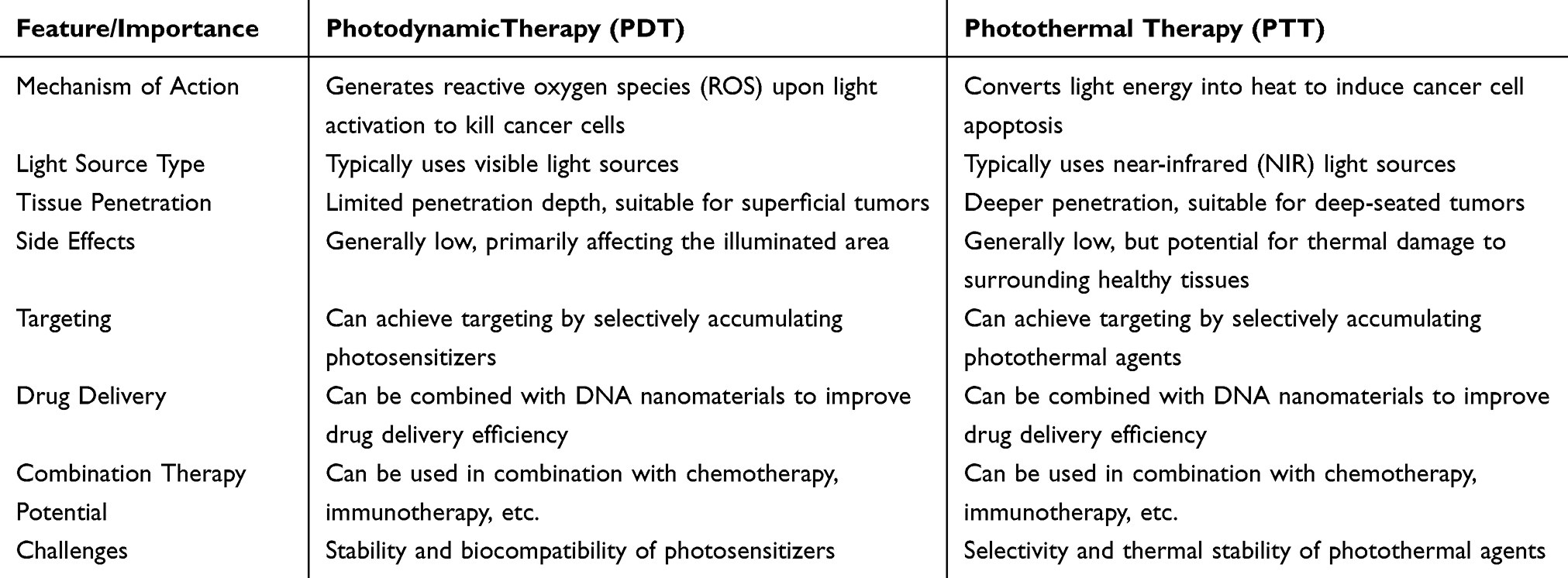 |
Table 1 Key Features and Significance of PDT and PTT in Oncological Treatment |
PDT is particularly notable for its non-invasive approach to tumor treatment, minimizing damage to surrounding tissues and organs.10–12 The effectiveness of PDT depends on photosensitizers, which, after laser irradiation, interact with molecular oxygen to produce reactive oxygen species (ROS) or singlet oxygen, resulting in the targeted destruction of tumor cells.13–16 Moreover, photosensitizers can be efficiently loaded onto nanomaterials, which can selectively deliver them to tumor sites, enhancing the precision of PDT.17–21 Several reviews have highlighted the critical role that PDT plays in cancer management.22,23
Advances in PTT have demonstrated its ability to convert light energy into thermal energy, leading to cancer cells’ targeted destruction and apoptosis with minimal damage to surrounding healthy tissue.24,25 The development of photothermal agents (PTAs) and nanomaterials capable of absorbing and converting light energy into heat has been instrumental in achieving precise and controlled tumor eradication, significantly improving the safety and efficacy of PTT in clinical settings.26–30
Nanomaterials can act as sensitizer carriers for both PDT and PTT, providing additional selectivity and tumor enrichment through the enhanced permeability and retention (EPR) effect. This accumulation leads to increased tissue heating and the generation of ROS. For instance, nanoparticles engineered with polydopamine (PDA) as a photothermal agent, loaded with hemoglobin (to supply oxygen) and photosensitizers, can synergistically combat tumors through PTT/PDT when subjected to laser irradiation.31 Certain nanomaterials, such as black phosphorus-based manganese ferrate nanocomposites (RGD-BPNS@SMFN), exhibit enhanced intrinsic catalase (CAT) activity at elevated temperatures. This increase in temperature leads to higher oxygen content, which alleviates hypoxia and subsequently improves PDT. Additionally, the free radicals generated by PDT disrupt the expression of heat shock proteins, thereby enhancing photothermal therapy (PTT). This interplay realizes the mutual promotion and synergistic enhancement of the anti-tumor effects of both PDT and PTT.32
At the intersection of nanotechnology and cancer therapy, deoxyribonucleic acid (DNA) nanomaterials have gained prominence due to their programmable sequence specificity and the ability to guide the assembly of nanostructures with high precision.33–35 These properties make DNA nanomaterials highly suitable for biomedical applications, including biosensing, drug delivery, and imaging.36,37 DNA nanomaterials can be used directly for PTT38 or as effective carriers to deliver photosensitizers in PDT39 (Figure 1).
 |
Figure 1 The application of DNA nanomaterials in cancer treatment, including the integrated mechanism of PTT, PDT combined with four major strategies of chemotherapy, immunotherapy, gene therapy and precision therapy. |
This review is designed to offer provide a comprehensive analysis of the latest advancements in DNA nanoplatforms for phototherapy, with an emphasis on their combination with synergistic therapies to boost the efficacy and precision of cancer treatment. We will scrutinize representative examples and explore prospective research avenues. Our analysis not only encapsulates the current research milestones but also identifies the challenges inherent in the application of DNA nanomaterials for cancer phototherapy. Furthermore, we propose future research directions, including enhancing biocompatibility and therapeutic efficacy.
Single Phototherapies Based on DNA Nanomaterials
DNA nanomaterials capitalize on the principle of DNA’s complementary base pairing to facilitate self-assembly into intricate structures. This inherent specificity confers a distinct advantage to DNA nanomaterials, enabling the construction of highly precise nanostructures and the execution of sophisticated biological functions. In the realm of cancer therapy, these nanomaterials can augment the precision and efficacy of phototherapy, minimize harm to adjacent healthy tissues, and ultimately, enhance treatment outcomes. Phototherapy, including PTT and PDT, represents a pivotal advance in oncological treatments. PTT utilizes the conversion of near-infrared light NIR into localized heat by DNA nanomaterials, thus inducing necrosis and apoptosis in cancer cells.39–43 Similarly, PDT uses the interaction between photosensitizers, light, and molecular oxygen to generate ROS, destroying tumor tissues and cells.44–46 Both modalities have significantly contributed to cancer treatment, and improving the efficacy of tumor cell eradication through phototherapy has become a significant research focus. In this context, DNA nanomaterials, which serve as carriers for photosensitizers, have emerged as particularly promising vectors.47–49 However, challenges such as in vivo uncertainties and potential off-target effects persist.50,51 Consequently, there is an urgent need to develop and refine novel DNA nanomaterials to ensure safer and more effective activation of PDT and PTT. This effort has created diverse DNA nanomaterials designed to improve phototherapeutic outcomes (Table 2).
 |
Table 2 DNA Nanomaterials for Phototherapy Applications |
DNA Nanomaterials for PDT Applications
PDT is a sophisticated therapeutic modality that employs external laser irradiation to induce the generation of ROS, facilitating tumor ablation. However, the efficacy of PDT is often limited by challenges related to the transport of photosensitizers and their unintended damage to healthy tissues.60–62 In this regard, DNA nanomaterials have attracted significant interest as carriers of hydrophobic anticancer drugs, providing innovative strategies to improve the effectiveness of PDT in tumor treatment.63
To address the persistent issue of photosensitizer-induced skin photosensitization and tissue damage, Li et al pioneered the development of PcC4-MSN-O1.52 This nanoplatform integrates a zinc (II) phthalocyanine derivative (PcC4) within mesoporous silica nanoparticles (MSNs), cloaked with DNA (O1) serving as a biogate. The mechanism uses a dual protein-driven sequential reaction mechanism to selectively activate photoactivity. PcC4-MSN-O1 demonstrated selective cytotoxicity towards Hale cells without affecting normal HEK-293 cells in vitro, highlighting its potential for targeting cancer cells. In vivo studies with Hale mice models showed precise regulation of PcC4-MSN-O1 activation and subsequent excretion, minimizing photosensitizer-associated side effects in tissues.
To further improve targeted PDT and mitigate side effects, He et al introduced Zr(IV)-based porphyrin metal-organic frameworks (MOFs) (Figure 2A),53 utilizing DNA templates to orchestrate the core structure of MOF-UCNPs. Under 980 nm irradiation, this design achieved significant cytotoxic singlet oxygen (1O2) production, specifically targeting MDA-MB-468 cells overexpressing EGFR. Integrating an EGFR antibody as a ligand improved tumor cell specificity, with confocal imaging showing pronounced cell death after irradiation, underscoring its potential for enhanced PDT.
 |
Figure 2 (A) The core-satellite MOF-UCNP superstructures, assembled through DNA sequences, generate more singlet oxygen under 980-nm laser irradiation compared to free UCNPs, enhancing the efficacy of photodynamic therapy. (B) Uniform NaYF4 nanoparticles co-doped with Yb3+ and Er3+ were synthesized by a solvothermal method, forming a core-shell structure. PEI was used as a surface-modifying agent, while salmon DNA (smDNA) formed a DNA-UCNP-Au hydrogel with cationic UCNP-Au nanoparticles through electrostatic complexation, creating a compact, confined network that increased the stiffness of the hydrogel. After peritumoral injection, NIR laser irradiation generated temperatures high enough to eliminate cancer cells by melting cell membranes and denaturing proteins. Elevated temperature also induced vascular spasms, leading to intravascular thrombosis, further triggering apoptosis and necrosis in tumor tissue. (C) The GRS-DNA-CuS nanostructure consists of a core (GRS) and a shell (CuS photothermal agent), modified with single-stranded DNA (ssDNA) and cross-linked via hydrogen bonds. GRS acts as a Raman nanoprobe, while CuS serves as a photothermal converter. Under NIR laser irradiation, the hydrogen bonds in the GRS-DNA-CuS complex break as the temperature exceeds the DNA melting point, causing the rapid dissociation of the complex into GRS and ultra-small CuS photothermal agents. These CuS agents achieve uniform intratumoral distribution (UITD), enhancing the efficiency of photothermal therapy. (D) The IR780@Td nanoparticles are fabricated by assembling a tetrahedral DNA (Td) structure from four single-stranded DNAs (S1-S4), followed by conjugation of the hydrophobic photosensitizer IR780 to Td by electrostatic interactions and π-π stacking. The nanoparticles, with an optimal size of approximately 200 nm, facilitate tumor targeting through the enhanced permeability and retention (EPR) effect. Upon NIR laser irradiation, IR780@Td nanoparticles significantly suppress tumor growth, demonstrating a synergistic effect of photothermal therapy (PTT) and photodynamic therapy (PDT) in cancer treatment. |
In their innovative study, Jayme and his team engineered DNA polymer films (DNA-PFs) conjugated with aluminium chloride phthalocyanine (AlClPc), creating a highly biocompatible material known as DNA-PFs-AlClPc. This material was adept at anchoring MCF7 cells, which were integrated into the same structure as the AlClPc and supported within the DNA-PFs-AlClPc framework. The classical photodynamic therapy (PDT) protocol was employed, utilizing visible light photoactivation on the DNA-PFs-AlClPc. This treatment led to a significant reduction in cell viability and an increase in cell death percentage. Furthermore, they simulated the tumor microenvironment using a 3D organoid system, which accurately mirrored early-stage breast cancer progression in humans. Under light exposure, cell viability in monolayer cultures was reduced by over 80%, while 3D organoid cell cultures exhibited a reduction of approximately 50%, highlighting the potential effectiveness of this approach in breast cancer therapy.54 Su et al developed G-quadruplex/porphyrin conjugated gold/persistent luminescent nanocomplexes (PLNPs) with DNA sequences rich in G-quadruplexes and designed to include the AS1411 aptamer. This aptamer targets cytosolic nucleolar proteins that are overexpressed in cancer cells. The DNA-conjugated Au-PLNPs containing the AS1411 aptamer were rapidly synthesized using a freezing method. The encapsulated PLNPs, with their high DNA content and excellent water stability, offer significant advantages for PDT. The nanoprobe combines persistent luminescent nanoparticles, gold coatings, specific DNA sequences, and photosensitizers, enabling the targeted photodynamic therapy of cancer cells through specific binding facilitated by the AS1411 aptamer.55
DNA Nanomaterials for PTT Applications
PTT uses NIR irradiation to induce thermal changes in the target tissues, demonstrating remarkable efficacy in eradicating tumor cells and addressing the recurrence issues commonly associated with traditional surgical interventions.64,65 However, applying PTT is hindered by challenges such as suboptimal photothermal conversion efficiency and the heterogeneous distribution of photothermal agents.66–69 Recent advances in DNA nanotechnology have attracted significant attention because novel materials offer minimal invasiveness and superior deep-tissue penetration, significantly improving the efficacy of PTT and enhancing tumor suppression.
Liu et al have significantly contributed to the field by developing DNA-UCNP-Au hydrogels exhibiting a potent photothermal effect (Figure 2B).56 The hierarchical structure of the UCNP-Au nanoparticles, decorated with smaller Au nanoparticles and integrated into hydrogels via electrostatic complexation with DNA, forms a porous, interconnected network. This structure facilitates photothermal conversion-evidenced by a substantial absorption peak at 540 nm under 808 nm laser irradiation-and elevates the photothermal conversion efficiency to 42.7%, a notable improvement over conventional UCNP-Au nanoparticle (32.4%). In vitro and in vivo studies demonstrate a marked cytotoxic effect in T24 bladder cancer cells and promising tumor inhibition in BALB/c nude mice, with minimal observed toxicity, showing the therapeutic potential of DNA-UCNP-Au in PTT. Zhang et al have developed a NIR light-activated probe based on Pt@Au nanorings for targeted photothermal therapy (PTT) of cancer cells. This probe features a Pt framework (Pt@AuNR) coated with an Au nanoring, which serves as a photosensitizer, and a functionalized double-stranded DNA (dsDNA) heterodimer that acts as a sensor. The cellular subtype-specific recognition and fluorescence imaging are controlled by NIR light irradiation, which induces dsDNA de-hybridization and localized temperature increase on the PAD probes. This approach enables excellent targeted PTT of cancer cells under NIR light control. After incubation with the dual PAD probes and subsequent artificial laser irradiation, the majority of the target tumor cells were effectively killed through photothermal action.58
However, NIR-triggered PTT is often insufficient to eradicate all tumor cells due to the inhomogeneous intra-tumor distribution of photothermal agents. To address this limitation, Zhang et al engineered a DNA nanoplatform resembling a dandelion structure (Figure 2C).57 The GRS-DNA-CuS nanoparticles, featuring a core of DNA-modified Raman nanoprobes surrounded by a shell of complementary single-stranded DNA-modified CuS photothermal agents, utilize hydrogen bond dynamics to enhance local PTA concentration and diffusion upon NIR irradiation. This innovative design improves the spatial distribution of PTAs and demonstrates pronounced antitumor effects in mice loaded with A549, with no discernible organ damage after treatment. These findings highlight the potential of hydrogen-bonding-based, DNA-assembled nanoplatforms in overcoming the limitations of traditional PTT and advancing the field of photothermal oncology.
In photothermal therapy, precise temperature monitoring of the treated tissue is essential for ensuring therapeutic efficacy and safety. Xueke Wu and his team have designed AuNBPs-DNA-TR, leveraging the unique surface plasmon resonance (SPR) effect and tunable synthesis capabilities of gold nanobipyramids (AuNBPs). The researchers conjugated temperature-responsive DNA stem loops labeled with Texas Red (TR) to the surface of AuNBPs. The significant photothermal effect of AuNBPs can elevate the temperature during PTT. As the temperature rises, the DNA stem loops assembled on the AuNBPs disassemble, increasing the distance between the TR and the AuNBPs, which in turn modulates the fluorescence intensity (FI) of the TR. This change in FI can be utilized to monitor the temperature during PTT. Irradiation of cancer cells cultured with AuNBPs-DNA-TR using an 808 nm laser led to a significant increase in system temperature, inactivating the cells due to the substantial photothermal effect of AuNBPs-DNA-TR. Concurrently, the fluorescence of AuNBPs-DNA-TR, used for temperature measurements, was markedly enhanced. This indicates that the fluorescence change can serve as a reliable indicator to monitor the effectiveness of PTT.59
DNA Nanomaterials for Combination Therapy
Phototherapy has emerged as a revolutionary approach, addressing the limitations of traditional cancer treatments while minimizing collateral damage to healthy tissues. This advancement has attracted significant attention for its potential to redefine oncological care. PDT relies on the interaction between photosensitizers, light and molecular oxygen to generate ROS, but its effectiveness is often limited by the delivery of photosensitizers and the potential for damage to healthy tissue. PTT, while capable of converting light energy into heat, is limited in its effectiveness by the efficiency of photothermal conversion and the inhomogeneous distribution of photothermal agents.Combination therapeutic strategies provide an effective solution to overcome the challenges of single-agent therapy. For example, the combination of chemotherapy and phototherapy can improve therapeutic efficacy while reducing the side effects of chemotherapeutic agents. In addition, by integrating gene therapy and immunotherapy with phototherapy, more precise cancer treatment can be achieved.70–74 Although chemotherapy remains the predominant choice for cancer treatment due to its ability to eliminate cancer cells through cytotoxic drugs and inhibit their proliferation, its associated side effects and the growing demand for more targeted therapies have fueled the exploration of alternative modalities.75–80
Gene therapy represents a promising frontier, offering the potential for long-term, tissue-specific expression of therapeutic proteins. By introducing therapeutic genes directly into cancer cells, gene therapy enables targeted cell death and inhibits tumor growth, positioning it as an innovative approach to cancer treatment.81–84 However, while these therapeutic strategies- phototherapy, chemotherapy, and gene therapy-have received substantial interest, they are not without challenges. Issues such as limited tissue penetration in PDT, prolonged treatment durations in PTT, the adverse effects of chemotherapeutic agents, and the need for efficient delivery systems in gene therapy demonstrate the complexities of cancer treatment.85–87
DNA nanomaterials have emerged as potential game-changers due to their exceptional drug-carrying capabilities and biocompatibility.88 The integration of these diverse therapeutic modalities into a unified DNA nanoplatform offers a strategic avenue to overcome individual limitations. The development of multifunctional and synergistic DNA nanoplatforms has shown great promise in enhancing the efficacy of phototherapy by combining PTT and PDT modalities, marking the beginning of a new era in coordinated and advanced cancer treatment strategies (Table 3).
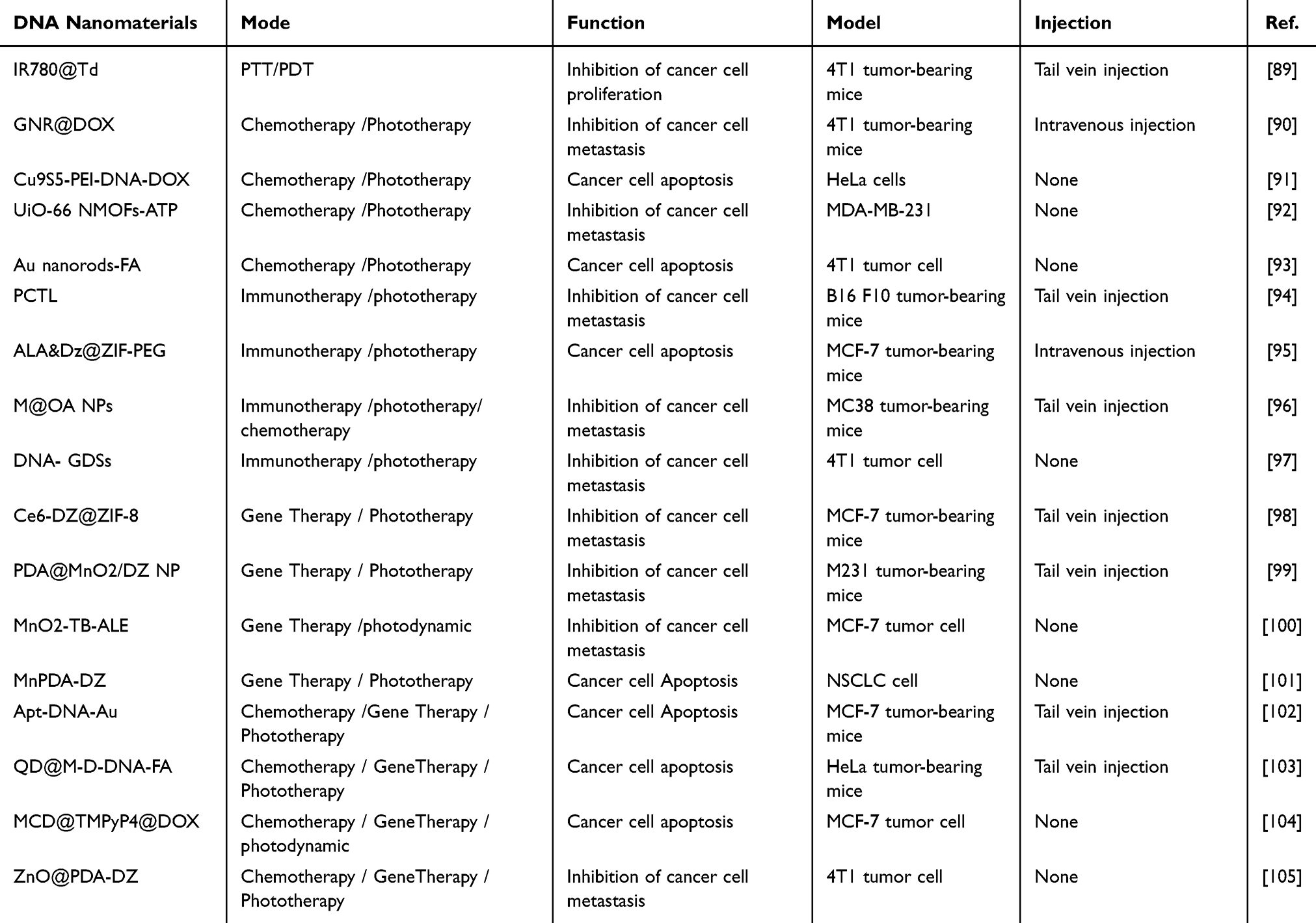 |
Table 3 DNA Nanomaterials for Phototherapy Combined With Other Therapeutic Strategies |
DNA Nanomaterials for PDT/PTT Applications
There is a concerted effort to harness synergistic therapies by integrating PTT and PDT, which requires suitable carriers for effective delivery. DNA nanostructures, known for their facile assembly, robust stability, and substantial drug-carrying capacity, are at the forefront of innovations in cancer therapy. These nanostructures improve photosensitizers’ stability and targeting precision, playing a crucial role in the combination of PTT and PDT for cancer treatment.106
In phototherapy, photosensitizers are essential in converting ambient light into localized heat and transferring energy to surrounding oxygen under targeted laser irradiation. This process generates ROS, which is key to cancer cell ablation. Among various photosensitizers, heptamethine cyanine IR-780 has emerged as a promising candidate. However, its poor stability and water insolubility pose significant challenges for clinical application.107,108 To address these limitations, Wang et al developed an IR780@Td nanoplatform (Figure 2D),89 using tetrahedral DNA (Td) nanostructures for the efficient loading of IR-780 through electrostatic interactions and π-π stacking. This innovation ensures targeted delivery to tumor sites via the enhanced EPR effect. Upon laser irradiation, the IR780@Td nanoplatform significantly elevates local temperatures above physiological levels, inducing tumor cell ablation and demonstrating potent photo-induced cytotoxicity. This approach enhances both the aqueous solubility and stability of IR-780, resulting in a synergistic enhancement of PTT and PDT effects.
Chemotherapy/Phototherapy
The synergistic potential of combining conventional phototherapy with chemotherapy has introduced a novel dimension to cancer treatment, demonstrating enhanced antitumor efficacy.109–111 Despite this progress, reports on integrating chemotherapeutic agents with phototherapy within DNA nanoplatforms remain limited. This section explores several pioneering examples in which chemotherapeutic drugs are loaded onto DNA nanoplatforms to augment phototherapy. These innovative DNA nanoplatforms have significant potential to advance antitumor combination therapy.
Wang et al engineered a multifunctional nanoplatform, GNR@DOX,90 by loading adriamycin (DOX) onto DNA-coated gold nanorods. On NIR irradiation, the gold nanorods (GNR) convert NIR light into localized heat, effectively targeting cancer cells within blood vessels. This process facilitates the controlled release of DOX and demonstrates remarkable PTT efficacy. The synergistic action of DOX and PTT mediated by GNR@DOX induces apoptosis in primary cancer cells, presenting a promising strategy to mitigate primary tumor metastases and enhance the overall efficacy of DNA nanoplatform-based phototherapy. Zhang and his team have crafted a novel nanomaterial leveraging UiO-66 metal-organic frameworks (NMOFs) that are gated by ATP-aptamer or VEGF-aptamer functionalized DNA tetrahedra and loaded with the chemotherapeutic agent DOX. These nanostructures are designed to release the drug in response to the presence of ATP or VEGF, molecules that are prevalent in cancer cells. Moreover, these nanostructures facilitate light-induced photosensitization, enabling the generation of reactive oxygen species (ROS), which contributes to the effective photodynamic therapy (PDT) treatment of cancer cells.92
Addressing the limitations of noble metal nanomaterials, such as their non-biodegradability and high cost, Liang et al developed an alternative multifunctional DNA nanoplatform, Cu9S5-PEI-DNA-DOX. This construct uses semiconductor copper sulfide nanoparticles (Cu9S5), polyethyleneimine (PEI), double-stranded DNA fragments (dsDNA), and DOX (Figure 3A).91 The strategic incorporation of DNA reduces the positive charge of the system, making PEI less cytotoxic and improving biocompatibility. The stability of the dsDNA helical structure under physiological conditions, combined with its NIR-induced disassembly, ensures safe intrabody delivery of DOX, triggering cancer cell apoptosis. Moreover, the Cu9S5 component efficiently absorbs infrared light, converting it into localized heat that, together with DOX, effectively eradicates cancer cells. Consequently, the Cu9S5-PEI-DNA-DOX nanoplatform exemplifies a potent synergy of chemotherapy and PTT, marking a new era of combined therapeutic strategies. Furthermore, Shanmugam et al have employed 5′-thiol-terminated single-stranded DNA assembled on gold nanorods, which upon hybridization, forms double-stranded DNA. This process assists in embedding DOX into the double-stranded DNA structure and immobilizes the platinum [Pt(IV)] precursor by forming amide bonds with the acid groups on the axial ligands to bind folic acid (FA). Irradiation at 880 nm triggers drug activation and photothermal ablation of cancer cells, offering a dual-modality therapeutic approach.93
 |
Figure 3 (A) Hydrophobic Cu9S5 nanoparticles (NPs) are modified for aqueous phase compatibility through a polyethyleneimine (PEI) coating. For loading, Doxorubicin (DOX) molecules are intercalated into the GC base pairs of double-stranded DNA (dsDNA). Upon NIR light irradiation, the photothermal effect of Cu9S5 NPs increases the local temperature, causing dsDNA denaturation and releasing the loaded DOX molecules. This nanocomposite demonstrates a synergistic effect of chemo-photothermal therapy under NIR irradiation, significantly enhancing cytotoxicity toward tumor cells. (B) The chemotherapeutic agent oxaliplatin (OXA) is encapsulated within the core of a metal-organic framework (MOF) nanoparticle, with the surface modified by a PD-L1 aptamer to form M@O-A. This system induces immunogenic cell death (ICD) through a combination of photodynamic therapy (PDT) and chemotherapy, triggering an immune response against the tumor. The M@O-A nanoparticle offers a non-invasive strategy for the precise delivery of the immune checkpoint inhibitor aptPD-L1, allowing simultaneous PDT, chemotherapy, and immunotherapy upon light irradiation. (C) A core-shell nanoplatform, PDA@MnO2, was developed using polydopamine (PDA) as a photothermal agent and coated with a heat shock protein 70 (HSP70) silencing DNAzyme (DZ). The MnO2 shell responds to endogenous stimuli such as glutathione (GSH), releasing manganese ions (Mn2+), which act as a metal cofactor to activate the DNAzyme. This activation cleaves the HSP70 mRNA, reducing HSP70 expression and improving photothermal therapy’s (PTT) efficacy. |
Immunotherapy/Phototherapy
Cancer immunotherapy is widely recognized for its mechanisms involving checkpoint inhibitors and manipulating the immune system to recognize and attack cancer cells.112,113 It has become a powerful clinical strategy for cancer treatment.114 However, due to limitations such as insufficient tumor immunogenicity and reduced response rates, immunotherapy is increasingly combined with phototherapy to improve the effectiveness of tumor treatment.115 In this context, we have summarized examples of synergistic applications of DNA-based nanoplatforms in immunotherapy and phototherapy, highlighting their potential to enhance therapeutic outcomes.
Although immunotherapeutic approaches are effective, they are often associated with overactivation of the immune response and autoimmune side effects. To enhance the immunogenicity of membrane-targeted PDT while reducing systemic toxicity risks, Wang et al developed a membrane-targeted photodynamic immune-DNA nanomedicine, AptPDL1/CpG@DNF-TMPyP4-pHLIP (PCTL).94 This nanoplatform contains a cyclic DNA template with sequences complementary to the PDL1 aptamer, a CpG oligodeoxynucleotide (ODN), and a functional G-quadruplex sequence for the RCA response, while embedding the porphyrin photosensitizer TMPyP4. The DNA nanostructure self-decomposes to release internal DNA immunomodulators, including CpG ODN for dendritic cell (DC) maturation and the PDL1 aptamer for immune checkpoint inhibition. In vitro experiments demonstrated that photoactivated PCTL effectively promotes T cell activation, improves immunotherapy, and exhibits strong tumor suppression in a 4T1 mouse model. This nanoplatform highlights the advantages of combining PDT with immune activation in tumor therapy. Kim and his research group have developed multifunctional DNA superstructures capable of adsorbing and locally enriching gold ions. By inducing local reduction, these ions are transformed into gold nanoclusters that are synergistically activated through the STING and TLR9 pathways. These DNA sponges not only trigger a synergistic and potent type I interferon response but also reverse the immunosuppressive tumor microenvironment (TME). In the context of poorly immunogenic breast cancer, these immunostimulatory DNA sponges have been shown to enhance the therapeutic efficacy and immune response rate of immune checkpoint blockade (ICB) therapy. Furthermore, the gold DNA superstructures (GDSs) are capable of absorbing light energy and converting it into heat energy upon light exposure, generating a photothermal effect that effectively kills cancer cells.97
Chen et al pioneered the development of a ZIF-PEG-based nanoplatform (ALA&Dz@ZIF-PEG) for targeted immunophotodynamic therapy.95 This system ingeniously incorporates 5-aminolevulinic acid (ALA) and DNAzyme. ALA, a precursor of the photosensitizer protoporphyrin IX (PpIX), exhibits mitochondrial targeting capabilities and minimal phototoxicity, making it an optimal photosensitizer for PDT. However, the biosynthesis efficiency of PpIX is closely related to oxygen concentration, which is often compromised in hypoxic tumor environments, hindering the conversion of ALA to PpIX. Flow cell assays revealed that Zn2+ ions released from the ALA&Dz@ZIF-PEG nanoplatform alleviate hypoxia, improving the conversion of ALA to PpIX. Confocal laser scanning microscopy (CLSM) confirmed the biotransformation of ALA in 4T1 cells, leading to increased intracellular accumulation of PpIX. The ALA&Dz@ZIF-PEG nanoplatform demonstrated potent photodynamic therapeutic effects across various cell lines, including MCF-7, 4T1, and CT26, exhibiting significant cytotoxicity. PDT mediated by ALA&Dz@ZIF-PEG resulted in the release of mitochondrial DNA (mtDNA) and activating the cGAS-STING pathway, an innate immune response triggered by mtDNA. In vivo studies using a 4T1 xenograft mouse model showed an 84% reduction in tumor size without notable adverse effects, underscoring the profound therapeutic potential of the ALA&Dz@ZIF-PEG nanoplatform for clinical applications in targeted cancer treatment.
Immunotherapy can induce organ-specific toxicity and hematologic dysfunction. To address this, Zhang et al developed M@OA NPs, a nanoplatform that generates large amounts of ROS upon targeted binding to MC38 cells (Figure 3B).96 This system effectively induces cell death and combines PDT and chemotherapy to promote immunogenic cell death (ICD), eliciting an anti-tumor immune response. M@OA NPs significantly inhibited primary tumors and prolonged survival in MC38-Luc tumor-bearing mouse models. This nanoplatform, combining chemotherapy, PDT, and ICD treatment, synergistically enhances immune efficiency by improving effector T-cell infiltration, representing a promising approach in cancer immunotherapy.
Gene Therapy/Phototherapy
Combining gene therapy and phototherapy is a promising therapeutic strategy, particularly in tumor treatment.116 Gene therapy aims to correct or compensate for diseases caused by defective or abnormal genes by introducing exogenous normal genes into target cells. At the same time, phototherapy uses specific wavelengths of light to activate photosensitizers, which in turn kill tumor cells.
Wang et al pioneered the development of the Ce6-DNAzyme@ZIF-8 nanoplatform, designed for synergistic gene-photodynamic therapy.98 This innovative platform incorporates chlorin e6-modified DNAzyme (DZ) within the ZIF-8 structure, leveraging the straightforward synthesis of DZ@ZIF-8. Upon administration to MCF-7 cells, nucleotide substitution in the DZ core did not affect its affinity for EGR-1 mRNA, maintaining its functionality. Utilizing an inactive DZ as a control demonstrated the specificity of the targeted mechanism: cells treated with DZ@ZIF-8 exhibited a marked decrease in EGR-1 mRNA levels, reduced clonogenicity, and lower cell viability compared to control-treated cells, as assessed by MTT assays.
The photosensitizer Ce6, embedded within the porous ZIF-8 structure, facilitates the effective integration of Ce6-DZ@ZIF-8, significantly enhancing therapeutic outcomes. Comparative analyses between light-irradiated Ce6-DZ@ZIF-8 and its non-irradiated counterparts or those containing Ce6-cDNAzyme demonstrated that the former achieved superior antiproliferative effects and induced early apoptosis in cells, significantly increasing the efficacy of photodynamic therapy. Following intravenous injection into MCF-7-bearing mice, the Ce6-DZ@ZIF-8 nanoplatform induced pronounced tumor cell apoptosis, attributed to the synergistic combination of gene and photodynamic therapy. This synergism involves DZ-mediated inhibition of EGR-1 expression and ROS generation, effectively promoting tumor cell apoptosis. This study highlights the therapeutic potential of the Ce6-DNAzyme@ZIF-8 nanoplatform and sets a precedent for the concurrent application of gene silencing and photodynamic therapy in cancer treatment. Wang and his team have engineered a GSH-glutathione responsive delivery system. This system serves as a carrier that is sensitive to intracellular GSH levels, encapsulating TB (a photosensitizer) and a DNAzyme, which acts as a catalyst for the degradation of EGR-1 mRNA to achieve gene silencing. TB, utilized as a photosensitizer, possesses aggregation-induced emission (AIE) characteristics, making it suitable for photodynamic therapy (PDT). In its aggregated state, TB emits intense fluorescence, and upon exposure to light, it generates singlet oxygen (1O2), which inhibits tumor cell growth.100
Another notable advancement is the PDA@MnO2/DZ NPs nanoplatform (Figure 3C).99 Constructed with a PDA@MnO2 core and shell structure, this nanoplatform facilitates the adsorption of DZ on its surface, with MnO2 catalyzing the release of Mn2+ ions to activate DZ for the targeted down-regulation of HSP70. Additionally, PDA is a photothermal agent activated by laser irradiation to deliver PTT. Strategic modifications with high cation concentrations reduce adsorption rejection, ensuring that DZ presence does not impede photothermal conversion efficiency.
Upon 500 seconds of irradiation, the PDA@MnO2/DZ NPs exhibited a notable temperature increase, sufficient for tumor ablation, achieving a photothermal conversion efficiency of 36.4%, significantly exceeding that of traditional nanomaterials such as gold nanorods. Quantitative PCR and Western blot analyses confirmed the down-regulation of HSP70 by PDA@MnO2/DZ NPs, mitigating PTT resistance. The suppression of HSP70 simultaneously induced caspase-3 expression, sensitizing tumor cells to thermotherapy and precipitating apoptosis after PTT. In vivo studies also demonstrated proficient targeting and photothermal conversion, with temperatures reaching 56°C in mice, effectively eradicating tumor cells without inducing acute toxicity or pathological changes in organs, underscoring the excellent biocompatibility of the system. The PDA@MnO2/DZ NPs nanoplatform not only inhibits HSP70 but also significantly enhances the PTT effect, representing a novel contribution to the self-activated gene therapy nanosystems field. Feng and his team have designed polydopamine-Mn2+ nanoparticles (MnPDA) with multifaceted capabilities. These nanoparticles can release free Mn2+ ions in situ, which act as cofactors for DNAzymes, effectively triggering the catalytic cleavage of mRNA and demonstrating gene silencing. Moreover, MnPDA carriers exhibit high photothermal conversion efficiency and stability in photothermal treatment and degradation within complex environments. Thanks to their excellent stability in photothermal treatment and degradation in complex settings, this vector is capable of both gene regulation and tumor therapy through the photothermal effect.101
Chemotherapy/Gene Therapy/Phototherapy
Gene therapy represents a pioneering approach in cancer treatment, demonstrating significant potential over conventional modalities.117–119 A critical aspect of gene therapy, whether in vivo or in vitro, is selecting an effective gene delivery system. DNA nanomaterials, celebrated for their superior properties, emerge as prime candidates for this role, enabling the targeted delivery of therapeutic genes to cancer cells.120,121 Furthermore, DNA nanomaterials allow for the concurrent localization of chemotherapeutic agents and phototherapeutic interventions on a singular DNA nanoplatform, offering a synergistic approach to cancer treatment. The Apt-DNA-Au multifunctional DNA nanoplatform, developed by Zhou et al exemplifies this innovation.102 This nanoplatform integrates chemotherapeutic drugs (doxorubicin, Dox), therapeutic genes (antisense DNA), photosensitizers (zinc phthalocyanine, ZnPc), and gold nanoparticles (AuNPs). It is specifically activated in the presence of TK1 mRNA, a known tumor growth biomarker, through a toehold-mediated strand displacement (TMSD) reaction. Upon activation, Apt-DNA-Au releases DOX and antisense DNA, aggregates fluorophores and AuNPs, and generates ROS from the photosensitizers upon irradiation. This multifunctional action not only achieves targeted imaging and inhibition of surviving, a protein that prevents apoptosis but also leverages the synergistic therapeutic potentials of PTT and PDT.
In the presence of TK1 mRNA, the Apt-DNA-Au nanomachine is activated, resulting in the controlled release of chemotherapeutic agents and antisense DNA. DOX intercalates into the double-stranded DNA region, inhibiting DNA replication, while the antisense DNA specifically binds to the survivin mRNA, preventing its translation into protein. Reduction of survivin protein levels enhances the chemotherapeutic effect and promotes tumor cell apoptosis. Wang and his team have crafted a sophisticated DNA nanomedicine, MCD@TMPyP4@DOX, tailored for PDT. This nanocarrier is equipped with a P-glycoprotein (P-gp) DNAzyme that selectively cleaves substrates and silences MDR1 mRNA with the assistance of the Mg2+ cofactor, thereby reducing the expression of P-gp. It also degrades single-stranded DNA, initiating the self-decomposition of MCD@TMPyP4@DOX and releasing the chemotherapeutic drug DOX and DNAzyme. Upon exposure to light, this system generates reactive oxygen species (ROS), which are crucial for the efficacy of PDT.104
Li et al further advanced this concept by conjugating desthiobiotin (db) to folic acid-modified silver sulfide@mesoporous silica core-shell nanoparticles, encapsulating DOX within nanopores, and attaching antisense oligonucleotides (db-DNA) to the exterior via affinity interaction to inhibit survivin expression.103 Integrating Ag2S quantum dots as cores for PTT enhances the system’s therapeutic efficacy. This configuration ensures targeted delivery and improved tumor cell eradication through folic acid receptor-mediated endocytosis, enhanced by the effect of PTT on cell membrane permeability. The released DOX intercalates into the double-stranded DNA region, inhibiting DNA replication and RNA transcription. In contrast, the released antisense oligonucleotide binds specifically to survivin mRNA, preventing its translation into protein. This process reduces survivin protein levels, enhances the chemotherapeutic effect, and promotes tumor cell apoptosis. Additionally, the strategic use of avidin to prevent DOX leakage via the desthiobiotin-avidin interaction further enhances the biosafety of the platform. This novel therapeutic DNA nanoplatform holds significant promise for effective cancer treatment, demonstrating the profound potential of integrating gene therapy with chemotherapeutic and phototherapeutic strategies at the nanoscale. Liu et al have developed a core-shell nanosystem that integrates zinc oxide (ZnO) nanocores with polydopamine (PDA) shells, creating a multimodal therapy platform, ZnO@PDA. This nanosystem combines DOX chemotherapy, gene therapy, and photothermal therapy into a single nanomaterial. The PDA shell layer possesses a robust capacity to absorb near-infrared (NIR) light and exhibits a high photothermal conversion efficiency, making it an excellent candidate for photothermal therapy. The ZnO nanocore serves as a metal cofactor reservoir, designed to release Zn2+ in response to intracellular stimuli. This release triggers the activation of DZ for gene silencing and simultaneously releases DOX to eliminate cancer cells.105
DNA Nanoreactor for Tumor Imaging and Phototherapy
New advances have been made in tumor treatment, with light therapy gaining wide recognition for its low side effects and favorable therapeutic outcomes.122–124 However, achieving precise treatment through most light therapies remains challenging, and tumor imaging is urgently needed to support treatment strategies.125–127 Early and accurate tumor diagnosis is a critical focus in the medical field, and imaging techniques such as photoacoustic imaging (PA), magnetic resonance imaging (MRI), and photothermal imaging based on DNA nanoplatforms have been used to enable accurate diagnosis and treatment.128 DNA nanotechnology offers the potential to integrate diagnostic and therapeutic platforms and various DNA nanoplatforms have been developed to synergistically combine tumor imaging and phototherapy.
MRI Imaging
MRI, an advanced radiowave imaging technique, plays a crucial role in diagnostic localization and has recently gained prominence in biomedical nanomaterial applications.129,130 MRI offers high resolution, non-invasiveness, and non-radiation exposure,131 making it an ideal candidate for integrating DNA nanomaterials on a single platform for tumor diagnosis and treatment.132 When combining MRI with DNA nanomaterials, researchers have developed platforms that synergize with light therapy, enhancing diagnostic and therapeutic outcomes.
The efficiency of PDT is highly dependent on precise diagnostics, and imaging probes are critical in this process. The researchers developed DNA template-based AgNC/Porphyrin/MnO2 nanoplatforms133 to address this. These platforms facilitate the intracellular MnO2-triggered release of silver nanoclusters (AgNCs), with endocytosed Mn2+ nanosheets transferring DNA probes to the cytoplasm to prevent degradation. Zn2+ triggers a cascade reaction within the DNA enzyme, amplifying the signal for Zn2+ detection while utilizing released Mn2+ as an MRI contrast agent. In vivo, studies in MCF-7 mouse models demonstrated that P-AgNCs-MnO2 exhibited superior diagnostic and therapeutic efficacy compared to controls without significant pathological changes in major organs. This underscores its potential to improve PDT through precise diagnosis.
To address the long-term retention of heavy metal ions by contrast agents, Sun et al developed PDA-DNA-DTPA/Gd nanoprobes (Figure 4A).134 In this design, DNA is bound to PDA nanosurfaces, while diethylenetriaminepentaacetic acid dianhydride (DTPA-DA) is attached to the ends of the nanoprobes. Gadolinium (Gd) ions are subsequently bound to the PDA surface, forming PDA-DNA-DTPA/Gd. Under 808 nm laser irradiation, the nanoprobes demonstrated effective photothermal conversion, raising the temperature of the PDA-DNA-DTPA/Gd tumor tissue to a DNA melting temperature of 50°C. The T1 imaging analysis revealed that the distribution of the Gd elements decreased significantly after heating, indicating the release of Gd. In 4T1 hormonal mouse models, laser-irradiated regions treated with DA-DNA-DTPA/Gd showed increased temperatures, and the PDA-DTPA/Gd was eliminated within 24 hours, with small Gd molecules excreted by renal filtration as DTPA/Gd complexes.
 |
Figure 4 (A) Utilizing polydopamine (PDA) as a platform, DNA chains are modified to conjugate magnetic resonance imaging (MRI) reporter genes containing gadolinium (Gd) for MRI imaging and the drug molecule doxorubicin (DOX). PDA, known for its high photothermal conversion efficiency, rapidly converts light energy into heat under near-infrared (NIR) irradiation. This heat controls the melting of DNA and the release of Gd. The CG base pairs in the DNA serve as binding sites for the drug, enabling controlled drug release through the photothermal effect, enhancing the therapeutic effect on tumors. (B) Gold nanoparticles (AuNPs) are modified with complementary DNA strands on their surface, linked to DOX via a thermally labile linker, and coated with polyethylene glycol (PEG) through a matrix metalloproteinase (MMP)-cleavable peptide. In tumor tissues with high MMP expression, the PEG layer is rapidly cleaved, exposing the complementary DNA strands on the surface of AuNPs, which induces rapid aggregation of the nanoparticles through DNA hybridization. Local increase in temperature triggers the cleavage of the thermally labile linker, leading to the fast release of DOX from the nanoparticles, achieving a synergistic effect with photothermal therapy (PTT). The aggregation of AuNPs in response to MMPs enhances the photoacoustic (PA) imaging signal. (C) Gold nanorod dimers (NR dimers, core) and upconversion nanoparticles (UCNPs, NaGdF4, satellite) attached to chlorin e6 (Ce6) photosensitizers are hierarchically assembled through complementary DNA sequence pairing. Ce6 generates cytotoxic singlet oxygen or reactive oxygen species (ROS), causing localized cell death and tissue destruction. Additionally, multimodal imaging of tumors is achieved using upconversion luminescence (UCL), MRI, PA, and computed tomography (CT) imaging techniques. |
Adriamycin was piggybacked onto the probe for adjuvant tumor treatment, significantly improving mouse survival rates. This nanoplatform exhibited excellent MRI imaging capabilities, while its high photothermal conversion efficiency enhanced the PTT therapeutic effect. Additionally, synergistic chemotherapy further contributed to tumor eradication, highlighting the clinical potential of PDA-DNA-DTPA/Gd for tumor treatment.
PA Imaging
Photoacoustic imaging (PA) has gained significant attention as a promising imaging technique in recent years.135,136 As a non-invasive and non-ionizing biomedical imaging modality, PA offers the advantages of high resolution and high contrast in optical absorption imaging, allowing for deep tissue optical contrast imaging with ultrasound-defined spatial resolution, which surpasses that of purely optical imaging techniques.137,138 The integration of photoacoustic imaging on DNA nanoplatforms for adjuvant phototherapy holds great potential, and several examples of DNA nanoplatform-based PA imaging applications in phototherapy are discussed in this section.
Conventional imaging techniques often do not provide the precision required for effective treatment due to their low sensitivity, poor specificity, limited spatial resolution, and low tissue penetration. DNA-origami gold-nanorod hybrids were developed to address these challenges, creating nanoplatforms by assembling gold nanorods (AuNR) on the surface of DNA-origami structures (D-AuNR).139 Due to gold nanoparticles’ unique light absorption and photochromic properties, these hybrids serve as nanocontrast agents in photoacoustic imaging. In contrast, the photothermal conversion properties of D-AuNRs trigger therapeutic activity and enable activation monitoring. After intravenous injection, D-AuNRs demonstrated more excellent permeability than conventional AuNRs, with PA signals predominantly located in the central hypoxic region of the tumor, providing excellent localization capacity. In contrast, the PA signal from traditional AuNRs was mainly distributed in the outer area of the cancer. The photothermal therapeutic efficacy of D-AuNRs was validated in nude mice bearing 4T1-fLuc tumors, demonstrating that D-AuNRs, as a PA-guided photothermal therapeutic platform, effectively inhibited tumor growth, highlighting the potential of the nanoplatform for clinical translation.
To further enhance the enhanced EPR effect for prolonged circulation and increased tumor accumulation, smaller AuNPs (10–100 nm) offer significant advantages in cancer imaging and therapeutic enhancement. Yang et al developed a tumor-targeted PA-guided photothermal therapy and drug delivery nanoplatform based on matrix metalloproteinase (MMP)-responsive gold nanoparticles (AuNPs)140 (Figure 4B). This system enabled enhanced NIR absorption through DNA hybridization-induced nanoprobe assembly. Polyethylene glycol (PEG) was attached to Dox-AuNPs to prevent non-specific aggregation, followed by PEG 5000-COOH conjugation with NH2-GPLGVRGC-SH, and subsequent attachment to Dox-AuNPs via C-terminal cysteine (PEG-pep-Dox-AuNPs). The chemotherapeutic drug doxorubicin was loaded together with the PA imaging agent and could be rapidly released upon local temperature elevation, triggered by the photothermal effect. Experiments conducted in SCC-7 tumor-bearing mice demonstrated precise tumor inhibition without toxic side effects. This nanoplatform not only shows promise for tumor-targeted imaging and therapy but also for advancing precision diagnostics in cancer treatment, thereby potentially revolutionizing the field.
Phototherapeutic Multimodal Imaging DNA Nanoplatform
Single imaging techniques often have limitations such as insufficient tissue penetration and a narrow perspective.141 In contrast, integrating multimodal imaging using DNA nanomaterials combined with phototherapy has emerged as a promising trend for future developments. This approach includes incorporating upconversion luminescence imaging (UCL),142 which offers the advantages of deep tissue penetration and a low autofluorescence background. Additionally, computed tomography (CT) imaging is employed,143 utilizing X-ray scanning and computer reconstruction technology to acquire high-resolution, three-dimensional images of internal body structures.
Sun et al developed a novel self-assembled core-satellite probe (Figure 4C).144 This design utilizes aptamer DNA nanostructures to construct a platform with a gold nanorod dimer core and chlorin e6 (Ce6)-attached upconversion nanoparticles (UCNPs) (NaGdF4) as satellites. The nanoplatform integrates multiple high-resolution biomedical imaging modalities, including UCL, CT, PA, and MR. Based on the principle of base complementary pairing, the hierarchical assembly is activated under 808 nm laser excitation. The core of the gold NR dimer generates a strong photothermal therapy effect, while energy transfer from UCNPs to Ce6 efficiently produces singlet oxygen (1O2) for PDT. Enhanced by the EPR effect for tumor targeting, this multifunctional nanoplatform demonstrates exceptional tumor eradication at safe doses, marking a new frontier for combining PTT and PDT in cancer therapy.
While the potential of nanomaterials in treating inflammatory diseases is promising, their clinical application is contingent upon their ability to accurately target inflamed areas to reduce damage to healthy tissues,145,146 Current targeting techniques may fall short in precision, sometimes leading to non-specific distribution and side effects that can trigger inflammatory responses or toxicity, thus limiting their use in clinics. DNA nanomaterials, however, leverage the base-pairing principle of DNA for precise sequence recognition, enabling their design to specifically target inflammatory cells or molecules. Furthermore, these nanomaterials can be labeled and equipped with various imaging probes for real-time monitoring of drug delivery and inflammatory processes, enhancing the precision and efficacy of therapies. For instance, the tetrahedral framed DNA-based miRNA delivery system (BiRDS), which incorporates three miRNAs and a nucleic acid core, maximizes loading capacity while retaining the properties of tetrahedral frames. BiRDS exhibits high stability, excellent cell and tissue penetration, and good biocompatibility. It selectively inhibits heparin-binding epidermal growth factor (HB-EGF), thereby suppressing the NF-κB pathway and modulating the PTEN/Akt pathway to facilitate the transition of wound healing from inflammation to the proliferative phase,147 Additionally, using mannose-binding lectin (MBL) in conjunction with nanoparticle tracking analysis (NTA) technology, Juul-Madsen et al discovered that the super-oligomeric structure of MBL can form nanoparticles with DNA. This formation correlates with an inverse relationship between the concentration of MBL super-oligomers and antibodies to double-stranded DNA, indicating that MBL-DNA interactions have pathological significance in systemic lupus erythematosus (SLE).148 Ma et al developed nanomedicines based on triangular DNA origami nanostructures modified with folic acid to create FA-tDONs154. These structures actively target M1-type macrophages, which are pivotal in the development of rheumatoid arthritis (RA). FA-tDONs are capable of efficiently neutralizing reactive oxygen species (ROS) and nitric oxide (NO), converting pro-inflammatory M1-type macrophages into anti-inflammatory M2-type macrophages.149
Zhang and his team developed a strategy using Nitrophenyl tetramethyldioxaborole benzylcarbamate (NBC)-modified DNase I, which was incorporated into DNA nanogels based on DNase-degradable aptamers. This approach facilitated nanogel degradation and drug release in response to intracellular ROS levels,150 thereby indirectly influencing the expression of inflammatory factors. Furthermore, they constructed a DNA/cerium oxide nanocomplex (DCNC) capable of dynamic intracellular assembly to form artificial peroxisomes (APs). Upon cellular entry, DCNC responds to the acidic environments in early endosomes and late endosomes/lysosomes, sequentially disassembling and reassembling to form APs within the cell. These APs emulate the ROS-scavenging function of peroxisomes through the action of cerium oxide, indirectly impacting the expression of inflammatory factors. By mimicking the ROS-scavenging function of peroxisomes, APs help to neutralize high concentrations of ROS, preventing cellular oxidative stress, which is crucial for maintaining intracellular ROS balance and safeguarding cells from oxidative damage.151
Conclusion and Future Challenges
DNA nanomaterials, characterized by their unique physical properties and programmability, have emerged as a focal point of interest in the biomedical field, particularly in developing multifunctional DNA nanoplatforms that integrate phototherapy with cancer treatment. These platforms have attracted significant attention, marking a new paradigm in cancer therapy research. This review examines DNA nanoplatforms that leverage various phototherapeutic modalities, such as photodynamic and photothermal therapies, to elucidate the synergistic relationship between DNA nanomaterials and phototherapy. Additionally, it explores the integration of chemotherapy, gene therapy, and phototherapy on a single nanoplatform, providing a foundational reference for future research. The review also highlights the role of functional DNA, including DNAzymes, in enhancing phototherapeutic applications, presenting innovative approaches to utilizing DNA nanomaterials in cancer treatment.
Long-term toxicity studies are crucial for evaluating the safety profile of DNA nanomaterials. These studies help assess the potential for prolonged immune activation, which could lead to chronic inflammation affecting tissue function and structure, as well as causing DNA damage or genetic mutations that may elevate the risk of cancer.152,153 Consequently, it is imperative to select DNA nanomaterial constructs that are inherently safe to reduce the likelihood of immune responses and toxicity. Examples of such constructs include DNA origami and gold nanoparticles, which have demonstrated lower toxicity profiles and are thus more suitable for biomedical applications.154,155 In clinical applications, to improve therapeutic efficacy and reduce side effects, DNA nanomaterials must be designed to specifically target tumour cells while avoiding interactions with normal cells, and stability and controllability in vivo are key to achieving effective treatment. This includes stability in the circulation and controlled release under specific conditions.
Despite the significant strides made in utilizing DNA nanomaterials for cancer phototherapy, several challenges must be addressed to optimize their therapeutic efficacy. Although many DNA nanomaterials have been developed to enhance tissue penetration and targeting while minimizing side effects, their exploration within tumor microenvironments (TME) remains limited, particularly under hypoxic conditions. This gap underscores the need for novel DNA nanomaterials specifically tailored to the complexities of the TME. For example, Yang et al developed the H-MnO2-PEG/C&D nanoplatform,156 designed not only for TME-specific imaging and on-demand drug delivery but also to modulate the hypoxic TME to improve cancer therapy. This represents a critical direction for future research. Although combination therapies using DNA nanomaterials have shown promise in tumor eradication,56,95,106 the biosafety and biocompatibility of these therapeutic strategies require further improvement. There is an urgent need for the rational design of DNA nanomaterials that enhance phototherapeutic efficacy and prioritize biocompatibility and biosafety. Gao et al developed a carrier-free nanotherapeutic agent (AIBME@IR780-APM NPs),157 demonstrating satisfactory biocompatibility and promising to create safer, more biocompatible DNA nanomaterials.
Future research should focus on developing functional DNA, such as DNAzymes and aptamers, that synergize with phototherapy to induce apoptosis in cancer cells. While the advancement of DNA nanomaterials for phototherapy poses complex challenges, the foundation established by existing materials provides valuable insights. It encourages the innovation of novel DNA nanomaterials for phototherapeutic applications. By addressing these challenges, the field can move closer to realizing the full potential of DNA nanomaterials in cancer phototherapy, paving the way for groundbreaking advances in treatment methodologies.
This review discusses several critical applications of DNA nanomaterials in phototherapy. Depending on the specific target, DNA nanomaterials can be designed to carry various photosensitizers, drugs for synergistic therapy, tumor imaging agents for precise diagnosis, and antisense DNA and other molecules for additional functions, such as gene therapy and immunotherapy. These diverse strategies highlight the significant potential of DNA nanomaterials to advance the complete clinical application of tumor therapy. While this review does not focus on a particular cancer type, this approach is intentional, reflecting the broad utility of DNA nanomaterials across cancer phototherapy. By showcasing the adaptability and versatility of DNA nanomaterials in phototherapy, we aim to offer a comprehensive guide for future research and applications, rather than confining our scope to a single cancer type. We are convinced that embracing this expansive view will foster the advancement of DNA nanomaterials in oncological treatments and pave the way for the development of effective therapies for a diverse array of cancers.
Abbreviations
PTT, Photothermal Therapy; PDT, Photodynamic Therapy; NIR, Near-Infrared; ROS, Reactive Oxygen Species; EPR, Enhanced Permeability and Retention; PDA, Polydopamine; RGD, Arginine-Glycine-Aspartic Acid; CAT, CAT - Catalase; MOF, Metal-Organic Framework; DNA, Deoxyribonucleic Acid; RNA, Ribonucleic Acid; APT, Aptamer; DOX, Doxorubicin; UCNPS, Upconversion Nanoparticles; Gd, Gadolinium; PA, Photoacoustic Imaging; MRI, Magnetic Resonance Imaging; UCL, Upconversion Luminescence; CT, Computed Tomography; ALE, Aggregation-Induced Emission; FA, Folic Acid; PDL-1, Programmed Death-Ligand 1; ICD, Immunogenic Cell Death; HSP70, Heat Shock Protein 7; GSH, Glutathione; MMP, Matrix Metalloproteinase; PEG, Polyethylene Glycol; TMZ, Temozolomide; TMZ, Targeted Magnetic Nanoparticles; MBL, Mannose-Binding Lectin; NTA, Nanoparticle Tracking Analysis; SLE, Systemic Lupus Erythematosus; tFNA, Tetrahedral Framework Nucleic Acid; AP, Artificial Peroxisomes; DCNC, DNA/Cerium Oxide Nanocomplex; NBC, Nitrophenyl Tetramethyldioxaborole; MBP, Mannose Binding Protein; TME, Tumor Microenvironment.
Data Sharing Statement
No data was used in the research and all related researches were referenced.
Acknowledgments
This study was supported by grants from the Hunan Provincial Natural Science Foundation Project (2024JJ5560).
Author Contributions
All authors made a significant contribution to the work reported, whether that is in the conception, study design, execution, acquisition of data, analysis and interpretation, or in all these areas; took part in drafting, revising or critically reviewing the article; gave final approval of the version to be published; have agreed on the journal to which the article has been submitted; and agree to be accountable for all aspects of the work.
Funding
Youth Innovation Science and Technology Program of Shandong Provincial Universities (2022KJ187) and Special Foundation for Taishan Scholars Program of Shandong Province (No. tsqn202306373).
Disclosure
The authors report no conflicts of interest in this work.
References
1. Siegel RL, Giaquinto AN, Jemal A. Cancer statistics, 2024. Ca A Cancer J Clinicians. 2024;74(1):12–49. doi:10.3322/caac.21820
2. Fan W, Yung B, Huang P, Chen X. Nanotechnology for Multimodal Synergistic Cancer Therapy. Chem Rev. 2017;117(22):13566–13638. doi:10.1021/acs.chemrev.7b00258
3. Mehlen P, Puisieux A. Metastasis: a question of life or death. Nat Rev Cancer. 2006;6(6):449–458. doi:10.1038/nrc1886
4. Dong K, Liu Z, Li Z, Ren J, Qu X. Hydrophobic anticancer drug delivery by a 980 nm laser-driven photothermal vehicle for efficient synergistic therapy of cancer cells in vivo. Adv Mater. 2013;25(32):4452–4458. doi:10.1002/adma.201301232
5. van Deurzen CH, de Boer M, Monninkhof EM, et al. Non-sentinel lymph node metastases associated with isolated breast cancer cells in the sentinel node. J National Cancer Inst. 2008;100(22):1574–1580. doi:10.1093/jnci/djn343
6. Miao L, Guo S, Zhang J, Kim WY, Huang L. Nanoparticles with Precise Ratiometric Co-Loading and Co-Delivery of Gemcitabine Monophosphate and Cisplatin for Treatment of Bladder Cancer. Adv Funct Mater. 2014;24(42):6601–6611. doi:10.1002/adfm.201401076
7. Patel NR, Pattni BS, Abouzeid AH, Torchilin VP. Nanopreparations to overcome multidrug resistance in cancer. Adv Drug Delivery Rev. 2013;65(13–14):1748–1762. doi:10.1016/j.addr.2013.08.004
8. Celli JP, Spring BQ, Rizvi I, et al. Imaging and photodynamic therapy: mechanisms, monitoring, and optimization. Chem Rev. 2010;110(5):2795–2838. doi:10.1021/cr900300p
9. Shen S, Tang H, Zhang X, et al. Targeting mesoporous silica-encapsulated gold nanorods for chemo-photothermal therapy with near-infrared radiation. Biomaterials. 2013;34(12):3150–3158. doi:10.1016/j.biomaterials.2013.01.051
10. Fan W, Lu N, Xu C, et al. Enhanced Afterglow Performance of Persistent Luminescence Implants for Efficient Repeatable Photodynamic Therapy. ACS nano. 2017;11(6):5864–5872. doi:10.1021/acsnano.7b01505
11. Dolmans DEJGJ, Fukumura D, Jain RK. Photodynamic therapy for cancer. Nat Rev Cancer. 2003;3(5):380–387. doi:10.1038/nrc1071
12. Qi J, Chen C, Zhang X, et al. Light-driven transformable optical agent with adaptive functions for boosting cancer surgery outcomes. Nat Commun. 2018;9(1):1848. doi:10.1038/s41467-018-04222-8
13. Li X, Lovell JF, Yoon J, Chen X. Clinical development and potential of photothermal and photodynamic therapies for cancer. Nat Rev Clin Oncol. 2020;17(11):657–674. doi:10.1038/s41571-020-0410-2
14. Li X, Lee D, Huang JD, Yoon J. Phthalocyanine-Assembled Nanodots as Photosensitizers for Highly Efficient Type I Photoreactions in Photodynamic Therapy. Angew Chem. 2018;57(31):9885–9890. doi:10.1002/anie.201806551
15. Li D, Wang XZ, Yang LF, et al. Size-Tunable Targeting-Triggered Nanophotosensitizers Based on Self-Assembly of a Phthalocyanine-Biotin Conjugate for Photodynamic Therapy. ACS Appl Mater Interfaces. 2019;11(40):36435–36443. doi:10.1021/acsami.9b13861
16. Lan M, Zhao S, Liu W, Lee CS, Zhang W, Wang P. Photosensitizers for Photodynamic Therapy. Adv Healthcare Mater. 2019;8(13):e1900132. doi:10.1002/adhm.201900132
17. Liang C, Zhang X, Wang Z, Wang W, Yang M, Dong X. Organic/inorganic nanohybrids rejuvenate photodynamic cancer therapy. J Mater Chem B. 2020;8(22):4748–4763. doi:10.1039/d0tb00098a
18. Gadzhimagomedova Z, Zolotukhin P, Kit O, Kirsanova D, Soldatov A. Nanocomposites for X-Ray Photodynamic Therapy. Int J mol Sci. 2020;21(11):4004. doi:10.3390/ijms21114004
19. Kim J, Jo YU, Na K. Photodynamic therapy with smart nanomedicine. Arch Pharmacal Res. 2020;43(1):22–31. doi:10.1007/s12272-020-01214-5
20. Lucky SS, Soo KC, Zhang Y. Nanoparticles in photodynamic therapy. Chem Rev. 2015;115(4):1990–2042. doi:10.1021/cr5004198
21. Yin M, Li Z, Liu Z, Ren J, Yang X, Qu X. Photosensitizer-incorporated G-quadruplex DNA-functionalized magnetofluorescent nanoparticles for targeted magnetic resonance/fluorescence multimodal imaging and subsequent photodynamic therapy of cancer. Chem commun. 2012;48(52):6556–6558. doi:10.1039/c2cc32129g
22. Usuda J, Kato H, Okunaka T, et al. Photodynamic therapy (PDT) for lung cancers. J Thoracic Oncol. 2006;1(5):489–493.
23. Correia JH, Rodrigues JA, Pimenta S, Dong T, Yang Z. Photodynamic Therapy Review: principles, Photosensitizers, Applications, and Future Directions. Pharmaceutics. 2021;13(9):1332. doi:10.3390/pharmaceutics13091332
24. Liu Y, Bhattarai P, Dai Z, Chen X. Photothermal therapy and photoacoustic imaging via nanotheranostics in fighting cancer. Chem Soc Rev. 2019;48(7):2053–2108. doi:10.1039/c8cs00618k
25. Kim J, Nah Y, Kim S, Kim WJ. Transformation of nanoparticles via the transition of functional DNAs responsive to pH and vascular endothelial growth factor for photothermal anti-tumor therapy. Biomater Sci. 2024;12(4):1031–1041. doi:10.1039/d3bm01968c
26. Zhang L, Qin Y, Zhang Z, et al. Dual pH/reduction-responsive hybrid polymeric micelles for targeted chemo-photothermal combination therapy. Acta Biomater. 2018;75:371–385. doi:10.1016/j.actbio.2018.05.026
27. Wang H, Chang J, Shi M, Pan W, Li N, Tang B. A Dual-Targeted Organic Photothermal Agent for Enhanced Photothermal Therapy. Angew Chem. 2019;58(4):1057–1061. doi:10.1002/anie.201811273
28. Xu Y, Wang S, Chen Z, et al. Highly stable organic photothermal agent based on near-infrared-II fluorophores for tumor treatment. J Nanobiotechnol. 2021;19(1):37. doi:10.1186/s12951-021-00782-y
29. Alamdari SG, Amini M, Jalilzadeh N, et al. Recent advances in nanoparticle-based photothermal therapy for breast cancer. J Control Rel. 2022;349:269–303. doi:10.1016/j.jconrel.2022.06.050
30. Xiong Y, Rao Y, Hu J, Luo Z, Chen C. Nanoparticle-Based Photothermal Therapy for Breast Cancer Noninvasive Treatment. Adv Mater. 2023;2023:e2305140. doi:10.1002/adma.202305140
31. Wang Y, Luo S, Wu Y, et al. Highly Penetrable and On-Demand Oxygen Release with Tumor Activity Composite Nanosystem for Photothermal/Photodynamic Synergetic Therapy. ACS nano. 2020;14(12):17046–17062. doi:10.1021/acsnano.0c06415
32. Song L, Chen B, Qin Z, et al. Temperature-Dependent CAT-Like RGD-BPNS@SMFN Nanoplatform for PTT-PDT Self-Synergetic Tumor Phototherapy. Adv Healthcare Mater. 2022;11(8):e2102298. doi:10.1002/adhm.202102298
33. Chidchob P, Sleiman HF. Recent advances in DNA nanotechnology. Curr Opin Chem Biol. 2018;46:63–70. doi:10.1016/j.cbpa.2018.04.012
34. Becerril HA, Woolley AT. DNA-templated nanofabrication. Chem Soc Rev. 2009;38(2):329–337. doi:10.1039/b718440a
35. He L, Mu J, Gang O, Chen X. Rationally Programming Nanomaterials with DNA for Biomedical Applications. Adv Sci. 2021;8(8):2003775. doi:10.1002/advs.202003775
36. Hu Q, Li H, Wang L, Gu H, Fan C. DNA Nanotechnology-Enabled Drug Delivery Systems. Chem Rev. 2019;119(10):6459–6506. doi:10.1021/acs.chemrev.7b00663
37. Jiang D, England CG, Cai W. DNA nanomaterials for preclinical imaging and drug delivery. J Control Rel. 2016;239:27–38. doi:10.1016/j.jconrel.2016.08.013
38. Malekzadeh R, Mortezazadeh T, Abdulsahib WK, et al. Nanoarchitecture-based photothermal ablation of cancer: a systematic review. Environ Res. 2023;236(Pt 1):116526. doi:10.1016/j.envres.2023.116526
39. Zhuang X, Ma X, Xue X, et al. A Photosensitizer-Loaded DNA Origami Nanosystem for Photodynamic Therapy. ACS nano. 2016;10(3):3486–3495. doi:10.1021/acsnano.5b07671
40. Dong H, Du SR, Zheng XY, et al. Lanthanide Nanoparticles: from Design toward Bioimaging and Therapy. Chem Rev. 2015;115(19):10725–10815. doi:10.1021/acs.chemrev.5b00091
41. Yan S, Zeng X, Tang Y, Liu BF, Wang Y, Liu X. Activating Antitumor Immunity and Antimetastatic Effect Through Polydopamine-Encapsulated Core-Shell Upconversion Nanoparticles. Adv Mater. 2019;31(46):e1905825. doi:10.1002/adma.201905825
42. Liu Y, Zhen W, Wang Y, et al. One-Dimensional Fe(2) P Acts as a Fenton Agent in Response to NIR II Light and Ultrasound for Deep Tumor Synergetic Theranostics. Angew Chem. 2019;58(8):2407–2412. doi:10.1002/anie.201813702
43. Liang Y, Wang C, Yu S, et al. IOX1 epigenetically enhanced photothermal therapy of 3D-printing silicene scaffolds against osteosarcoma with favorable bone regeneration. Mater Today Bio. 2023;23:100887. doi:10.1016/j.mtbio.2023.100887
44. Sharman WM, Allen CM, van Lier JE. Role of activated oxygen species in photodynamic therapy. Methods Enzymol. 2000;319:376–400. doi:10.1016/s0076-6879(00)19037-8
45. Li X, Kim CY, Shin JM, et al. Mesenchymal stem cell-driven activatable photosensitizers for precision photodynamic oncotherapy. Biomaterials. 2018;187:18–26. doi:10.1016/j.biomaterials.2018.09.041
46. Cao Y, Meng X, Wang D, et al. Intelligent MnO(2)/Cu(2- x)S for Multimode Imaging Diagnostic and Advanced Single-Laser Irradiated Photothermal/Photodynamic Therapy. ACS Appl Mater Interfaces. 2018;10(21):17732–17741. doi:10.1021/acsami.8b05050
47. Long X, Zhang X, Chen Q, et al. Nucleus-Targeting Phototherapy Nanodrugs for High-Effective Anti-Cancer Treatment. Front Pharmacol. 2022;13:905375. doi:10.3389/fphar.2022.905375
48. Shen F, Zhang C, Cai Z, et al. Carbon Nanohorns/Pt Nanoparticles/DNA Nanoplatform for Intracellular Zn(2+) Imaging and Enhanced Cooperative Phototherapy of Cancer Cells. Anal Chem. 2020;92(24):16158–16169. doi:10.1021/acs.analchem.0c03880
49. Cui M, Tang D, Wang B, Zhang H, Liang G, Xiao H. Bioorthogonal Guided Activation of cGAS-STING by AIE Photosensitizer Nanoparticles for Targeted Tumor Therapy and Imaging. Adv Mater. 2023;35(52):e2305668. doi:10.1002/adma.202305668
50. Park J, Jiang Q, Feng D, Mao L, Zhou HC. Size-Controlled Synthesis of Porphyrinic Metal-Organic Framework and Functionalization for Targeted Photodynamic Therapy. J Am Chem Soc. 2016;138(10):3518–3525. doi:10.1021/jacs.6b00007
51. Yu Z, Ge Y, Sun Q, et al. A pre-protective strategy for precise tumor targeting and efficient photodynamic therapy with a switchable DNA/upconversion nanocomposite. Chem Sci. 2018;9(14):3563–3569. doi:10.1039/c8sc00098k
52. Li X, Fan H, Guo T, et al. Sequential Protein-Responsive Nanophotosensitizer Complex for Enhancing Tumor-Specific Therapy. ACS nano. 2019;13(6):6702–6710. doi:10.1021/acsnano.9b01100
53. He L, Brasino M, Mao C, et al. DNA-Assembled Core-Satellite Upconverting-Metal-Organic Framework Nanoparticle Superstructures for Efficient Photodynamic Therapy. Small. 2017;13(24). doi:10.1002/smll.201700504
54. Jayme CC, Pires AF, Fernandes DS, Bi H, Tedesco AC. DNA polymer films used as drug delivery systems to early-stage diagnose and treatment of breast cancer using 3D tumor spheroids as a model. Photodiagn Photodyn Ther. 2022;37:102575. doi:10.1016/j.pdpdt.2021.102575
55. Su YB, Zhao X, Chen LJ, Qian HL, Yan XP. Fabrication of G-quadruplex/porphyrin conjugated gold/persistent luminescence theranostic nanoprobe for imaging-guided photodynamic therapy. Talanta. 2021;233:122567. doi:10.1016/j.talanta.2021.122567
56. Liu B, Sun J, Zhu J, et al. Injectable and NIR-Responsive DNA-Inorganic Hybrid Hydrogels with Outstanding Photothermal Therapy. Adv Mater. 2020;32(39):e2004460. doi:10.1002/adma.202004460
57. Zhang Y, Cui Y, Li M, et al. DNA-assembled visible nanodandelions with explosive hydrogen-bond breakage achieving uniform intra-tumor distribution (UITD)-guided photothermal therapy. Biomaterials. 2022;282:121381. doi:10.1016/j.biomaterials.2022.121381
58. Zhang H, Wang Y, Zhong H, Li J, Ding C. Near-Infrared Light-Activated Pt@Au Nanorings-Based Probe for Fluorescence Imaging and Targeted Photothermal Therapy of Cancer Cells. ACS Appl Bio Mater. 2019;2(11):5012–5020. doi:10.1021/acsabm.9b00712
59. Wu X, Mu L, Chen M, et al. Bifunctional Gold Nanobipyramids for Photothermal Therapy and Temperature Monitoring. ACS Appl Bio Mater. 2019;2(6):2668–2675. doi:10.1021/acsabm.9b00344
60. Huang Z. A review of progress in clinical photodynamic therapy. Technol Cancer Res Treat. 2005;4(3):283–293. doi:10.1177/153303460500400308
61. Wang Y, Lin Y, Zhang HG, Zhu J. A photodynamic therapy combined with topical 5-aminolevulinic acid and systemic hematoporphyrin derivative is more efficient but less phototoxic for cancer. J Cancer Res Clin Oncol. 2016;142(4):813–821. doi:10.1007/s00432-015-2066-3
62. Kan JL, Jiang Y, Xue A, et al. Surface Decorated Porphyrinic Nanoscale Metal-Organic Framework for Photodynamic Therapy. Inorg Chem. 2018;57(9):5420–5428. doi:10.1021/acs.inorgchem.8b00384
63. Liu J, Ding G, Chen S, et al. Multifunctional Programmable DNA Nanotrain for Activatable Hypoxia Imaging and Mitochondrion-Targeted Enhanced Photodynamic Therapy. ACS Appl Mater Interfaces. 2021;13(8):9681–9690. doi:10.1021/acsami.0c21681
64. De melo-diogo D, Pais-Silva C, Dias DR, Moreira AF, Correia IJ. Strategies to Improve Cancer Photothermal Therapy Mediated by Nanomaterials. Adv Healthcare Mater. 2017;6(10). doi:10.1002/adhm.201700073
65. Zhou C, Zhang L, Sun T, et al. Activatable NIR-II Plasmonic Nanotheranostics for Efficient Photoacoustic Imaging and Photothermal Cancer Therapy. Adv Mater. 2021;33(3):e2006532. doi:10.1002/adma.202006532
66. Zhu X, Feng W, Chang J, et al. Temperature-feedback upconversion nanocomposite for accurate photothermal therapy at facile temperature. Nat Commun. 2016;7:10437. doi:10.1038/ncomms10437
67. Yan F, Duan W, Li Y, et al. NIR-Laser-Controlled Drug Release from DOX/IR-780-Loaded Temperature-Sensitive-Liposomes for Chemo-Photothermal Synergistic Tumor Therapy. Theranostics. 2016;6(13):2337–2351. doi:10.7150/thno.14937
68. Jung HS, Verwilst P, Sharma A, Shin J, Sessler JL, Kim JS. Organic molecule-based photothermal agents: an expanding photothermal therapy universe. Chem Soc Rev. 2018;47(7):2280–2297. doi:10.1039/c7cs00522a
69. Sharifi S, Behzadi S, Laurent S, Forrest ML, Stroeve P, Mahmoudi M. Toxicity of nanomaterials. Chem Soc Rev. 2012;41(6):2323–2343. doi:10.1039/c1cs15188f
70. Torres AE, Lyons AB, Hamzavi IH, Lim HW. Role of phototherapy in the era of biologics. J Am Acad Dermatol. 2021;84(2):479–485. doi:10.1016/j.jaad.2020.04.095
71. Kozma E, Bojtár M, Kele P. Bioorthogonally Assisted Phototherapy: recent Advances and Prospects. Angew Chem. 2023;62(33):e202303198. doi:10.1002/anie.202303198
72. Sun J, Xing F, Braun J, et al. Progress of Phototherapy Applications in the Treatment of Bone Cancer. Int J mol Sci. 2021;22(21):11354. doi:10.3390/ijms222111354
73. Merlin JPJ, Crous A, Abrahamse H. Nano-phototherapy: favorable prospects for cancer treatment. Wiley Interdiscip Rev Nanomed Nanobiotechnol. 2024;16(1):e1930. doi:10.1002/wnan.1930
74. Zhi D, Yang T, O’Hagan J, Zhang S, Donnelly RF. Photothermal therapy. J Control Rel. 2020;325:52–71. doi:10.1016/j.jconrel.2020.06.032
75. Ahmed Z, LoGiudice K, Mays G, et al. Increasing Chemotherapeutic Efficacy Using pH-Modulating and Doxorubicin-Releasing Injectable Chitosan-Poly(ethylene glycol) Hydrogels. ACS Appl Mater Interfaces. 2023;15(39):45626–45639. doi:10.1021/acsami.3c09733
76. Cao J, Huang D, Peppas NA. Advanced engineered nanoparticulate platforms to address key biological barriers for delivering chemotherapeutic agents to target sites. Adv Drug Delivery Rev. 2020;167:170–188. doi:10.1016/j.addr.2020.06.030
77. Zhai K, Mazurakova A, Koklesova L, Kubatka P, Büsselberg D. Flavonoids Synergistically Enhance the Anti-Glioblastoma Effects of Chemotherapeutic Drugs. Biomolecules. 2021;11(12):1841. doi:10.3390/biom11121841
78. Moen EL, Godley LA, Zhang W, Dolan ME. Pharmacogenomics of chemotherapeutic susceptibility and toxicity. Genome Med. 2012;4(11):90. doi:10.1186/gm391
79. Yadav A, Singh S, Sohi H, Dang S. Advances in Delivery of Chemotherapeutic Agents for Cancer Treatment. AAPS Pharm Sci Tech. 2021;23(1):25. doi:10.1208/s12249-021-02174-9
80. Mittal S, Khunger N, Kataria SP. Nail Changes With Chemotherapeutic Agents and Targeted Therapies. Indian Dermatol Online J. 2022;13(1):13–22. doi:10.4103/idoj.IDOJ_801_20
81. Brody H. Gene therapy. Nature. 2018;564(7735):S5. doi:10.1038/d41586-018-07639-9
82. Friedmann T. A brief history of gene therapy. Nat Genet. 1992;2(2):93–98. doi:10.1038/ng1092-93
83. Pederson T. Gene Therapy Now? FASEB J. 2018;32(4):1731–1732. doi:10.1096/fj.180401ufm
84. Barzel A, Flotte TR. New Horizons for Immune Gene Therapy. Human Gene Therapy. 2019;30(4):379–380. doi:10.1089/hum.2019.29085.aba
85. Chu Y, Xu XQ, Wang Y. Ultradeep Photothermal Therapy Strategies. J Phys Chem Lett. 2022;13(41):9564–9572. doi:10.1021/acs.jpclett.2c02642
86. Hou YJ, Yang XX, Liu RQ, et al. Pathological Mechanism of Photodynamic Therapy and Photothermal Therapy Based on Nanoparticles. Int J Nanomed. 2020;15:6827–6838. doi:10.2147/ijn.S269321
87. Raguram A, Banskota S, Liu DR. Therapeutic in vivo delivery of gene editing agents. Cell. 2022;185(15):2806–2827. doi:10.1016/j.cell.2022.03.045
88. Jiang Q, Liu S, Liu J, Wang ZG, Ding B. Rationally Designed DNA-Origami Nanomaterials for Drug Delivery In Vivo. Adv Mater. 2019;31(45):e1804785. doi:10.1002/adma.201804785
89. Wang S, Liu Z, Tong Y, et al. Improved cancer phototheranostic efficacy of hydrophobic IR780 via parenteral route by association with tetrahedral nanostructured DNA. J Control Rel. 2021;330:483–492. doi:10.1016/j.jconrel.2020.12.048
90. Wang D, Xu Z, Yu H, et al. Treatment of metastatic breast cancer by combination of chemotherapy and photothermal ablation using doxorubicin-loaded DNA wrapped gold nanorods. Biomaterials. 2014;35(29):8374–8384. doi:10.1016/j.biomaterials.2014.05.094
91. Liang S, Xie Z, Wei Y, Cheng Z, Han Y, Lin J. DNA decorated Cu(9)S(5) nanoparticles as NIR light responsive drug carriers for tumor chemo-phototherapy. Dalton Trans. 2018;47(24):7916–7924. doi:10.1039/c8dt01174e
92. Zhang P, Fischer A, Ouyang Y, et al. Aptamer-modified DNA tetrahedra-gated metal-organic framework nanoparticle carriers for enhanced chemotherapy or photodynamic therapy. Chem Sci. 2021;12(43):14473–14483. doi:10.1039/d1sc04229g
93. Shanmugam V, Chien YH, Cheng YS, et al. Oligonucleotides--assembled Au nanorod-assisted cancer photothermal ablation and combination chemotherapy with targeted dual-drug delivery of Doxorubicin and Cisplatin prodrug. ACS Appl Mater Interfaces. 2014;6(6):4382–4393. doi:10.1021/am5000905
94. Wang D, Liu J, Duan J, et al. Photocontrolled Spatiotemporal Delivery of DNA Immunomodulators for Enhancing Membrane-Targeted Tumor Photodynamic Immunotherapy. ACS Appl Mater Interfaces. 2022;14(39):44183–44198. doi:10.1021/acsami.2c12774
95. Zhao X, Cheng H, Wang Q, et al. Regulating Photosensitizer Metabolism with DNAzyme-Loaded Nanoparticles for Amplified Mitochondria-Targeting Photodynamic Immunotherapy. ACS nano. 2023;17(14):13746–13759. doi:10.1021/acsnano.3c03308
96. Zhang J, Li W, Qi Y, et al. PD-L1 Aptamer-Functionalized Metal–Organic Framework Nanoparticles for Robust Photo-Immunotherapy against Cancer with Enhanced Safety. Angewandte Chemie International Edition. 2023;62(5):e202214750. doi:10.1002/anie.202214750
97. Kim D, Kim S-J, Jeong J, et al. Multimodal Golden DNA Superstructures (GDSs) for Highly Efficient Photothermal Immunotherapy. ACS nano. 2024;18(2):1744–1755. doi:10.1021/acsnano.3c12535
98. Wang H, Chen Y, Wang H, Liu X, Zhou X, Wang F. DNAzyme-Loaded Metal–Organic Frameworks (MOFs) for Self-Sufficient Gene Therapy. Angewandte Chemie International Edition. 2019;58(22):7380–7384. doi:10.1002/anie.201902714
99. Xi Y, Xie X, Peng Y, Liu P, Ding J, Zhou W. DNAzyme-adsorbed polydopamine@MnO 2 core–shell nanocomposites for enhanced photothermal therapy via the self-activated suppression of heat shock protein 70. Nanoscale. 2021;13(9):5125–5135. doi:10.1039/d0nr08845e
100. Wang X, Dai J, Wang X, et al. MnO2-DNAzyme-photosensitizer nanocomposite with AIE characteristic for cell imaging and photodynamic-gene therapy. Talanta. 2019;202:591–599. doi:10.1016/j.talanta.2019.05.003
101. Feng J, Xu Z, Liu F, et al. Versatile Catalytic Deoxyribozyme Vehicles for Multimodal Imaging-Guided Efficient Gene Regulation and Photothermal Therapy. ACS nano. 2018;12(12):12888–12901. doi:10.1021/acsnano.8b08101
102. Yu S, Zhou Y, Sun Y, et al. Endogenous mRNA Triggered DNA-Au Nanomachine for In Situ Imaging and Targeted Multimodal Synergistic Cancer Therapy. Angewandte Chemie International Edition. 2021;60(11):5948–5958. doi:10.1002/anie.202012801
103. Li C, Yang X-Q, Zhang M-Z, et al. In vivo Imaging-Guided Nanoplatform for Tumor Targeting Delivery and Combined Chemo-, Gene- and Photothermal Therapy. Theranostics. 2018;8(20):5662–5675. doi:10.7150/thno.28241
104. Wang D, Yi H, Geng S, et al. Photoactivated DNA Nanodrugs Damage Mitochondria to Improve Gene Therapy for Reversing Chemoresistance. ACS nano. 2023;17(17):16923–16934. doi:10.1021/acsnano.3c04002
105. Liu M, Peng Y, Nie Y, et al. Co-delivery of doxorubicin and DNAzyme using ZnO@polydopamine core-shell nanocomposites for chemo/gene/photothermal therapy. Acta Biomater. 2020;110:242–253. doi:10.1016/j.actbio.2020.04.041
106. Udomprasert A, Kangsamaksin T. DNA origami applications in cancer therapy. Cancer Sci. 2017;108(8):1535–1543. doi:10.1111/cas.13290
107. Wang L, Niu C. IR780-based nanomaterials for cancer imaging and therapy. J Mater Chem B. 2021;9(20):4079–4097. doi:10.1039/d1tb00407g
108. Alves CG, Lima-Sousa R, Melo BL, et al. Poly(2-ethyl-2-oxazoline)-IR780 conjugate nanoparticles for breast cancer phototherapy. Nanomedicine. 2022;17(27):2057–2072. doi:10.2217/nnm-2022-0218
109. Zhu YX, Jia HR, Duan QY, Wu FG. Nanomedicines for combating multidrug resistance of cancer. Wiley Interdiscip Rev Nanomed Nanobiotechnol. 2021;13(5):e1715. doi:10.1002/wnan.1715
110. Li M, Zhao M, Li J. Near-infrared absorbing semiconducting polymer nanomedicines for cancer therapy. Wiley Interdiscip Rev Nanomed Nanobiotechnol. 2023;15(3):e1865. doi:10.1002/wnan.1865
111. Zeng Q, Jiang X, Chen M, Deng C, Li D, Wu H. Dual chemodynamic/photothermal therapeutic nanoplatform based on DNA-functionalized prussian blue. Bioorg Chem. 2024;143:106981. doi:10.1016/j.bioorg.2023.106981
112. Xu P, Liang F. Nanomaterial-Based Tumor Photothermal Immunotherapy. Int J Nanomed. 2020;15:9159–9180. doi:10.2147/ijn.S249252
113. Liu Y, Guo J, Huang L. Modulation of tumor microenvironment for immunotherapy: focus on nanomaterial-based strategies. Theranostics. 2020;10(7):3099–3117. doi:10.7150/thno.42998
114. Ding B, Chen H, Tan J, et al. ZIF-8 Nanoparticles Evoke Pyroptosis for High-Efficiency Cancer Immunotherapy. Angewandte Chemie International Edition. 2023;62(10):e202215307. doi:10.1002/anie.202215307
115. Gong N, Sheppard NC, Billingsley MM, June CH, Mitchell MJ. Nanomaterials for T-cell cancer immunotherapy. Nat Nanotechnol. 2021;16(1):25–36. doi:10.1038/s41565-020-00822-y
116. Wang Z, Pang S, Liu X, et al. Chitosan- and hyaluronic acid-based nanoarchitectures in phototherapy: combination cancer chemotherapy, immunotherapy and gene therapy. Int J Biol Macromol. 2024;273(Pt 1):132579. doi:10.1016/j.ijbiomac.2024.132579
117. Experimentation H. Gene therapy deserves a fresh chance. Nature. 2009;461(7268):1173. doi:10.1038/4611173a
118. Wilson JM. The technology of gene therapy. Human Gene Therapy. 2011;22(2):117. doi:10.1089/hum.2011.1092
119. Barton PJ. Gene therapy -- the state of play. Eur Heart J. 1996;17(9):1295–1296. doi:10.1093/oxfordjournals.eurheartj.a015057
120. Angell C, Xie S, Zhang L, Chen Y. DNA Nanotechnology for Precise Control over Drug Delivery and Gene Therapy. Small. 2016;12(9):1117–1132. doi:10.1002/smll.201502167
121. Chen B, Mei L, Wang Y, Guo G. Advances in intelligent DNA nanomachines for targeted cancer therapy. Drug Discovery Today. 2021;26(4):1018–1029. doi:10.1016/j.drudis.2020.11.006
122. Li J, Cheng F, Huang H, Li L, Zhu -J-J. Nanomaterial-based activatable imaging probes: from design to biological applications. Chem Soc Rev. 2015;44(21):7855–7880. doi:10.1039/c4cs00476k
123. Zhang W, Shi Y, Abd Shukor S, et al. Phase-shift nanodroplets as an emerging sonoresponsive nanomaterial for imaging and drug delivery applications. Nanoscale. 2022;14(8):2943–2965. doi:10.1039/d1nr07882h
124. Zhang W, Zang Y, Lu Y, Han J, Xiong Q, Xiong J. Photothermal Effect and Multi-Modality Imaging of Up-Conversion Nanomaterial Doped with Gold Nanoparticles. Int J mol Sci. 2022;23(3):1382. doi:10.3390/ijms23031382
125. Wang Q, Xia G, Li J, et al. Multifunctional Nanoplatform for NIR-II Imaging-Guided Synergistic Oncotherapy. Int J mol Sci. 2023;24(23):16949. doi:10.3390/ijms242316949
126. Rosenkrans ZT, Ferreira CA, Ni D, Cai W. Internally Responsive Nanomaterials for Activatable Multimodal Imaging of Cancer. Adv Healthcare Mater. 2021;10(5):e2000690. doi:10.1002/adhm.202000690
127. Augustine R, Mamun AA, Hasan A, et al. Imaging cancer cells with nanostructures: prospects of nanotechnology driven non-invasive cancer diagnosis. Adv Colloid Interface Sci. 2021;294:102457. doi:10.1016/j.cis.2021.102457
128. Yan T, Su M, Wang Z, Zhang J. Second Near-Infrared Plasmonic Nanomaterials for Photoacoustic Imaging and Photothermal Therapy. Small. 2023;19(30):e2300539. doi:10.1002/smll.202300539
129. Greer MC, Voss SD, States LJ. Pediatric Cancer Predisposition Imaging: focus on Whole-Body MRI. Clin Cancer Res. 2017;23(11):e6–e13. doi:10.1158/1078-0432.Ccr-17-0515
130. Kuhl CK. Abbreviated Magnetic Resonance Imaging (MRI) for Breast Cancer Screening: rationale, Concept, and Transfer to Clinical Practice. Ann Rev Med. 2019;70:501–519. doi:10.1146/annurev-med-121417-100403
131. Anani T, Rahmati S, Sultana N, David AE. MRI-traceable theranostic nanoparticles for targeted cancer treatment. Theranostics. 2021;11(2):579–601. doi:10.7150/thno.48811
132. Hu Y, Chen Z, Zhang H, et al. Development of DNA tetrahedron-based drug delivery system. Drug Delivery. 2017;24(1):1295–1301. doi:10.1080/10717544.2017.1373166
133. Yao Y, Li N, Zhang X, et al. DNA-Templated Silver Nanocluster/Porphyrin/MnO(2) Platform for Label-Free Intracellular Zn(2+) Imaging and Fluorescence-/Magnetic Resonance Imaging-Guided Photodynamic Therapy. ACS Appl Mater Interfaces. 2019;11(15):13991–14003. doi:10.1021/acsami.9b01530
134. Sun W, Wang Z, Liu J, et al. On-demand degradable magnetic resonance imaging nanoprobes. Sci Bull. 2021;66(7):676–684. doi:10.1016/j.scib.2020.10.020
135. Manohar S, Gambhir SS. Clinical photoacoustic imaging. Photoacoustics. 2020;19:100196. doi:10.1016/j.pacs.2020.100196
136. Choi W, Park EY, Jeon S, Kim C. Clinical photoacoustic imaging platforms. Biomed Eng Lett. 2018;8(2):139–155. doi:10.1007/s13534-018-0062-7
137. Sobhanan J, Anas A, Biju V. Nanomaterials for Fluorescence and Multimodal Bioimaging. Chem Rec. 2023;23(3):e202200253. doi:10.1002/tcr.202200253
138. Zang Y, Wei Y, Shi Y, Chen Q, Xing D. Chemo/Photoacoustic Dual Therapy with mRNA-Triggered DOX Release and Photoinduced Shockwave Based on a DNA-Gold Nanoplatform. Small. 2016;12(6):756–769. doi:10.1002/smll.201502857
139. Du Y, Jiang Q, Beziere N, et al. DNA-Nanostructure-Gold-Nanorod Hybrids for Enhanced In Vivo Optoacoustic Imaging and Photothermal Therapy. Adv Mater. 2016;28(45):10000–10007. doi:10.1002/adma.201601710
140. Yang K, Liu Y, Wang Y, et al. Enzyme-induced in vivo assembly of gold nanoparticles for imaging-guided synergistic chemo-photothermal therapy of tumor. Biomaterials. 2019;223:119460. doi:10.1016/j.biomaterials.2019.119460
141. Khan S, Falahati M, Cho WC, et al. Core-shell inorganic NP@MOF nanostructures for targeted drug delivery and multimodal imaging-guided combination tumor treatment. Adv Colloid Interface Sci. 2023;321:103007. doi:10.1016/j.cis.2023.103007
142. Yao J, Huang C, Liu C, Yang M. Upconversion luminescence nanomaterials: a versatile platform for imaging, sensing, and therapy. Talanta. 2020;208:120157. doi:10.1016/j.talanta.2019.120157
143. Flohr T, Petersilka M, Henning A, Ulzheimer S, Ferda J, Schmidt B. Photon-counting CT review. Phys Med. 2020;79:126–136. doi:10.1016/j.ejmp.2020.10.030
144. Sun M, Xu L, Ma W, et al. Hierarchical Plasmonic Nanorods and Upconversion Core-Satellite Nanoassemblies for Multimodal Imaging-Guided Combination Phototherapy. Adv Mater. 2016;28(5):898–904. doi:10.1002/adma.201505023
145. Padmanabhan J, Kyriakides TR. Nanomaterials, inflammation, and tissue engineering. Wiley Interdiscip Rev Nanomed Nanobiotechnol. 2015;7(3):355–370. doi:10.1002/wnan.1320
146. Farahmand L, Darvishi B, Majidzadeh AK. Suppression of chronic inflammation with engineered nanomaterials delivering nuclear factor κB transcription factor decoy oligodeoxynucleotides. Drug Delivery. 2017;24(1):1249–1261. doi:10.1080/10717544.2017.1370511
147. Lyu X, Wu H, Xu M, et al. A Bioswitchable MiRNA Delivery System: tetrahedral Framework DNA-Based miRNA Delivery System for Applications in Wound Healing. ACS Appl Mater Interfaces. 2024;16(26):33192–33204. doi:10.1021/acsami.4c06460
148. Juul-Madsen K, Troldborg A, Wittenborn TR, et al. Characterization of DNA-protein complexes by nanoparticle tracking analysis and their association with systemic lupus erythematosus. Proc Natl Acad Sci USA. 2021;118(30). doi:10.1073/pnas.2106647118
149. Ma Y, Lu Z, Jia B, et al. DNA Origami as a Nanomedicine for Targeted Rheumatoid Arthritis Therapy through Reactive Oxygen Species and Nitric Oxide Scavenging. ACS nano. 2022;16(8):12520–12531. doi:10.1021/acsnano.2c03991
150. Zhang X, Zhang P, Xiao C, Chen X. ROS-Responsive Self-Degradable DNA Nanogels for Targeted Anticancer Drug Delivery. ACS Macro Lett. 2023;12(10):1317–1323. doi:10.1021/acsmacrolett.3c00442
151. Yao C, Xu Y, Tang J, Hu P, Qi H, Yang D. Dynamic assembly of DNA-ceria nanocomplex in living cells generates artificial peroxisome. Nat Commun. 2022;13(1):7739. doi:10.1038/s41467-022-35472-2
152. Madannejad R, Shoaie N, Jahanpeyma F, Darvishi MH, Azimzadeh M, Javadi H. Toxicity of carbon-based nanomaterials: reviewing recent reports in medical and biological systems. Chem Biol Interact. 2019;307:206–222. doi:10.1016/j.cbi.2019.04.036
153. Nag S, Mitra O, Tripathi G, et al. Nanomaterials-assisted photothermal therapy for breast cancer: state-of-the-art advances and future perspectives. Photodiagn Photodyn Ther. 2024;45:103959. doi:10.1016/j.pdpdt.2023.103959
154. Sameiyan E, Bagheri E, Ramezani M, Alibolandi M, Abnous K, Taghdisi SM. DNA origami-based aptasensors. Biosens Bioelectron. 2019;143:111662. doi:10.1016/j.bios.2019.111662
155. Li W, Cao Z, Liu R, et al. AuNPs as an important inorganic nanoparticle applied in drug carrier systems. Artif Cells Nanomed Biotechnol. 2019;47(1):4222–4233. doi:10.1080/21691401.2019.1687501
156. Yang G, Xu L, Chao Y, et al. Hollow MnO(2) as a tumor-microenvironment-responsive biodegradable nano-platform for combination therapy favoring antitumor immune responses. Nat Commun. 2017;8(1):902. doi:10.1038/s41467-017-01050-0
157. Gao D, Shi Y, Ni J, et al. NIR/MRI-Guided Oxygen-Independent Carrier-Free Anti-Tumor Nano-Theranostics. Small. 2022;18(36):e2106000. doi:10.1002/smll.202106000
 © 2025 The Author(s). This work is published and licensed by Dove Medical Press Limited. The
full terms of this license are available at https://www.dovepress.com/terms.php
and incorporate the Creative Commons Attribution
- Non Commercial (unported, 3.0) License.
By accessing the work you hereby accept the Terms. Non-commercial uses of the work are permitted
without any further permission from Dove Medical Press Limited, provided the work is properly
attributed. For permission for commercial use of this work, please see paragraphs 4.2 and 5 of our Terms.
© 2025 The Author(s). This work is published and licensed by Dove Medical Press Limited. The
full terms of this license are available at https://www.dovepress.com/terms.php
and incorporate the Creative Commons Attribution
- Non Commercial (unported, 3.0) License.
By accessing the work you hereby accept the Terms. Non-commercial uses of the work are permitted
without any further permission from Dove Medical Press Limited, provided the work is properly
attributed. For permission for commercial use of this work, please see paragraphs 4.2 and 5 of our Terms.