")
Back to Journals » International Journal of Nanomedicine » Volume 20
N-2-Hydroxypropyl Trimethyl Ammonium Chloride Chitosan-Aluminum Nano-Adjuvant Elicit Strong Immune Responses in Porcine Epidemic Diarrhea Inactivated Vaccine
Authors Jin Z, Liu J, Guo S, Xu S, Gong X, Zhang C, Zhao K
Received 13 September 2024
Accepted for publication 15 January 2025
Published 31 January 2025 Volume 2025:20 Pages 1321—1334
DOI https://doi.org/10.2147/IJN.S496077
Checked for plagiarism Yes
Review by Single anonymous peer review
Peer reviewer comments 3
Editor who approved publication: Professor Eng San Thian
Zheng Jin,1 Jiali Liu,1 Sihan Guo,2 Shangen Xu,1 Xiaochen Gong,1,3 Chunjing Zhang,3 Kai Zhao1,2
1Zhejiang Provincial Key Laboratory of Plant Evolutionary Ecology and Conservation, Taizhou Key Laboratory of Biomedicine and Advanced Dosage Forms, School of Life Sciences, Taizhou University, Taizhou, Zhejiang, 318000, People’s Republic of China; 2Engineering Research Center of Agricultural Microbiology Technology, Ministry of Education, Heilongjiang University, Harbin 150080, China; Key Laboratory of Microbiology, College of Heilongjiang Province, School of Life Sciences, Heilongjiang University, Harbin, Heilongjiang, 150080, People’s Republic of China; 3School of Medical Technology, Qiqihar Medical University, Qiqihar, Heilongjiang, 161006, People’s Republic of China
Correspondence: Kai Zhao; Chunjing Zhang, Email [email protected]; [email protected]
Background: Porcine epidemic diarrhea virus (PEDV) inactivated vaccine lacks an effective vaccine adjuvant as an immune activator. The aim of this study was to develop N-2-HACC-Al nano-adjuvant as a high immune-enhancing adjuvant and to make the vaccine suitable for intramuscular and oral administration.
Methods: N-2-HACC-Al nano-adjuvant was prepared by ion crosslinking method using the N-2-hydroxypropyl trimethyl ammonium chloride chitosan (N-2-HACC). The N-2-HACC-Al nano-adjuvant was characterised, and its safety was determined by analysing the cytotoxicity and hemolysis. PED inactivated vaccine (N-2-HACC-Al/PEDV) was prepared by electrostatic adsorption method, and mice were inoculated by intramural injection and orally to evaluate the immune enhancement effect and application potential of the N-2-HACC-Al/PEDV.
Results: The hemolysis rate was 3.89 ± 0.12% and the activity of PK15 cells was 77.40 ± 1.74%, indicating that the N-2-HACC-Al/PEDV had good biosafety. The levels of PEDV antibodies induced by the N-2-HACC-Al/PEDV were higher than those of commercially available vaccines, both by intramural injection and oral administration. Except for the serum IgG1 levels in the N-2-HACC-Al/PEDV injection group, which were similar to those in the commercial PEDV group, the serum IgG1, IgG2a, IgG2c and sIgA levels in the injection, and the oral groups were significantly higher than those in the commercial group. These results indicated and that N-2-HACC-Al nano-adjuvant significantly enhanced cellular immunity and N-2-HACC-Al nano-adjuvant could deliver PEDV antigen across the mucosal layer of the intestine and induced a strong mucosal immune response.
Conclusion: N-2-HACC-Al nano-adjuvant is safe and can efficiently induce humoral, cellular and mucosal immunity efficiently, which provides a new idea for the development of oral mucosal vaccine adjuvant.
Keywords: N-2-Hydroxypropyl trimethyl ammonium chloride chitosan, nano-adjuvant, vaccine, porcine epidemic diarrhea, immune effect
Graphical Abstract:

Introduction
Porcine epidemic diarrhea (PED) is a highly contagious intestinal disease caused by porcine epidemic diarrhea virus (PEDV). Pigs of all ages are susceptible to PEDV, with morbidity and mortality rates as high as 50–100% in newborn piglets.1,2 It has caused significant economic losses to the global pig industry.3 PEDV is mainly transmitted through feces and saliva.4 PEDV enters the small intestine through the digestive tract and proliferates in the intestinal villous epithelial cells of animals, causing diarrhea, dehydration and even death in pigs.5 Vaccination is currently the most effective method on the market, but existing vaccines are not effective in controlling and preventing PEDV,6,7 mainly due to the lack of safe vaccine antigen delivery carriers and adjuvants that can stimulate the body to simultaneously produce cellular, humoral and mucosal immune responses.8
PEDV is infected by invading porcine intestinal villous epithelial cells, so mucosal immunity is considered to play an important role in PEDV control, and mucosal delivery of vaccine is more effective than parenteral vaccination in inducing mucosal immunity. Due to the destruction of antigen proteins by the gastrointestinal environment, an effective oral delivery system is usually prepared using polymer carriers such as coated microspheres and hydrogels to protect PEDV antigens from the complex gastrointestinal environment.9–11 Adjuvants such as MF59, AS01 and AS03 have been successfully used in human vaccines.12 However, the wide application of these adjuvants in animal vaccines still has technical difficulties and price problems to be overcome, and it is necessary to develop inexpensive, safe and effective animal vaccine adjuvants.
Aluminum salt adjuvants, the earliest adjuvants approved for use, have been used by humans for more than 80 years13 and have been approved for human use by the US Food and Drug Administration (FDA).14 Aluminum salt adjuvants can enhance vaccine-induced high titer IgG levels and have a longer immune time, which has an effective protective effect on the immunity of extracellular pathogens.15,16 Studies have shown that aluminum salt adjuvant can significantly increase the level of IgG antibodies induced by lysed influenza A (H1N1) vaccine, effectively improve vaccine immunological efficacy, and reduce vaccine dose.17,18 However, aluminum salt adjuvants induce only humoral immunity and weaker cellular immunity.19,20 By modifying the traditional aluminum salt adjuvant, it is an effective idea to develop a cheap, safe and effective animal vaccine adjuvant, improve its immune effect, and make it have good humoral immunity, cellular immunity and even mucosal immunity.21–23 Studies have shown that the Th1 response of the human immune system can be enhanced by transferring aluminum salt adjuvants from the micron scale to the nanoscale.24 By embedding aluminum salt adjuvant in β-glucan particles or combining aluminum salt adjuvant with chitosan (CS),25 the body can stimulate cellular immune response.
CS has the advantages of good biodegradability, good biocompatibility, non-toxic, and easy modification, and has been widely used in the field of vaccines.26 However, the CS is not charged, insoluble in water at neutral pH. We have prepared N-2-hydroxypropyl trimethylammonium chloride chitosan (N-2-HACC), which is a soluble cation at neutral pH.27 We have prepared a variety of chitosan derivative nanoparticles and mesoporous silica nanoparticles.28–30 Through intramuscular injection, nasal drops and oral vaccination of PEDV, OVA and BSA antigens, we demonstrated that nanoparticle activation significantly enhanced the immune response to the antigens. Our studies have shown that compared to chitosan, N-2-HACC has a higher positivity at different pH, especially under neutral or alkaline conditions, making it more capable of transporting antigens across the intestinal mucosa in the small intestine. N-2-HACC is more effective than CS in activating antigen presenting cells (APCs), inducing cytokine stimulation, generating an effective immune response and promoting a Th1/Th2 response balance.28
In this study, N-2-HACC modified aluminum salt adjuvant was used to prepare N-2-HACC-Al nano-adjuvant to solve the problem of no cellular immunity and mucosal immune reaction of aluminum salt adjuvant. N-2-HACC-Al nano-adjuvant was used as vaccine adjuvant, the morphology of N-2-HACC-Al nano-adjuvant was characterized, PED inactivated vaccine was prepared by electrostatic adsorption method, and the immune effect of prepared PED inactivated vaccine was studied to evaluate the immune effect and application potential of N-2-HACC-Al nano-adjuvant (Figure 1). This study is of great significance for developing novel animal vaccine adjuvants and improving the protective efficacy of PEDV vaccine.
 |
Figure 1 Schematic diagram of PEDV inactivated antigen immunization strategy based on N-2-HACC-Al nano-adjuvant. |
Materials and Methods
Ethics Statement
Mice (5–6 weeks, 325 ± 25 g) were purchased from the Harbin Songbei District Xianglin Co., Ltd. (SCXK (Hei) 2016–002), and were uniformly raised in the animal room of Harbin Pharmaceutical Group Bio Vaccine Co., Ltd. All the animal studies were approved by the Institutional Animal Care and Use Committee of Heilongjiang University (Ethics number 20190304001, Heilongjiang, China). The care of laboratory animals and all animal experiments were in accordance with the “National Research Council’s Guide for the Care and Use of Laboratory Animals”. Animals were fasted for 24 h before the experiment but were allowed free access to water.
Preparation of the N-2-HACC-Al Nano-Adjuvant and CS-Al NPs
The N-2-HACC with a 60% substitution degree was synthesized.26 The N-2-HACC-Al nano-adjuvant were prepared from the N-2-HACC and Al2(SO4)3 (Tianjin Tianli Chemical Reagent Co., Ltd, Tianjin, China). Briefly, 0.125 g of the N-2-HACC was added to 100 mL of 25 mmol/L sodium acetate buffer (pH 6.0) and completely dissolved; then, 100 mL of 6.5g/L Al2(SO4)3 solution was quickly poured into the mixture, stirred at 800 r/min for 20s, and incubated at room temperature for 1 h. The precipitate was collected by centrifugation at 6500 r/min at 4°C for 30 min. The precipitate was washed repeatedly with distilled water 3 times, and the precipitate was freeze-dried under vacuum to obtain N-2-HACC-Al nano-adjuvant solid powder. CS (deacetylation degree 80%, molecular weight 71.3 kDa, Tianjin Tianli Chemical Reagent Co., Ltd, Tianjin, China) was used instead of N-2-HACC to repeat the above steps to obtain CS-Al NPs.
Structural Characterization of the N-2-HACC-Al Nano-Adjuvant
Scanning electron microscopy (S-4800, Hitachi, Tokyo, Japan) was used to examine the morphology of the N-2-HACC-Al nano-adjuvant. The particle size and Zeta potential of the N-2-HACC-Al nano-adjuvant were determined by a laser particle size analyzer (ZEN3690/Nano ZS90, Malvern Instruments, Melbourne, UK). The structures of the N-2-HACC and N-2-HACC-Al nano-adjuvant in the range of 4000 cm−1 to 500 cm−1 were recorded with a Fourier transform infrared spectrometer (IS10, Nicolet, Madison, USA).
Cytotoxicity of the N-2-HACC-Al Nano-Adjuvant
The cell viability of N-2-HACC-Al nano-adjuvant on PK15 was determined using the MTT kit (Shanghai Enzyme Link Biotechnology Co., Ltd, Shanghai, China). Briefly, 5 μL suspension of the N-2-HACC-Al nano-adjuvant at different concentrations (400 μg/mL, 200 μg/mL, 100 μg/mL, 50 μg/mL and 25 μg/mL) was added to the 96-well plate and incubated for 24 h. Then, 10 μL MTT solution was added to each well and incubated in an incubator for 4 h, 100 μL formazan solution was added to each well and incubated in an incubator for 3–4 h, and the absorbance was measured at a wavelength of 550 nm. Cell viability was calculated according to formula (1). Where As was empty cells and N-2-HACC-Al nano-adjuvant and MTT and formazan lysate, Ab was empty cell medium, Ac was empty cells and MTT and formazan lysate (without addition of N-2-HACC-Al nano-adjuvant).

Hemolysis Test
1mL fresh blood was added to 2mL 0.9% normal saline, diluted and centrifuged at 1000 r/min for 10 min to obtain red blood cells. And then red blood cells were diluted with 10mL normal saline to obtain a red blood cell solution. 2 mg of the N-2-HACC-Al nano-adjuvant was added to 10 mL of saline to prepare a N-2-HACC-Al nano-adjuvant solution with a concentration of 0.2 mg/mL. Physiological saline was the negative control, and deionized water was the positive control. 0.8 mL of N-2-HACC-Al nano-adjuvant solution, physiological saline, and distilled water were added to 0.2 mL of the above erythrocyte solution and then centrifuged at 10,000 r/min for 3 min in a water bath at 37°C for 60 min. Two hundred microliter of the supernatant was aspirated and OD570 was measured. The hemolysis degree was calculated by the formula (2).

Preparation of the N-2-HACC-Al/PEDV and CS-Al/PEDV
Using the N-2-HACC-Al nano-adjuvant as a vaccine adjuvant and PED inactivated virus (PEDV-SZ, 107.0 TCID50/mL, Harbin Pharmaceutical Group Bio-Vaccine Co., Ltd, Harbin, China) as antigen, the inactivated vaccine (N-2-HACC-Al/PEDV) was prepared by electrostatic adsorption. Briefly, the N-2-HACC-Al nano-adjuvant suspension was incubated with the PEDV inactivated solution at a ratio of 1:1, and the incubation time was 5 min to obtain the N-2-HACC-Al/PEDV inactivated vaccine. CS-Al NPs is used instead of N-2-HACC-Al nano-adjuvant to repeat the above steps to obtain CS-Al/PEDV inactivated vaccine.
In vivo Safety Evaluation of N-2-HACC-Al Nano-Adjuvants
To confirm the in vivo biosafety of N-2-HACC-Al nano-adjuvant, 12 healthy male BALB/c mice were divided into 4 groups, N-2-HACC-Al nano-adjuvant i.m. group, N-2-HACC-Al nano-adjuvant P.O. group, control i.m. group and control P.O. group. The mice were orally (or intramuscularly) administered 7 times consecutively, once every 2 days (0.2 mL dose). After the last administration, all the mice were sacrificed, and the tissues of liver, kidney and spleen were collected and preserved in 4% paraformaldehyde for H&E staining.
Immune Effect of N-2-HACC-Al/PEDV and CS-Al/PEDV
A total of 36 mice with negative PEDV serum antibodies were randomly divided into 6 groups (PBS group, N-2-HACC-AL nano-adjuvant group, CS-Al NPs group, commercial PEDV vaccine group, N-2-HACC-Al/PEDV inactivated vaccine group, and CS-Al/PEDV inactivated vaccine group). There were 6 mice in each group, of which 3 were immunised intramuscularly (i.m.) and 3 were immunised orally (P.O.) in a 0.2 mL immunisation dose.
The second immunization was performed with the same immunization mode and immunization dose (0.2mL) 2 weeks after the first immunization. Cardiac blood was collected 1 d before the first immunization and weekly post the first immunization, and the serum was separated to determine the contents of mouse PEDV-specific antibodies, IgG, IgG1, IgG2a, IgG2c, IL-4 and IFN-γ in serum with kits (Shanghai Enzyme Link Biotechnology Co., Ltd, Shanghai, China); at the same time, one day before the first immunization, Feces were collected every week after the first immunization, and the sIgA content in mouse feces was determined.
Collect Cardiac Blood
With the mouse restrained in the supine position, palpated with the left hand, the site of the heart beat, generally at the left edge of the sternum 4–6 ribs to find the most obvious site. Disinfect the skin with iodine and ethanol to ensure a sterile procedure. Using our left hand to hold the heart, holding the syringe in our right hand and puncture the heart vertically. When the needle is properly inserted into the heart, blood will jump into the syringe at the heart. The blood is withdrawn quickly to prevent it from clotting in the syringe. Blood should be withdrawn quickly to reduce the time the needle remains in the heart. When we have taken the required amount of blood, we withdraw the needle and press the needle hole with dry cotton wool for a moment to prevent bleeding.
Statistical Analysis
Data were confirmed by repeating 3 times and presented as mean ± standard deviation (SD). Two-way ANOVA statistical tests were performed using GraphPad Prism 8 (GraphPad Software Institute, San Diego, CA) to determine the significance of differences between groups. P<0.05 was considered significant.
Results
Characterization of the N-2-HACC-Al Nano-Adjuvant and CS-Al NPs
As shown in Figure 2A-B, the N-2-HACC-Al nano-adjuvant had regular shape, all of which are spherical, and the size of N-2-HACC-Al nano-adjuvant is about 100 nm, The particle size was relatively uniform and the dispersion was good. The particle size, PDI and zeta potential of the N-2-HACC-Al nano-adjuvant in the solution were 367.9 ± 2.78 nm, 0.14 ± 0.03 and 33.4 ± 0.66 mV, respectively (Figure 2C). From the difference of particle diameter results between SEM and particle size analysis, it can be concluded that there will be 2–3 N-2-HACC-Al nano-adjuvant clustered together in the solution. The size of CS-Al NPs is about 50 nm, the shape is irregular, and it is easy to agglomerate together (Figure 2F). The particle size, PDI and zeta potential of the CS-Al NPs were 156.8 ± 5.75 nm, 0.47 ± 0.02 and 29.1 ± 3.57 mV, respectively (Figure 2E). Based on the difference in particle diameter results between SEM and particle size analysis, it can be concluded that there will be 2–3 CS-Al NPs clustered together in the solution. In the FTIR spectrum of N-2-HACC (Figure 2D), 1484 cm−1 was the trimethylene peak of N-2-HACC, 3384 cm−1 was the absorption peak of N-H and O-H absorption peaks, 2916 cm−1 was the absorption peak of -NH2, and 1657 cm−1 was the absorption peak of amide I stretching vibration. In the FTIR spectrum of N-2-HACC-Al nano-adjuvant, it could be seen that the trimethyl characteristic peak of N-2-HACC at 1484 cm−1 was still present, indicating that the N-2-HACC-Al nano-adjuvant contained N-2-HACC, and the absorption peak of -NH2 at 2916 cm−1 was reduced, probably due to the binding of N-2-HACC with Al2(SO4)3.
 |
Figure 2 Physicochemical properties of N-2-HACC-Al nano-adjuvant. (A) SEM image of the N-2-HACC-Al nano-adjuvant; (B) SEM image of the N-2-HACC-Al nano-adjuvant; (C) Particle size and Zeta potential of the N-2-HACC-Al nano-adjuvant; (D) FTIR of the N-2-HACC-Al nano-adjuvant; (E) Particle size and Zeta potential of the CS-Al NPs; (F) SEM image of the CS-Al NPs. |
Impact of the N-2-HACC-Al Nano-Adjuvant Solution Concentration on Cell Viability
It is of great importance to evaluate the safety of nanoparticles in cultured cells in vitro. When the concentration of N-2-HACC-Al nano-adjuvant reached 400 μg/mL, the activity of PK15 cells decreased to 77.40 ± 1.74%, which was higher than the FDA standard of 75% (Figure 3A), indicating that N-2-HACC-Al nano-adjuvant still had good biosafety at this concentration.
 |
Figure 3 In vitro safety of the N-2-HACC-Al nano-adjuvant and physicochemical properties of N-2-HACC-Al/PEDV. (A) The effect of the N-2-HACC-Al nano-adjuvant solution concentration on cell viability; (B) Hemolysis test of the N-2-HACC-Al nano-adjuvant; (C) Particle size and Zeta potential of theN-2-HACC-Al/PEDV; SEM image of theN-2-HACC-Al/PEDV; (E) Particle size and Zeta potential of the CS-Al NPs/PEDV; (F) SEM image of the CS-Al NPs/PEDV. *** P<0.001; **** P<0.0001. |
Impact of N-2-HACC-Al Nano-Adjuvant on Hemolysis of Red Blood Cells
Measuring the hemolytic potential of nanomaterials in red blood cells is an alternative method to test biological properties in vivo.27 Hemolysis occurs when cells expand to a critical volume to disrupt the cell membrane. In the present study, rat erythrocytes were exposed to the N-2-HACC-Al nano-adjuvant, there was no significant lysis of erythrocytes in the N-2-HACC-Al nano-adjuvant solution, and the hemolysis rate was 3.89 ± 0.12% (Figure 3B). According to the standard test method for the analysis of hemolysis products of nano-adjuvant (ASTM E2524-08), materials with hemolysis rates below 5% are considered safe.31 It was shown that the N-2-HACC-Al nano-adjuvant showed very low in vitro toxicity and negligible hemolytic activity, and the N-2-HACC-Al NP did not cause hemolysis and was biosafe.
Characterization of the N-2-HACC-Al/PEDV and CS-Al NPs/PEDV
As shown in Figure 3C, the particle size, PDI and zeta potential of the N-2-HACC-Al/PEDV in solution were 320 ± 5 nm, 0.087 ± 0.065 and 24.6 ± 1.4 mV, respectively. N-2-HACC-Al/PEDV is relatively uniform and has good dispersion, and the nano-adjuvant size of N-2-HACC-Al is about 100 nm (Figure 3D). Although no obvious virus can be seen from Figure 3D, the particle size and zeta potential of N-2-HACC-Al/PEDV in solution are significantly reduced compared to N-2-HACC-Al nano-adjuvant. The zeta potential of CS-Al NPs/PEDV decreased to 19.4 ± 0.96 mV, and the interparticle repulsive force was greatly reduced, so that the particle size of CS-Al NPs/PEDV in solution reached 574.6 ± 14.5 nm (PDI 0.16 ± 0.05) (Figure 3E). It can also be seen by SEM that CS-Al NPs/PEDV are more likely to agglomerate together than CS-Al NPs (Figure 3F).
In vivo Safety Evaluation of N-2-HAC-Al Nano-Adjuvants
The external shape and color of the heart, liver, spleen and kidneys of the mice in the N-2-HACC-Al nanoadjuvant group were normal regardless of injection or oral administration, and no lesions were observed by naked eye (Figure 4).
 |
Figure 4 Photographs and H&E staining images of mouse hearts, livers, spleens and kidneys (Magnification 20×). Scale bar=100 μm. |
Pathological sections of the mouse’s heart, liver, spleen and kidneys were re-examined (Figure 4). In the pathological section of heart tissue, the N-2-HACC-Al nano-adjuvant group was consistent with the control group, the morphology of cardiomyocytes was normal, the interstitial inflammatory infiltration was not observed, and no pathological changes were observed. In the histopathologic section of liver, the control liver, the tissue structure of the N-2-HACC-Al nano-adjuvant group was consistent with that of the control group. The cells were closely arranged, and the liver cords radially arranged with the central vein as the center and the irregular liver blood sinuses between the liver cords were complete, dense and clearly oriented, without any pathological changes.
In the pathological sections of spleen, the tissue structure of the N-2-HACC-Al nano-adjuvant group was consistent with that of the control group, and the white and red pulp areas and trabeculae were clearly visible. The structure of the splenic corpuscle was complete and clear, and the central artery was visible with a complete germinal center. No pathological changes were observed.
In kidney histopathological sections, the tissue structure of the N-2-HACC-Al nano-adjuvant group was consistent with that of the control group, with normal cell morphology, normal glomerular and renal tubule structures, clear balloon lumen, para-bulbar cells and macula densa, which were closely arranged on the side adjacent to the vascular pole of the glomeruli. No pathological changes were observed in renal tissue.
Immune Effect of the N-2-HACC-Al/PEDV
Mice were immunized by injection and oral administration, and the immune effect of nano-aluminum induced mice was studied. Among them, in addition to activating the expression of peripheral homing receptors in mucosal B cells, oral immune antigens can also enter the circulation from mucosal epithelium through blood or lymph to induce systemic immune response. In this study, antibodies to porcine epidemic diarrhea virus (PEDV Ab) and immunoglobulin G (IgG) were measured in the serum of mice. The results were shown that in Figure 5, whether injected or orally, N-2-HACC nano-aluminum adjuvant group induced antibodies to porcine epidemic diarrhea virus (Figure 5A and B) and IgG (Figure 5C and D) have higher content than commercial vaccine.
 |
Figure 5 Mouses were respectively immunized PBS group, N-2-HACC-AL nano-adjuvant group, CS-Al NPs group, PEDV commercial vaccine group, N-2-HACC-Al /PEDV inactivated vaccine group and CS-Al/PEDV inactivated vaccine group. (A) Content of PEDV antibody in intramuscular injection; (B) Content of PEDV antibody in oral administration; (C) Content of IgG in intramuscular injection; (D) Content of IgG in oral administration. * P<0.05; ** P<0.01; *** P<0.001; **** P<0.0001. |
IgG1, IgG2a, and IgG2c were further evaluated to evaluate the effect of the N-2-HACC-Al/PEDV as an oral adjuvant on systemic humoral and cellular immune responses (Figure 6). IgG1 is humoral immunity, IgG2a and IgG2c represent cellular immune response levels. IgG1, IgG2a, and IgG2c of N-2-HACC-AL /PEDV were similar to those of CS-Al/PEDV and commercial vaccine groups (Figure 6A, C and E). IgG1, IgG2a, IgG2c and CS-Al/PEDV in the oral N-2-HACC-Al/PEDV group had significant advantages over those in the commercial vaccine group (Figure 6B, D and F).
 |
Figure 6 Mouses were respectively immunized PBS group, N-2-HACC-AL nano-adjuvant group, CS-Al NPs group, PEDV commercial vaccine group, N-2-HACC-Al /PEDV inactivated vaccine group and CS-Al/PEDV inactivated vaccine group. (A) Content of IgG1 in intramuscular injection; (B) Content of IgG1 in oral administration; (C) Content of IgG2a in intramuscular injection; (D) Content of IgG2a in oral administration; (E) Content of IgG2c in intramuscular injection; (F) Content of IgG2c in oral administration. * P<0.05; ** P<0.01; *** P<0.001; **** P<0.0001. |
The IgG1/IgG2a ratio in all groups was greater than 1 (Figure 7A and B), indicating that immunity mainly induced humoral response, whether oral or injectable. However, IgG1/IgG2a values were lower in the N-2-HACC-Al/PEDV group than in the commercial vaccine, indicating an improved th1 type immune response.
 |
Figure 7 Mouses were respectively immunized PBS group, N-2-HACC-AL nano-adjuvant group, CS-Al NPs group, PEDV commercial vaccine group, N-2-HACC-Al /PEDV inactivated vaccine group and CS-Al/PEDV inactivated vaccine group. (A) Content of IgG1/IgG2a in intramuscular injection; (B) Content of IgG1/IgG2a in oral administration. ** P<0.01; *** P<0.001; **** P<0.0001. |
Cytokines released by spleen cells were then further measured to investigate the effect of Th2/Th1 polarization of the nanoparticle aluminum adjuvant on the immune response (Figure 8). Cytokine interleukin 4 (IL-4) secretion is associated with Th2 type immune response and IgG1 antibody stimulation, while IgG2a is the main antibody isotype stimulated by cytokine interferon gamma (INF-γ) in Th1 type immune response. Both injection and oral N-2-HACC-AL/PEDV were effective in inducing Th2 humoral immune response and Th1 cellular immune response (Figure 8). The levels of Th2 IL-4 and Th1 IFN-γ after injection of N-2-HACC-AL/PEDV were slightly higher than those in the commercial group (Figure 8A and C), while the levels of IL-4 and IFN-γ in the N-2-HACC-AL/PEDV group were significantly higher than those in the commercial group after oral vaccination (Figure 8B and D).
 |
Figure 8 Mouses were respectively immunized PBS group, N-2-HACC-AL nano-adjuvant group, CS-Al NPs group, PEDV commercial vaccine group, N-2-HACC-Al /PEDV inactivated vaccine group and CS-Al/PEDV inactivated vaccine group. (A) Content of IFN-γ in intramuscular injection; (B) Content of IFN-γ in oral administration; (C) Content of IL-4 in intramuscular injection; (D) Content of IL-4 in oral administration. * P<0.05; ** P<0.01; *** P<0.001; **** P<0.0001. |
sIgA is one of the most important criteria for evaluating the mucosal immune response of vaccines. As shown in Figure 9, the sIgA of N-2-HACC-Al/PEDV group was the highest, which was significantly higher than that of PEDV commercial vaccine group and CS-Al/PEDV group, indicating that N-2-HACC-Al nano-adjuvant could carry PEDV antigen through the mucosal layer and cause strong mucosal immune response.
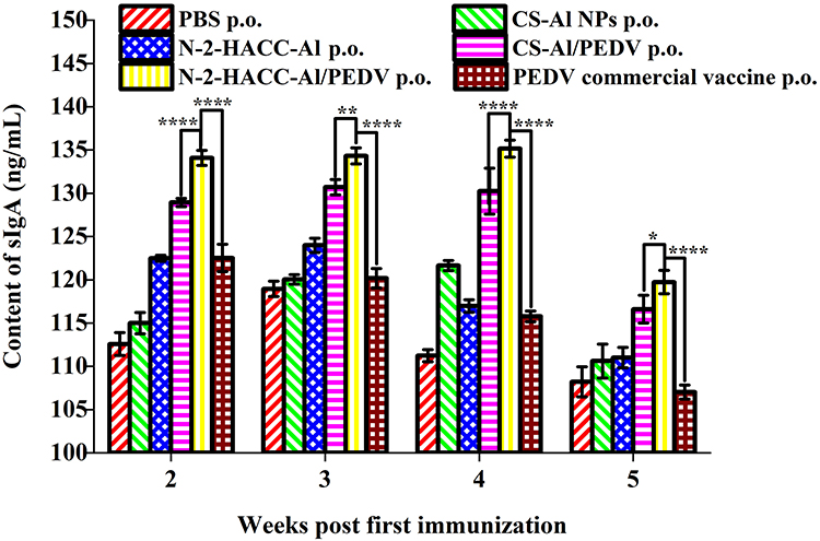 |
Figure 9 Content of sIgA in oral administration of mouses were respectively immunized PBS group, N-2-HACC-AL nano-adjuvant group, CS-Al NPs group, PEDV commercial vaccine group, N-2-HACC-Al /PEDV group and CS-Al/PEDV inactivated vaccine group. (A) Content of IFN-γ in intramuscular injection; (B) Content of IFN-γ in oral administration. * P<0.05; ** P<0.01; **** P<0.0001. |
Discussion
In licensed vaccines, two types of aluminum salt adjuvants are permitted: aluminum hydroxide (AH) and aluminum phosphate (AP).21 AH has a point of zero charge (PZC) of about 11.4 and has a positive charge at neutral pH.32 In recent years, a series of studies have shown that the adjuvant activity of alumina hydroxide nanoparticles (AH nano-adjuvant) at less than 500nm is significantly stronger than that of alumina hydroxide microparticles (AH MPs).33 Adjuvant particle size not only affects immune response level but also immune response type. It is generally believed that AH MPs mainly stimulate the Th2 response.20 Studies have shown that AH nano-adjuvant can stimulate the Th1 response and support cellular immunity.33 AH MPs are concentrated at the injection site and recruit innate immune cells, especially neutrophils. Compared with AH MPs, AH nano-adjuvant can increase antigen uptake by APCs. AH nano-adjuvant has a larger specific surface area and more antigen adsorption binding sites than MPs.34 Smaller nanoparticles (<500 nm) are absorbed by APCs through endocytosis, while larger nanoparticles and microparticles (>500 nm) require phagocyte transport from the injection site to the lymph nodes.35 AH nano-adjuvant tends to be internalized by the DC, while larger AH MPs may stick to the DC surface without being internalized. When the antigen is adsorbed on AH, the zeta potential decreases, which will affect the uptake of the antigen carried by the APC, so it is necessary to further improve the zeta potential of the nanoparticles.34
One effective solution for increasing the zeta potential of aluminum salt adjuvants is the introduction of cationic polymers, and one of the most effective ways for aluminum salt adjuvants to induce low level cellular immunity is to combine them with adjuvants that can enhance the Th1 type immune response.35 In our previous work, we found that N-2-HACC has the ability to induce dendritic cell maturation and antigen-specific Th1 response.29 The modification of CS into N-2-HACC not only improves the solubility of CS but also enhances the Zeta positivity of CS under neutral and alkaline conditions.27 Therefore, in this experiment, N-2-HACC and Al2(SO4)3 were combined by ion crosslinking method to prepare a composite nano-adjuvant N-2-HACC-Al nano-adjuvant.
The average particle size of the N-2-HACC-Al nano-adjuvant prepared in this experiment was 367.9 ± 2.78 nm, and the zeta potential was +33.4±0.66 mV. Nanoparticles smaller than 500 nm can be better internalised by APC and better eliminated through lymphatic vessels to activate inflammatory bodies and induce cellular immunity.36 Nanoparticles with a zeta potential greater than +30mV have better adsorption effect and stability. Protective antigens can still be carried in the intestine under neutral and alkaline conditions, so it has a better effect of enhancing mucosal immune response compared to CS-Al NPs.27,37 The results of immunisation experiments showed that the N-2-HACC-Al/PEDV group could simultaneously stimulate cellular immunity, humoral immunity and mucosal immunity, and achieve Th1/Th2 mixed immunity. In addition, the N-2-HACC-Al/PEDV vaccine is suitable for intramuscular and oral immunity, which greatly compensates for the type of immune response that is missing in the body induced by traditional single adjuvants.
Conclusions
In this study, we successfully prepared N-2-HACC-Al nano-adjuvant, which can be used for injection and oral inoculation to cause strong immune enhancement response and play an immune enhancement role, providing a new idea and strategy for the research and development of vaccine adjuvants. First, N-2-HACC-Al nano-adjuvant proved to be a safe and non-toxic vaccine adjuvant. Secondly, N-2-HACC-Al nano-adjuvant, as a vaccine adjuvant, can not only significantly enhance humoral immunity but also stimulate cellular immune response. At the same time, oral vaccination can also stimulate good mucosal immunity. In addition, the strategy is highly scalable and provides a valuable reference for the preparation of novel vaccine adjuvants.
Acknowledgments
This work was supported in part by the National Natural Science Foundation of China (32370987), “Pioneer” and “Leading Goose” R&D Program of Zhejiang (2025C04047), Natural Science Foundation of Heilongjiang Province (LH2024H076), Project of Qiqihar Academy of Medical Sciences (QMSI2024M-11), Agricultural Science and Technology Program in Taizhou (22nya04, 24nyb04 and 202410) and Industrial Science and Technology Program in Taizhou (23gya02).
Author Contributions
All authors made a significant contribution to the work reported, whether that is in the conception, study design, execution, acquisition of data, analysis and interpretation, or in all these areas; took part in drafting, revising or critically reviewing the article; gave final approval of the version to be published; have agreed on the journal to which the article has been submitted; and agree to be accountable for all aspects of the work.
Disclosure
The authors declare no competing interest in this work.
References
1. Zhang H, Zou C, Peng O, et al. Global dynamics of porcine enteric coronavirus PEDV epidemiology, evolution, and transmission. Mol Biol Evol. 2023;40(3):msad052. doi:10.1093/molbev/msad052
2. Zhang F, Chen Y, Ke Y, et al. Single chain fragment variable (scFv) antibodies targeting the spike protein of porcine epidemic diarrhea virus provide protection against viral infection in piglets. Viruses. 2019;11(1):11010058. doi:10.3390/v11010058
3. Yan Q, Liu X, Sun Y, et al. Swine enteric coronavirus: diverse pathogen-host interactions. Int J mol Sci. 2022;23(7):3953. doi:10.3390/ijms23073953
4. Gillespie T, Song Q, Inskeep M, Stone S, Murtaugh MP. Effect of booster vaccination with inactivated porcine epidemic diarrhea virus on neutralizing antibody response in mammary secretions. Viral Immunology. 2018;31(1):62–68. doi:10.1089/vim.2017.0023
5. Liu J, Shi H, Chen J, et al. A new neutralization epitope in the spike protein of porcine epidemic diarrhea virus. Int J mol Sci. 2022;23(17):9674. doi:10.3390/ijms23179674
6. Zhang Q, Hu R, Tang X, et al. Occurrence and investigation of enteric viral infections in pigs with diarrhea in China. Arch Virol. 2013;158(8):1631–1636. doi:10.1007/s00705-013-1659-x
7. Langel SN, Paim FC, Lager KM, Vlasova AN, Saif LJ. Lactogenic immunity and vaccines for porcine epidemic diarrhea virus (PEDV): historical and current concepts. Virus Res. 2016;226:93–107. doi:10.1016/j.virusres.2016.05.016
8. O’Hagan DT, Fox CB. Are we entering a new age for human vaccine adjuvants? Expert Rev Vaccines. 2015;14(7):909–911. doi:10.1586/14760584.2015.1043273
9. Zhang JH, Cui L, Zhang YL, et al. Oral administration of PEDV-dissolved Alg-CS gel induces high and sustained mucosal immunity in mice. J Gen Virol. 2024;105(4):001979. doi:10.1099/jgv.0.001979
10. Qin ZL, Nai Z, Li G, et al. The oral inactivated porcine epidemic diarrhea virus presenting in the intestine induces mucosal immunity in mice with alginate-chitosan microcapsules. Animals. 2023;13(5):889. doi:10.3390/ani13050889
11. Wen ZF, Xu ZC, Zhou QF, et al. Oral administration of coated PEDV-loaded microspheres elicited PEDV-specific immunity in weaned piglets. Vaccine. 2018;36(45):6803–6809. doi:10.1016/j.vaccine.2018.09.014
12. O’Hagan DT, Lodaya RN, Lofano G. The continued advance of vaccine adjuvants – ‘we can work it out’. Semin Immunopathol. 2020;50:101426. doi:10.1016/j.smim.2020.101426
13. Liu H, Jia Z, Yang C, et al. Aluminum hydroxide colloid vaccine encapsulated in yeast shells with enhanced humoral and cellular immune responses. Biomaterials. 2018;167:32–43. doi:10.1016/j.biomaterials.2018.03.014
14. Dong H, Wen ZF, Chen L, et al. Polyethyleneimine modification of aluminum hydroxide nanoparticle enhances antigen transportation and cross-presentation of dendritic cells. Int j Nanomed. 2018;13:3353–3365. doi:10.2147/IJN.S164097
15. Bo C, Wei X, Wang X, et al. Physicochemical properties and adsorption state of aluminum adjuvants with different processes in vaccines. Heliyon. 2023;9(8):e18800. doi:10.1016/j.heliyon.2023.e18800
16. Smith WJ, Thompson R, Egan PM, et al. Impact of aluminum adjuvants on the stability of pneumococcal polysaccharide-protein conjugate vaccines. Vaccine. 2023;41(35):5113–5125. doi:10.1016/j.vaccine.2023.05.059
17. Zhang T, He P, Guo D, Chen K, Hu Z, Zou Y. Research progress of aluminum phosphate adjuvants and their action mechanisms. Pharmaceutics. 2023;15(6):1756. doi:10.3390/pharmaceutics15061756
18. Ahuja R, Srichandan S, Meena J, Biswal BK, Panda AK. Immunogenicity evaluation of thermostable microparticles entrapping receptor binding domain of SARS-CoV-2 by single point administration. J Pharmaceut Sci. 2023;112(6):1664–1670. doi:10.1016/j.xphs.2023.01.024
19. Wang ZB, Xu J. Better adjuvants for better vaccines: progress in adjuvant delivery systems, modifications, and adjuvant-antigen codelivery. Vaccines. 2020;8(1):128. doi:10.3390/vaccines8010128
20. Kumru OS, Sanyal M, Friedland N, et al. Formulation development and comparability studies with an aluminum-salt adjuvanted SARS-CoV-2 spike ferritin nanoparticle vaccine antigen produced from two different cell lines. Vaccine. 2023;41(44):6502–6513. doi:10.1101/2023.04.03.535447
21. Kooijman S, Vrieling H, Verhagen L, et al. Aluminum hydroxide and aluminum phosphate adjuvants elicit a different innate immune response. J Pharmaceut Sci. 2022;111(4):982–990. doi:10.1016/j.xphs.2022.01.014
22. Aleebrahim-Dehkordi E, Molavi B, Mokhtari M, et al. T helper type (Th1/Th2) responses to SARS-CoV-2 and influenza A (H1N1) virus: from cytokines produced to immune responses. Transplant Immunology. 2022;70:101495. doi:10.1016/j.trim.2021.101495
23. Meena J, Singhvi P, Srichandan S, et al. RBD decorated PLA nanoparticle admixture with aluminum hydroxide elicit robust and long lasting immune response against SARS-CoV-2. Eur J Pharm Biopharm. 2022;176:43–53. doi:10.1016/j.ejpb.2022.05.008
24. Nazarizadeh A, Staudacher AH, Wittwer NL, Turnbull T, Brown MP, Kempson I. Aluminium nanoparticles as efficient adjuvants compared to their microparticle counterparts: current progress and perspectives. Int J mol Sci. 2022;23(9):4707. doi:10.3390/ijms23094707
25. Lebre F, Bento D, Ribeiro J, et al. Association of chitosan and aluminium as a new adjuvant strategy for improved vaccination. Int J Pharm. 2017;527(1–2):103–114. doi:10.1016/j.ijpharm.2017.05.028
26. Poonsuk K, Gimenez-Lirola LG, Magtoto RL, et al. The effect of chemical clarification of oral fluids on porcine epidemic diarrhea virus antibody responses. J Vet Diagn Invest. 2018;30(6):937–941. doi:10.1177/1040638718798672
27. Jin Z, Hu G, Zhao K. Mannose-anchored quaternized chitosan/thiolated carboxymethyl chitosan composite nano-adjuvant as mucoadhesive carrier for drug delivery. Carbohydr Polym. 2022;283:119174. doi:10.1016/j.carbpol.2022.119174
28. Li X, Xing R, Xu C, et al. Immunostimulatory effect of chitosan and quaternary chitosan: a review of potential vaccine adjuvants. Carbohydr Polym. 2021;264:118050. doi:10.1016/j.carbpol.2021.118050
29. Zhao K, Han J, Zhang Y, et al. Enhancing mucosal immune response of Newcastle disease virus DNA vaccine using N-2-hydroxypropyl trimethylammonium chloride chitosan and N,O-carboxymethyl chitosan nanoparticles as delivery carrier. Mol Pharmaceut. 2018;15(1):226–237. doi:10.1021/acs.molpharmaceut.7b00826
30. Meng N, Zhou NL. Synthesis and properties of PDMS/montmorillonite-cetyltrimethyl ammonium bromide-heparin films. Carbohydr Polym. 2014;105:70–74. doi:10.1016/j.carbpol.2014.01.052
31. Özçelik S, Yalçın B, Arda L, et al. Structure, magnetic, photocatalytic and blood compatibility studies of nickel nanoferrites prepared by laser ablation technique in distilled water. J Alloys Compd. 2021;854:157279. doi:10.1016/j.jallcom.2020.157279
32. Raponi A, Brewer JM, Garside P, Laera D. Nanoalum adjuvanted vaccines: small details make a big difference. Semin Immunopathol. 2021;56:101544. doi:10.1016/j.smim.2021.101544
33. Ruwona TB, Xu H, Li X, Taylor AN, Shi Y, Cui Z. Toward understanding the mechanism underlying the strong adjuvant activity of aluminum salt nanoparticles. Vaccine. 2016;34(27):3059–3067. doi:10.1016/j.vaccine.2016.12.047
34. Li X, Aldayel AM, Cui Z. Aluminum hydroxide nanoparticles show a stronger vaccine adjuvant activity than traditional aluminum hydroxide microparticles. J Control Release. 2014;173:148–157. doi:10.1016/j.jconrel.2013.10.032
35. Yan S, Gu W, Xu ZP. Re-considering how particle size and other properties of antigen–adjuvant complexes impact on the immune responses. J Colloid Interface Sci. 2013;395:1–10. doi:10.1016/j.jcis.2012.11.061
36. Khorshidvand Z, Khosravi A, Mahboobian MM, Larki-Harchegani A, Fallah M, Maghsood AH. Novel naltrexone hydrochloride nanovaccine based on chitosan nanoparticles promotes induction of Th1 and Th17 immune responses resulting in protection against Toxoplasma gondii tachyzoites in a mouse model. Int J Biol Macromol. 2022;208:962–972. doi:10.1016/j.ijbiomac.2022.03.146
37. Lin YH, Sun BN, Jin Z, Zhao K. Enhanced immune responses to mucosa by functionalized chitosan-based composite nanoparticles as a vaccine adjuvant for intranasal delivery. ACS Appl Mater Interfaces. 2022;14:52691–52701. doi:10.1021/acsami.2c17627
 © 2025 The Author(s). This work is published and licensed by Dove Medical Press Limited. The
full terms of this license are available at https://www.dovepress.com/terms.php
and incorporate the Creative Commons Attribution
- Non Commercial (unported, 3.0) License.
By accessing the work you hereby accept the Terms. Non-commercial uses of the work are permitted
without any further permission from Dove Medical Press Limited, provided the work is properly
attributed. For permission for commercial use of this work, please see paragraphs 4.2 and 5 of our Terms.
© 2025 The Author(s). This work is published and licensed by Dove Medical Press Limited. The
full terms of this license are available at https://www.dovepress.com/terms.php
and incorporate the Creative Commons Attribution
- Non Commercial (unported, 3.0) License.
By accessing the work you hereby accept the Terms. Non-commercial uses of the work are permitted
without any further permission from Dove Medical Press Limited, provided the work is properly
attributed. For permission for commercial use of this work, please see paragraphs 4.2 and 5 of our Terms.