")
Back to Journals » International Journal of Nanomedicine » Volume 20
Nano-Based Drug Delivery Systems for Managing Diabetes: Recent Advances and Future Prospects
Authors Liu M, Wang R, Hoi MPM, Wang Y, Wang S, Li G, Vong CT , Chong CM
Received 27 November 2024
Accepted for publication 5 February 2025
Published 16 May 2025 Volume 2025:20 Pages 6221—6252
DOI https://doi.org/10.2147/IJN.S508875
Checked for plagiarism Yes
Review by Single anonymous peer review
Peer reviewer comments 2
Editor who approved publication: Dr Kamakhya Misra
Meihan Liu,1 Rui Wang,2,3 Maggie Pui Man Hoi,1 Yitao Wang,1,4 Shengpeng Wang,1,4 Ge Li,2,3,5,6 Chi Teng Vong,1,4 Cheong-Meng Chong1
1State Key Laboratory of Quality Research in Chinese Medicine, Institute of Chinese Medical Sciences, University of Macau, Macau, People’s Republic of China; 2Guangdong Provincial Key Laboratory of Pathogenesis, Targeted Prevention and Treatment of Heart Disease, Guangdong Provincial People’s Hospital (Guangdong Academy of Medical Sciences), Guangzhou, People’s Republic of China; 3Department of Histology and Embryology, School of Basic Medical Science, Southern Medical University, Guangzhou, People’s Republic of China; 4Macau Centre for Research and Development in Chinese Medicine, Institute of Chinese Medical Sciences, University of Macau, Macau, People’s Republic of China; 5Guangzhou Key Laboratory of Cardiac Pathogenesis and Prevention, Guangdong Provincial People’s Hospital (Guangdong Academy of Medical Sciences), Guangzhou, People’s Republic of China; 6School of Medicine, South China University of Technology, Guangzhou, People’s Republic of China
Correspondence: Cheong-Meng Chong; Chi Teng Vong, N22 Research Building, Institute of Chinese Medical Sciences, University of Macau, Avenida de Universidade Taipa, Macau, People’s Republic of China, Tel +853 8822 4512; +853 8822 8063, Fax +853 2884 1358, Email [email protected]; [email protected]
Abstract: Diabetes mellitus is a chronic metabolic disorder, which is characterized by high blood glucose levels, and this can lead to serious diabetic complications. According to the World Health Organization, approximately 830 million adults worldwide are living with diabetes in 2024, with its prevalence continuing to rise steadily over the years. To treat this disease, researchers have developed a variety of first-line drugs, such as sulfonylureas and thiazolidinediones. Despite their long clinical use, there are still many drawbacks and limitations. One of the main drawbacks is low bioavailability, this causes the diabetic patients to take the drugs frequently to lower the blood glucose levels continuously. Some patients may have to take multiple drugs to increase the effectiveness of lowering blood glucose levels. To address these limitations, nano-based drug delivery systems have emerged to overcome these problems. It has emerged as a promising approach for diabetes management, which offers controlled and localized release of anti-diabetic drugs, thus enhancing therapeutic efficacy. This review discusses recent advances in the field of nano-based drug delivery systems for diabetes management, safety and toxicity profiles of anti-diabetic drugs, and future perspectives for the development of nanomedicine in diabetic treatment. Literature search was conducted using electronic databases, and only English literatures were used and published between 2014 and 2024. Recent advancements in nanotechnology have facilitated the development of various nanocarriers, such as polymeric carrier nanoparticles, nanoliposomes, nanocrystals, nanosuspension and inorganic nanoparticles, which enhance drug stability, bioavailability, and efficacy. These systems can deliver anti-diabetic drugs and natural compounds more effectively, thereby minimizing side effects and improving patient compliance. As the field continues to evolve, the successful clinical implementation of nanodrugs could revolutionize the management of diabetes and improve the quality of life for millions of diabetic patients worldwide.
Keywords: drug delivery system, diabetes, nanomedicine, anti-diabetic drugs, Nanomedicine
Graphical Abstract:

Introduction
Nanotechnology broadly refers to technologies applied at the nanoscale.1 In recent years, nanotechnology has emerged as a promising field in drug delivery, thus offering innovative solutions to overcome the limitations of traditional pharmaceutical approaches.2 Although the strict definition of nanoparticles (NPs) is between 1–100 nm, various studies in recent years have extended the upper size range to approximately 1,000 nm in the context of this application. Figure 1 represents the length of the relative size of nanomaterials in comparison to naturally occurring things. At the nanoscale, materials exhibit unique physical, chemical, and biological properties that can be utilized to enhance the efficacy, targeting, and delivery of therapeutic drugs. Similar to cancer therapies, the enhanced permeability and retention (EPR) effect allows NPs to accumulate in tissues with increased vascular permeability, such as inflamed or insulin-resistant tissues. In diabetes, this effect can help to target insulin, or therapeutic agents can be directed to insulin-sensitive tissues, like adipose tissue or muscle, to improve their efficacy. Nanomaterials can be engineered to interact with cell membranes more effectively through various means, for example, coating NPs with ligands or antibodies that target specific receptors can facilitate endocytosis.3 Besides, they can also be engineered for controlled drug release through stimuli-responsive systems, releasing drugs in response to specific biological triggers, such as changes in glucose levels.4 This helps to maintain stable drug levels in the bloodstream, thus reducing the need for frequent dosing and enhancing patient compliance. The development of nano-based drug delivery systems has progressed significantly over the past few decades. The concept of using liposomes as drug carriers was firstly introduced in the 1960s, paving the way for the development of lipid-based NPs.5 Subsequently, polymeric NPs, such as those made from biodegradable polymers like poly(lactic-co-glycolic acid) (PLGA), were explored for drug encapsulation and controlled release.6 In 2000s, a wide range of NP platforms were developed, including polymeric carriers,7 dendrimers8 and inorganic NPs.9 In addition, targeted drug delivery using NPs functionalized with ligands, antibodies, or other targeting moieties gained prominence in this field of research.10 The use of NPs for the delivery of biomacromolecules, such as proteins, peptides, and nucleic acids, also becomes an area of active research.11 Stimuli-responsive NPs that release drugs in specific conditions, such as pH, temperature and enzymes, were also designed.12 There are many reasons why nanomedicine delivery systems are so popular. One of the primary reasons is the ability to improve the solubility, bioavailability, and stability of drugs.2 NPs, such as polymeric NPs, liposomes, and micelles, can encapsulate hydrophobic drugs, thus protecting them from degradation and facilitating their transport across biological barriers, such as intestinal epithelium and blood-brain barrier. This helps to overcome a major challenge in traditional drug formulations, where poor solubility and low bioavailability often limit the efficacy of many therapeutic drugs. Besides, the unique size and surface properties of NPs can prolong the circulation time of drugs in the body, thereby leading to enhanced pharmacokinetic profiles.13 Furthermore, nanocarriers can be engineered to specifically target diseased or desired tissues. This can be achieved through the incorporation of targeting moieties, such as antibodies, peptides or ligands, on the surface of the NPs, which recognize and bind to receptors or markers that are expressed on the target cells or tissues.14 This targeted delivery approach improves the therapeutic effects by maximizing the drug concentration at the site of action and minimizing systemic exposure and off-target effects.
 |
Figure 1 Different types of particles and living things. A nanoparticle is a particle of matter with 1 to 1000 nm in diameter. At the lowest range, metal particles, that are smaller than 1 nm, are usually called atom clusters. Some nanoparticles containing biological materials can be hundreds of nm in diameter. |
The superior properties of nanodrugs enable them to have great potential in disease treatment. They are now used to treat cancer,15 neurodegenerative diseases,16 cardiovascular diseases,17 inflammation18 and diabetes.19 Among these, the global prevalence of Type 2 diabetes mellitus (T2DM) continues to rise, which poses a significant public health challenge.20 Conventional therapeutic approaches may fall short in achieving optimal glycemic control and preventing the progression of diabetic complications. Therefore, the emergence of nanodrugs has sparked new hope in revolutionizing the management of diabetes. NP-based insulin formulations can protect the drugs from degradation of insulin, prolong its action, and facilitate more consistent and controlled release, thereby potentially reducing the frequency of insulin injections.21 In addition, to overcome the challenges of poor absorption and enzymatic degradation in the gastrointestinal tract, NPs can enhance the bioavailability of orally administered insulin, thus improving patient compliance.13 NPs can also co-deliver insulin and other anti-diabetic drugs, such as metformin or glucagon-like peptide-1 (GLP-1) agonists, that can synergistically improve glycemic control and mitigate diabetic complications. Importantly, common first-line anti-diabetic drugs, such as metformin, GLP-1 receptor agonists, dipeptidyl peptidase-4 (DPP-4) inhibitors and sodium-glucose cotransporter-2 (SGLT2) inhibitors, are also being used to design nanomedicine delivery systems. Moreover, many natural compounds with anti-diabetic activities are also expected to be better applied to patients by being prepared into NPs.22 These advanced drug delivery systems, such as polymeric carriers, lipid carriers, nanocrystal, nanosuspension and inorganic carriers, hold the promise of enhancing therapeutic efficacy, improving patient adherence, and ultimately, transforming the therapeutic approaches for diabetic treatment and prevention of diabetic complications.
In this review, literature search was conducted using electronic databases, including PubMed, Google Scholar and Web of Science. A combination of the following keywords, including “Type 2 diabetes”, “nanomedicine”, “nanoparticles”, “drug delivery systems”, “antidiabetic agents” and their related terms, was used for the search of English literatures between 2014 and 2024. This review aims to summarize studies from the past decade on the use of nanomedicine in diabetes treatment, focusing on commonly used anti-diabetic drugs and natural compounds encapsulated in NPs, as well as NPs exhibiting anti-diabetic activities. We focus on the design of different types of NPs and their applications for the improvement of diabetic treatment, as well as their toxicity profiles. Finally, we discuss recent advances and future perspectives for the development of NPs as therapeutic drugs in diabetic treatment.
Current Anti-Diabetic Drugs and Their Limitations
Diabetes is one of the largest chronic diseases in the world. According to statistical data, the number of people with diabetes has been more than doubled in the past 20 years.23,24 Its main characteristics is hyperglycemia, which is accompanied by disturbances in carbohydrate, protein and fat metabolism. There are two types of diabetes commonly seen in clinical practice, Type 1 diabetes mellitus (T1DM) and T2DM.25 In T1DM, due to an autoimmune response, the body produces antibodies that destroy pancreatic β-cells and cause reduced insulin production.26 Therefore, insulin injection therapy is an extremely important way to maintain the health of patients with T1DM. T2DM accounts for 85–90% of diabetic patients,27 and the main causes include genetics, lifestyle and obesity.28,29 In T2DM, the insulin receptors on the cell membrane of insulin-responsive cells are poorly or even unresponsive to insulin, which is known as insulin resistance, thus the blood glucose levels are increased.30
Currently approved drugs for T2DM mainly include biguanides, sulfonylureas, thiazolidinediones, DPP-4 inhibitors, SGLT2 inhibitors and GLP-1 analogues.23 The clinical drugs for T2DM are mainly oral tablets, capsules and suspension. In addition, polyphenols, flavonoids and alkaloids derived from natural plants also have excellent anti-diabetic activities.31 Many natural product components can play a role in hypoglycemia by protecting pancreatic β-cells, promoting insulin secretion, reducing glucose absorption in the intestine, increasing the number of insulin receptors on target cells, and enhancing insulin sensitivity.32,33 However, diabetic patients may have suffered from the side effects of anti-diabetic drugs. Common sulfonylureas include glipizide, glibenclamide and gliclazide.34 They have a certain degree of hypoglycemia, gastrointestinal reactions and may destroy liver and kidney functions. Biguanides are used to treat T2DM by improving the sensitivity of peripheral tissues and liver to insulin, reducing intestinal glucose absorption and enhancing activating anaerobic peripheral tissue glycolysis.35 The main drug of this class used in the clinic is metformin, but it has adverse effects on liver, kidney and heart functions.36 On the other hand, α-glucosidase inhibitors reversibly inhibit the activity of α-glucosidase in small intestinal wall cells, thus delaying the degradation of carbohydrates and glucose absorption in the digestive tract. As a result, they effectively delay and reduce the time and process of postprandial glucose rise in diabetic patients, and help to control the development of diabetes.37 However, it also has gastrointestinal side effects. Most of these anti-diabetic drugs need to be taken for a long time and frequently to achieve the desired effect, which leads to the accumulation of drugs in the body, thereby resulting in various adverse reactions. In recent years, researchers have focused on new targets and new drugs. In addition, for these traditional anti-diabetic drugs, the current focus is to improve their bioavailability and dose reduction, and improve patient medication compliance.38
The use of nano-based drug delivery systems has become the first choice for researchers.39 Wrapping anti-diabetic drugs in nanocarrier system can fully protect drugs from gastrointestinal decomposition and reduce gastrointestinal adverse reactions. In addition, drugs can be released in a gradual and effective manner, thus improving the bioavailability of the drugs, reducing drug accumulation and administration times, and improving patient medication compliance. Over the past few years, many efforts have been made to manufacture suitable materials, systems, and nanostructures for drug delivery and biomedical applications. Several drug delivery systems that have been reported to improve the bioavailability and effective drug delivery for anti-diabetic drugs include polymerized NPs, lipids nanocarrier systems, nanocrystals, nanosuspensions and inorganic nanocarriers.
Nanomedicine for the Management of Diabetes
Nanotechnology has rapidly advanced across many disciplines in recent years.40,41 Among them, using nanotechnology to deliver the drugs becomes successful and has been used clinically.42 Nanomedicine can be broadly divided into nanocarrier drugs and nanostructured drugs. Nanocrystals are the most common type of nanostructured drug delivery systems. They have no nanomaterial used as a carrier and are synthesized by breaking the drugs down to the nanoscale.43 The reduction of particle size greatly improves water solubility, bioavailability and efficacy of the drugs. The other type of nanomedicine is nanocarrier drugs. Nanocarrier drugs utilize carrier materials to bind, adsorb, disperse, or encapsulate genes, proteins and drugs, thus enhancing their transport to the body and bioavailability. Nanocarriers can protect the drugs from degradation, increase their half-life, and improve the solubility of poorly water-soluble drugs. Therefore, they have significant potential in delivering drugs more efficiently for the treatment of diseases, including T2DM. The main types of nanocarrier drugs include natural polymer carrier, synthetic polymer carrier and combination of both.
Polymeric Nanoparticles
Polymeric NPs are composed of polymer materials that have excellent biocompatibility and possess controllable physical and chemical properties.44 Drugs can interact with polymeric NPs through processes, such as adsorption, encapsulation and covalent binding, thus allowing effective encapsulation within the NPs. Polymeric NPs can encase or physically encase the drugs within the polymer matrix, thereby reducing its interaction with healthy cells and reducing the toxicity of the drugs. The commonly used polymers include natural polymers, such as albumin, dextran, chitosan and hyaluronate, and synthetic polymers, including polyglutamic acid, polyglycolic acid, polyethylene glycol, polycaprolactone and polylactic acid (PLA).
Natural Polymeric Nanoparticles
Natural polymers used for anti-diabetic drug delivery include a wide range of gums, mucilages and polysaccharides.45 Their primary advantages over synthetic polymers are their non-toxicity and biocompatibility. They can also benefit from lower immune rejection and are biocompatible as all of them are derived from organisms, and their final products are polysaccharides or amino acids, which are easy to absorb and not prone to inflammation.46 Meanwhile, the problem of degradation can be solved by enzymatic hydrolysis. In addition, natural polymers often have their own biological activities. For example, some natural polymers have anti-bacterial, anti-inflammatory, wound healing effects, which make them to be promising for a wide range of applications in medical and bioengineering fields.45 They also have the advantages of stable performance, non-toxic, safe application, low cost, so scientists are paying more attention to the development of natural polymers for diabetic treatment. Table 1 summarizes the characteristics and therapeutic effects of natural polymeric NPs for diabetes management.
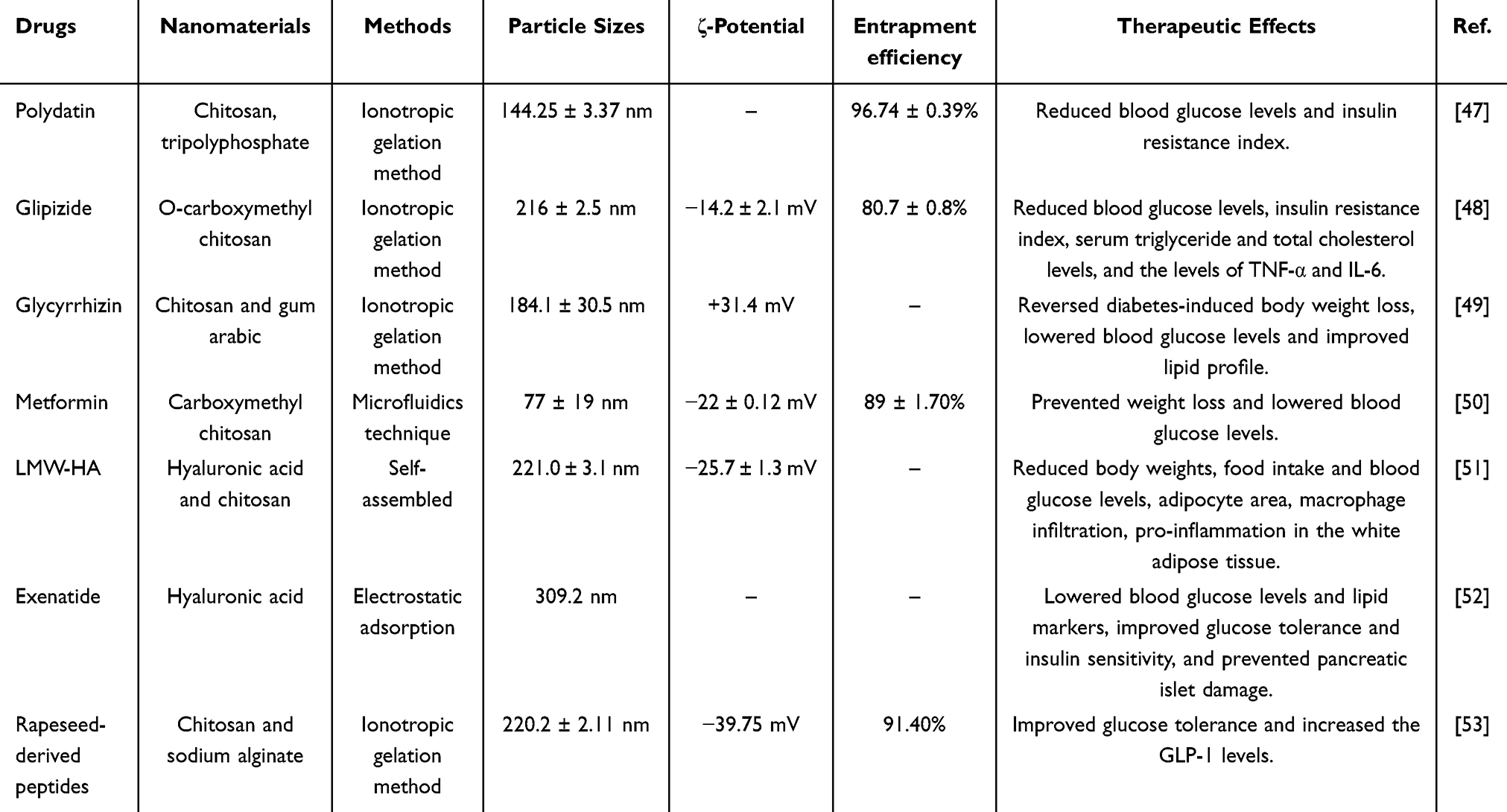 |
Table 1 Natural Polymeric Nanoparticles for Diabetes Management |
Chitosan
Chitosan belongs to one kind of natural polymers, which is the product of deacetylation of chitin.54 It consists of D-glucosamine units that are held together by a β-1,4-glucoside bond.55 The free active amino group in its structure provides favorable conditions for modification, and is often used to introduce other groups, such as alkyl, carboxylic and aromatic groups.54,56 Chitosan has the same good biocompatibility and biodegradability as chitin,57,58 and has unique biological effects, such as bio-adhesion, good safety, anti-bacterial, anti-tumor and anti-diabetic activities.59–62 It has been widely used to prepare polymeric NPs for diabetic treatment due to its anti-diabetic activity and advantages in drug delivery.
Polydain is a polyphenol and resveratrol derivative, and has various pharmacological effects, including anti-inflammatory and antioxidant effects.63 It is poorly water-soluble and has low bioavailability and high first-pass metabolism, which limits its clinical applications.64 Abdel-Moneim et al loaded polydain onto chitosan to synthesize polydain-loaded chitosan NPs using a modified ionic gelation method to improve the therapeutic effects of polydain.47 They added polydain into chitosan solution, and tripolyphosphate aqueous solution was then added to obtain polydatin-loaded chitosan NPs. The particle size of these NPs was 144.25 ± 3.37 nm. These NPs showed excellent in vitro sustained-release ability, which could continuously release polydain when compared with free polydain. Besides, the increased concentration of chitosan in the formulation could result in longer time of release. They suggested that the hydration of chitosan and the dissolution medium formed a gel-like substance, which delayed the release of polydain. Moreover, these NPs were found to prevent body weight loss, lower blood glucose levels and insulin resistance index, and increase plasma insulin levels in diabetic rats, demonstrating better anti-diabetic effects than free polydain. In another study, similar ionic gelation method was used to load glipizide with O-carboxymethyl chitosan (CMC) to synthesize glipizide-CMC-NPs with particle size of 216 ± 2.5 nm.48 The ζ-potential was −14.2 ± 2.1 mV. O-CMC is a soluble chitosan derivative with both amino and carboxyl groups that enables glipizide to have long-lasting effects on diabetic treatment. Glipizide is a T2DM drug that belongs to the class of sulfonylureas and has several anti-diabetic effects, including enhancing insulin secretion and sensitivity. It has fewer side effects, however, its half-life is short (2–4 h), which requires frequent administration.65 A more prolonged and superior release profile of these NPs was observed when compared with free glipizide. When the NPs were administered to T2DM mice, it was found that blood glucose levels, insulin resistance index, serum triglyceride and total cholesterol (TC) levels were reduced in the NPs group. Besides, the serum levels of the pro-inflammatory cytokines tumor necrosis factor (TNF)-α and interleukin (IL)-6 were also reduced and serum adiponectin levels were increased in the NPs group when compared with the free glipizide group, suggesting that better therapeutic effects were achieved after glipizide was prepared into chitosan NPs.
Glycyrrhizin is a major bioactive component of Licorice, however, its oral bioavailability is very low, and its absorption in the gastrointestinal tract is slow and incomplete.66 Therefore, glycyrrhizin-loaded NPs were prepared using chitosan and gum arabic as polymers by an ionotropic gelation method.49 The particle size was 184.1 ± 30.5 nm and ζ-potential was +31.4 mV. The in vitro release of these NPs was slow and sustained. Importantly, glycyrrhizin-loaded NPs only contained about a quarter of the amount that was used in the free glycyrrhizin group. These NPs reversed diabetes-induced body weight loss, lowered blood glucose levels and improved lipid profile in nicotinamide plus streptozotocin (STZ)-induced diabetic rats, demonstrating better therapeutic effects in T2DM.
In addition to the common ionic gelation method, there are other different methods used for the synthesis of NPs, such as emulsion, spray drying and precipitation. The preparation of chitosan-coated NPs by microfluidic method has become more and more popular in recent years. Studies showed that the use of microfluidic method enabled polymeric NPs become smaller and more uniform.67 Lari et al encapsulated metformin hydrochloride, a commonly used T2DM drug, into novel crosslinked CMC NPs using microfluidics technique.50 Moreover, CMC has been widely used for the synthesis of NPs due to good safety, water soluble and mucosal adhesion.68 However, this polymer has some disadvantages, including poor stability and rapid degradation. An ionic crosslinker, such as calcium chloride, can be added to enhance drug loading and control drug release.69 Metformin-loaded crosslinked CMC NPs were synthesized using calcium chloride as the ionic crosslinker and CMC as the polymer.50 The blood glucose level reduction of metformin-loaded crosslinked CMC NPs was 15% higher than free metformin in STZ-induced diabetic rats, and these NPs preserved pancreatic islet regeneration, revealing superior therapeutic effects.
Hyaluronic Acid (HA)
HA is a high molecular weight polymer and a linear polysaccharide that is composed of D-glucuronic acid and n-acetylglucosamine.70 Its molecular weight can reach as much as 25 kDa.71 As a natural polymer, HA is biocompatible, biodegradable and non-toxic. In addition, due to its structural particularity, it can be subjected to different chemical modifications to achieve different physical and chemical properties, such as conjugation, nanotubes, dendritic macromolecules, liposomes and self-assembled NPs.72 In recent years, HA has been widely studied for targeted and long-acting drug delivery in the biomedical research. It is worth noting that HA can bind and interact with CD44, which allows HA to exert therapeutic effects for the treatment of many diseases, such as diabetes, inflammatory diseases and cancer.51,73,74 HA has a negative charge under physiological pH conditions, so as a nanocarrier, it can enrich the negative charge and escape the capture of serum proteins.75 In addition, HA is hydrophilic, which can avoid the effects of opsonization. Meanwhile, hydrophilicity allows HA to introduce bound water, thus establishing its barrier and extending its circulation time in the body. Due to its rich functional groups and binding sites, the chemical modification of HA can also be varied.71 As a common nanocarrier material, drugs loaded with HA exhibit a longer biological half-life, which significantly enhanced cellular uptake. In the study of Bhujbal et al, extremely stable NPs were formed by loading HA with the common T2DM drug metformin, and their strong permeability was verified in Caco-2 intestinal epithelial cells.76 Besides, the NPs loaded with HA as the carrier also showed good therapeutic effects on diabetes in vivo (Figure 2A and B).51,52,77
 |
Figure 2 Polymeric nanoparticles (NPs) as drug delivery systems for the management of diabetes. (A) Schematic illustration of RMs-O/W-HA mucus penetration and CD44-mediated endocytosis to facilitate the uptake of Exenatide. Adapted from Eur J Pharm Biopharm, volume 191, Lu Y, Wu L, Lin M, et al. Double layer spherical nanoparticles with hyaluronic acid coating to enhance oral delivery of exenatide in T2DM rats. 205–218, Copyright 2023, with permission from Elsevier.52 (B) Schematic illustration of the therapeutic effects of self-assembled HA-NPs on adipose tissue inflammation and insulin resistance. Adapted from J Control Release, volume 279, Rho JG, Han HS, Han JH, et al. Self-assembled hyaluronic acid nanoparticles: implications as a nanomedicine for treatment of type 2 diabetes. 89–98, Copyright 2018, with permission from Elsevier.51 (C) Schematic illustration of chitosan-sodium alginate nanocarrier system for controlling the release of rapeseed-derived peptides and enhancing anti-diabetic efficacy. Adapted from Int J Biol Macromol, volume 265, Wang Q, Dong X, Castañeda-Reyes ED, et al. Chitosan and sodium alginate nanocarrier system: controlling the release of rapeseed-derived peptides and improving their therapeutic efficiency of anti-diabetes. 130713, copyright 2024, with permission from Elsevier.53 (D) Schematic diagram showing curcumin-loaded PLA-PEG copolymer NPs for the treatment of liver inflammation in streptozotocin-induced diabetic rats. Adapted from Colloids Surf B, volume 177, El-Naggar ME, Al-Joufi F, Anwar M, Attia MF, El-Bana MA. Curcumin-loaded PLA-PEG copolymer nanoparticles for treatment of liver inflammation in streptozotocin-induced diabetic rats. 389–398, copyright 2019, with permission from Elsevier.78 |
Due to the ability of HA in recognizing and binding to CD44, a study synthesized self-assembled amphiphilic HA-conjugated NPs with particle size of 221.0 ± 3.1 nm particle size and ζ-potential of −25.7 ± 1.3 mV.51 These NPs could aggregate in the adipose tissue and inhibit the binding of free low molecular weight (LMW) HA to CD44, thereby inhibiting the pro-inflammatory effects of LMW HA in mouse bone marrow-derived macrophages (BMDMs). IL-1β and TNF-α levels were significantly decreased in the HA NPs group of CD44 +/+ BMDMs, but not in CD44 -/- BMDMs, suggesting that the effects were through CD44. HA NPs successfully reduced body weight, food intake and blood glucose levels in high-fat diet (HFD)-induced obese mice, and also significantly reduced adipocyte area, macrophage infiltration and pro-inflammation in the white adipose tissue. Therefore, HA alone, without drug loading, showed significant therapeutic effects on diabetes. Moreover, HA is more widely used as an external coating of NPs with drug loading to achieve better therapeutic effects. In the study of Lu et al, a positively-charged reverse micelle self-emulsifying polypeptide drug delivery system encapsulated with exenatide, a T2DM drug, was firstly developed, and then HA was wrapped around the micelles by electrostatic adsorption to synthesize exenatide NPs.52 The particle size was 309.2 nm. HA coating improved the adhesion of NPs and enhanced CD44-mediated endocytosis to increase cellular uptake in Caco-2 intestinal epithelial cells. Besides, it also enhanced NP accumulation in the gastrointestinal tract of HFD + STZ-induced diabetic rats. Exenatide NPs coated with HA also showed better therapeutic effects than uncoated NPs in diabetic rats, including lowered blood glucose levels and lipid markers, improvement in glucose tolerance and insulin sensitivity, and prevention from pancreatic islet damage.
Sodium Alginate
Sodium alginate is a kind of natural hydrophilic polysaccharide extracted from brown algae. It is a polymer composed of d-mannuronic and l-guluronic acids.79,80 Sodium alginate, as the main component of the cell wall and extracellular matrix of brown algae, has certain mechanical strength and flexibility, which allows it to have the natural advantage for drug delivery.81 Due to its low extraction and separation cost and good biocompatibility, sodium alginate has been increasingly used in nanomedical drug delivery systems.82 Besides, Shilpa et al has shown that sodium alginate has the highest mucosal adhesion strength when compared with other polymers, such as CMC, polystyrene, PLA and chitosan.83
For the treatment of diabetes, natural peptides have been shown to exhibit excellent efficacy with fewer side effects.84 However, due to the action of gastrointestinal digestive enzymes and rapid metabolism in the body, the delivery of natural peptides has become a thorny problem. To solve this problem, Wang et al used chitosan and sodium alginate to deliver rapeseed-derived peptides.53 Sodium alginate was crosslinked with calcium to form a gel, and stable NPs were formed by electrostatic adsorption with chitosan under ultrasonic conditions, which could protect the polypeptides and achieve continuous drug release in the intestine (Figure 2C). The particle size was 220.2 ± 2.11 nm and ζ-potential was −39.75 mV. In Sprague Dawley rats, these NPs improved glucose tolerance, and GLP-1 levels were increased and peaked at 120 mins after administration. They also suggested that calcium-sensing receptor played an important role in regulating glucose homeostasis by these NPs.
Synthetic Polymeric Nanoparticles
Comparing to the inartificial polymers, synthetic polymers are easier to synthesize in large quantities, and there are no significant differences between batches. Synthetic polymers can also be modified according to the desired properties.85 These nanomaterials are stable in the blood, non-toxic, non-thrombotic, non-immunogenic, non-inflammatory, non-activating neutrophils, biodegradable.86 They can avoid the reticuloendothelial system and are suitable for encapsulating various molecules, such as drugs, proteins, peptides and nucleic acids. It is worth noting that we need to consider many factors for synthetic polymers, such as their structures, degree of deacetylation, molecular weights and solubility. The physical and chemical properties of polymers determine their absorption, distribution and metabolism in the body. The surface modified NPs have anti-adhesion properties due to the extended configuration of the particle surface, which acts as a spatial barrier, thereby reducing the clearance of circulating macrophages in the liver and increasing the possibility of enhanced osmotic processes. Besides, the release mechanism can be regulated by the molecular weight of the polymers. The higher the molecular weight of the polymers, the slower the release of the drugs.87 Biodegradable polymers, such as PLA and their co-polymers, poly-p-dioxanone, poly(L-lactide-co-ε-caprolactone) (PLCL) and co-polymers of trimethylene carbonate and glycolide, have been used in a number of clinical applications.88,89 Table 2 summarizes the characteristics and therapeutic effects of synthetic polymeric NPs for diabetes management.
 |
Table 2 Synthetic Polymeric Nanoparticles for Diabetes Management |
Polylactic Acid
PLA polymer is a biocompatible and biodegradable material that breaks down into lactic acid monomer units in the body. PLA NPs are mainly prepared by solvent evaporation, solvent displacement, salting out and solvent diffusion.94 Stevioside is an US Food and Drug Administration (FDA)-approved non-toxic, natural, non-caloric sweetener, which has been shown to display anti-diabetic activity. It regulates blood glucose levels by enhancing insulin secretion and reducing phosphoenolpyruvate carboxykinase levels.95 However, due to its low bioavailability, poor intestinal absorption and rapid metabolism, its therapeutic effect was not obvious. Barwal et al successfully encapsulated stevioside in Pluronic-F-68-PLA NPs by the precipitation method.90 The particle sizes were between 134.4 and 236.8 nm and ζ-potential was −28 mV. The structure of stevioside was successfully preserved without heating and ultrasonic damage during the preparation process. This study is helpful for further research on the application of stevioside for the treatment of diabetes. Similarly, PLA NPs loaded with stem extracts of Tinospora cordifolia was synthesized by double solvent evaporation using PLA polymer.91 The decreased blood glucose levels in NPs-treated diabetic rats was almost similar to that in positive control (glibenclamide)-treated rats. In addition, Tinospora cordifolia-loaded PLA NPs also significantly reduced plasma insulin, total cholesterol and triglyceride (TG) levels, and increased high-density lipoprotein (HDL) levels when compared with diabetic group, and exerted anti-oxidant activities.
Polyethylene Glycol (PEG)
In recent years, PLA is often combined with PEG for drug delivery.96 As we know, PEG, which is widely used due to its biodegradability and the possibility of multiple molar masses. The FDA has approved PEG for use in food, cosmetics and pharmaceuticals.97 The use of PEG prevents NPs from being quickly recognized and cleared by the immune system, thereby prolonging the circulation time of drugs in the body. As the molecular weight of PEG increases, its blood circulation time is prolonged and PEG attachment to tissues is enhanced, which reduces renal filtration.98 In addition, PEG with different molecular weights have been used to form additional coatings during the preparation and packaging of PLA NPs, thus the stability of the nanomaterials can be greatly improved. As PEG can form a hydration layer on the surface of the NPs, which increases its stability, reduces the direct contact between the drug and the blood, and decreases the rate of drug degradation. Besides, these coatings provide a protective barrier against widespread uptake of human monocytes.
El-Naggar et al designed and developed curcumin-loaded PLA-PEG NPs using nano-emulsification technique (Figure 2D).78 The particle size was 117 nm and ζ-potential was +35 mV. Curcumin-loaded PLA-PEG NPs were administered orally to STZ-induced diabetic rats, it significantly reduced hyperglycemia, increased plasma insulin levels when compared with free curcumin group. It also protected the liver by reducing liver inflammation. This was through inhibiting nuclear factor kappa-light-chain-enhancer of activated B cells (NF-κB) activation, down-regulating hepatic cyclooxygenase-2 (COX-2) and transforming growth factor-β (TGF-β) levels and up-regulating peroxisome proliferator-activated receptor-γ (PPAR-γ) expression.
Despite all the advantages of PEG, there are several issues that still need to be addressed. As the molar mass of the polymers increases, PEG tends to accumulate within certain organs, which must be overcome.99,100 The second concern is the possibility of immune responses due to complement C activation, which can lead to hypersensitivity reactions that result in anaphylactic shock. To solve these problems, the researchers made different modifications to the PEG. In the study of Moghimi et al, liposomes with a nonionic 1-O-phospholipid-mPEG conjugate were designed. They demonstrated that methylated PEG did not activate the complement in human and rat serum. This might be because methylation could prevent phosphate oxygen from binding to complement, or interfere with the complement to prevent PEG from binding to the complement.101
Poly(Lactic-Co-Glycolic Acid)
PLGA is a synthetic, biodegradable block copolymer obtained from lactic acid and glycolic acid, which has been approved by the FDA for human and nanomedicine use. The surface modification of PLGA, the encapsulation method and particle size of the drugs, the additive added during the preparation process, the molecular weight of the drugs, and the ratio of lactide to glycolactone have great influence on the release and effective reaction of the prepared nanomedical drugs. Due to its modified versatility, PLGA is one of the most useful synthetic, biodegradable copolymers for the design and development of controlled and targeted drug delivery systems.102
Silybin is a polyphenolic flavonoid isolated from Silybum marianum and Cynara scolymus with poor water solubility and bioavailability. It was formulated into chitosan-stabilized PLGA NPs.103 These NPs were shown to reduce blood glucose levels, improve glucose tolerance, increase plasma insulin levels, improve lipid profile and liver function in STZ-induced diabetic rats when compared with free silybin. In addition to being used to deliver insoluble natural compounds, PLGA is also used to deliver peptides and proteins. A novel multifunctional composite system has been designed for the dual delivery of GLP-1 and DPP-4 inhibitor.92 First, they prepared PLGA NPs loaded with GLP-1, and then modified PLGA NPs with chitosan and cell-penetrating peptides. Finally, it was encapsulated in a pH-sensitive polymer loaded with DPP-4 inhibitor, thus resulting in a dual-delivery composite system. The final particle size of this system was 286.7 ± 5.5 nm and ζ-potential was +34.7 ± 2.8 mV. This system demonstrated increased hypoglycemic effects and improved glucose tolerance when compared with free GLP-1-DPP-4 inhibitor solution in STZ and nicotinamide-induced diabetic rats. Blood glucose levels were reduced by 44% at 4 hours after glucose administration, and plasma insulin levels were increased at 6 hours after oral administration of this system.
Poly(L-Lactide-Co-Caprolactone)
Drugs are generally encapsulated inside polymer NPs or bound to the surface of NPs. Some studies have found that biodegradable polymer nanomaterials have a constant and slow degradation rate under fixed experimental conditions,87 which provides a new way to solve the problem that diabetic patients who take drugs frequently to achieve good therapeutic effects. A complex of a polymer with a drug or other active agents is taken up into the cells or tissues or blood, and then the drug is released from the nanomaterial in a controlled manner under pre-designed response condition.104 Naik et al designed a novel delivery system using PLCL as a carrier to maintain peak plasma levels of gliclazide.93 Gliclazide was encapsulated in the PLCL NP system rather than coupling to the surface. The release of gliclazide from NPs was divided into two stages, the early rapid release occurring within 2–5 hours (up to 75–80%) and the PLCL begins to degrade at the same time. The remaining drug was released slowly over the next 26 hours.
Inorganic Nanoparticles
Metals are essential components in the body and play important roles in physiological processes, such as glucose metabolism.105 Inorganic nanocarriers refer to the use of metals as nanocarriers in the drug delivery systems. The existing drugs have side effects and low bioavailability, and nanotechnology has emerged as a new strategy to be developed for enhancing target delivery and the bioavailability of drugs, and there are increasing interests in developing bioactive metallic NPs for the treatment of diseases. Metallic NPs can be used to deliver the drugs to target sides and reduce off-target effects, and release drugs in a low and sustained manner.106 These metallic NPs can be synthesized by biological sources, such as bacteria, fungi, yeast and medicinal plants, and their synthesis only requires environmental-friendly solvents, good reducing and stabilizing agents, so this is regarded as green synthesis. They also have the characteristics of small size with large surface area, which help to bind to targeting agents effectively on their surface for tissue-targeting delivery. Besides, they can also penetrate into the skin, cell membrane and nucleus to deliver and release the drugs to target sites and exert biological activities, thus increasing bioavailability and reducing side effects. Table 3 summarizes the characteristics and therapeutic effects of inorganic nanocarriers for diabetes management.
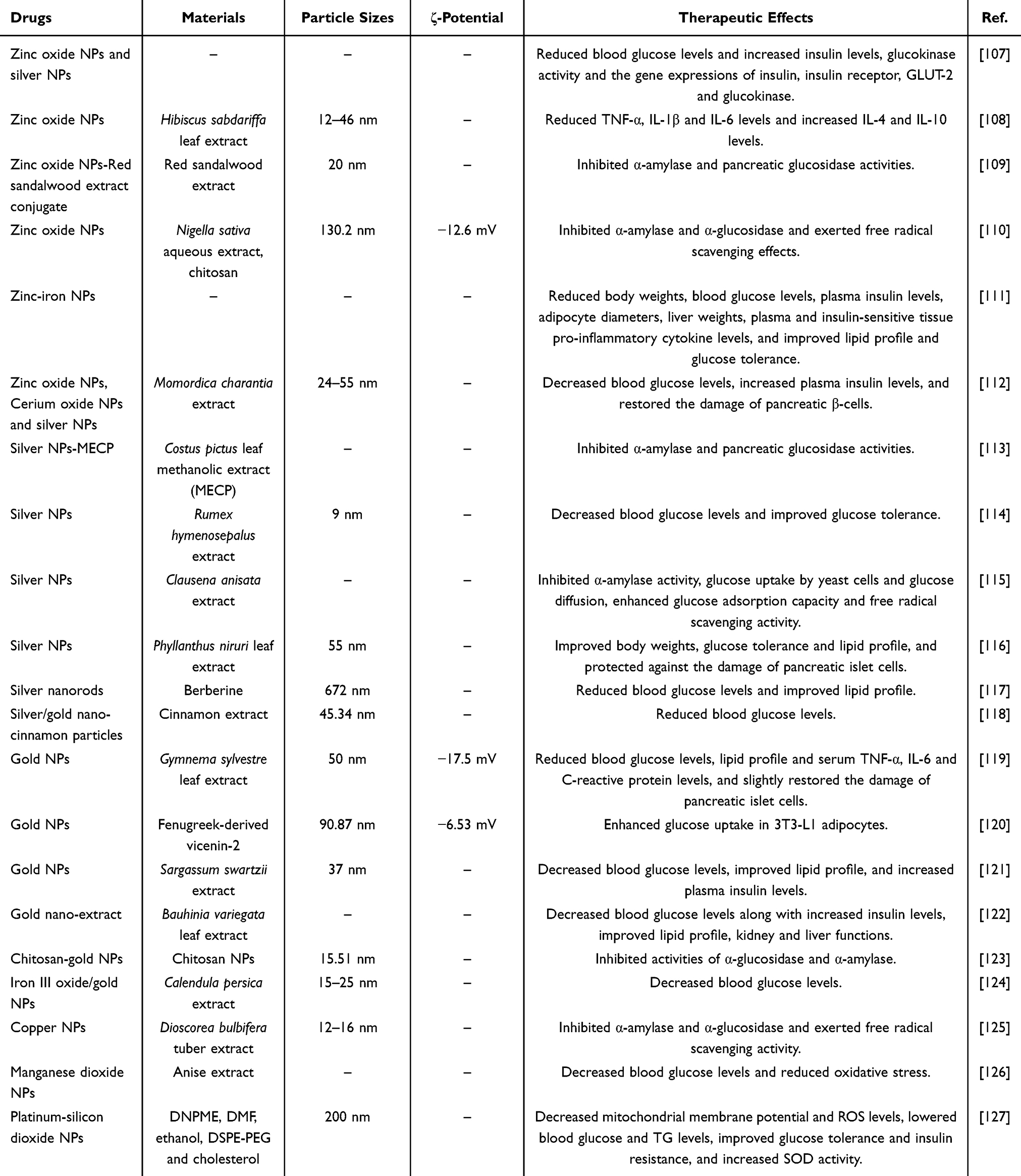 |
Table 3 Inorganic Nanocarriers for Diabetes Management |
Several metallic NPs were developed for the treatment of diabetes, including zinc, silver, copper and gold NPs. Zinc oxide NPs and silver NPs were used for the study of their anti-diabetic effects in STZ-induced diabetic rats.107 Zinc is an essential metal in the body, and plays a vital role in glucose metabolism and insulin synthesis and action,128 while silver is not involved in any physiological processes, but it displays potent anti-bacterial effects.129 They demonstrated that both zinc oxide NPs and silver NPs could reduce blood glucose levels and increase insulin levels in diabetic rats, and zinc oxide NPs were more potent than silver NPs.107 In the study of Bala et al, zinc oxide NPs were also synthesized using Hibiscus sabdariffa leaf extract, and exhibited anti-bacterial and anti-diabetic effects.108 The average particle sizes were between 12 and 46 nm. They decreased blood glucose levels over the period of the study in STZ-induced diabetic mice. As T1DM is associated with pancreatic inflammation, they also showed that zinc oxide NPs significantly reduced TNF-α, IL-1β and IL-6 levels and increased IL-4 and IL-10 levels in the pancreas of diabetic mice, suggesting that they exerted protective effects against T1DM. Interestingly, zinc oxide NPs were also synthesized and conjugated with red sandalwood (RSW) extract.109 RSW was shown to possess anti-diabetic, anti-inflammatory and antioxidant activities.130 The average size of this zinc oxide NPs-RSW conjugate was 20 nm, and the conjugation was through the carboxylic group of the components of RSW.109 Zinc oxide NPs showed higher inhibition of α-amylase activity than zinc oxide NPs-RSW conjugate, in contrast, this conjugate highly inhibited murine pancreatic glucosidase when compared with zinc oxide NPs, and both of them exhibited similar inhibition on murine intestinal glucosidase. These enzymes are responsible for breaking down polysaccharides into glucose, so this conjugate exhibited anti-diabetic effects. In addition, chitosan was used to coat on the surface of zinc oxide NPs for the enhancement of therapeutic efficacy.110 Chitosan is a natural polymer that is commonly used in the drug delivery system due to its biocompatibility and bioadhesive properties, and its coating on the surface of metallic NPs could make them become more biocompatible.131 This study used Nigella sativa aqueous extract to synthesize zinc oxide NPs, and then chitosan was coated on the surface of zinc oxide NPs to form NS-CS/ZnONPs.110 The final particle size was 130.2 nm and ζ-potential was −12.6 mV. They exerted potent inhibition on α-amylase and α-glucosidase and free radical scavenging effects, demonstrating significant anti-diabetic effects. Furthermore, zinc-iron NPs were synthesized for the controlled release of hydrogen molecules (H2) in the stomach and enhanced therapy in T2DM.111 H2 was shown to be an anti-inflammatory agent for inflammatory diseases such as metabolic diseases,132 and H2-rich water could improve glucose metabolism in T2DM patients,133 however, there are many issues for the efficient delivery of H2. The amount of H2 delivered to the body by water administration is relatively low. This study firstly synthesized zinc microparticles, and then iron was added to form zinc-iron NPs (Figure 3A).111 Oral administration of these NPs in water enhanced zinc hydrolysis and H2 generation in the stomach, and then H2 accumulation in insulin-sensitive tissues, including liver, adipose tissue and skeletal muscle (Figure 3B and C). The ratio of iron to zinc in zinc-iron NPs was 1:100, and H2 concentrations were increased rapidly in the insulin-sensitive tissues, peaked within 20 mins and remained detectable even after 6 h of treatment. Zinc-iron NPs significantly reduced body weights, blood glucose levels, plasma insulin levels, adipocyte diameters, liver weights, plasma and insulin-sensitive tissue pro-inflammatory cytokine levels, and improved lipid profile and glucose tolerance in ob/ob mice. Besides, these NPs exerted no toxicity in the stomach, small intestine, liver, kidney, spleen and heart of ob/ob mice. They suggested that oral administration of zinc-iron NPs could be a potential strategy for the clinical use with T2DM patients. Furthermore, zinc oxide NPs, cerium oxide NPs, and silver NPs were also synthesized using the simple green synthesis method.112 In STZ-induced diabetic rats, zinc oxide NPs, silver NPs and cerium oxide NPs reduced blood glucose levels and increased plasma insulin levels. Zinc oxide NPs and silver NPs demonstrated better anti-hyperglycemic effects than cerium oxide NPs. In addition, silver NPs also showed a better effect of restoring the damage of pancreatic β-cells.
 |
Figure 3 Inorganic nanoparticles (NPs) as drug delivery system for diabetes management. (A) Schematic illustration for the design of zinc (Zn)-iron (Fe) primary-battery micro-/nano-structure. (B) A new strategy of controlled hydrogen molecules (H2) release to match the time window of gastric emptying for maximizing the bioavailability and therapeutic outcome of H2. This enhanced the hydrolysis rate of Zn by constructing a Zn-Fe primary-battery micro-/nano-structure. (C) Schematic diagram showing the therapeutic effects of Zn-Fe micro-/nano-structure in ob/ob mice. Reprinted from Liu B, Lv P, Zhang X, et al. Zn-Fe primary battery-enabled controlled hydrogen release in stomach for improving insulin resistance in obesity-associated type 2 diabetes. Bioact Mater. 2024;33:242–250. Creative Commons111. |
In addition to zinc oxide NPs, silver NPs were also synthesized using the methanolic extract of Costus pictus leaves (MECP) for enhancing the dissolution rates and bioavailability of MECP.113 These silver NPs of MECP (MECPAgNPs) were shown to possess better anti-diabetic activities than MECP, including α-glucosidase and α-amylase inhibition. MECPAgNPs also enhanced the inhibition of glucose uptake in yeast cells when compared with MECP. Similarly, silver NPs with small particle size of 9 nm were synthesized using Rumex hymenosepalus extracts as reducing agents and exhibited decreased blood glucose levels in STZ-induced diabetic rats.114 They also improved glucose tolerance in diabetic rats, demonstrating anti-hyperglycemic effects. Moreover, silver NPs were also synthesized using the ethanolic root extract of Clausena anisata.115 They exerted a dose-dependent inhibition on α-amylase activity, glucose uptake by yeast cells and glucose diffusion, enhancement in glucose adsorption capacity, and free radical scavenging activity, suggesting that it could be a therapeutic agent for diabetes. In addition, silver NPs with particle size of 55 nm were also synthesized using the ethanolic extract of Phyllanthus niruri leaves.116 Their anti-diabetic effects were examined in STZ-induced diabetic rats. These silver NPs significantly decreased blood glucose levels, improved body weights, glucose tolerance and lipid profile, and protected against the damage of pancreatic islet cells in diabetic rats, suggesting that they have potent anti-diabetic effects. Furthermore, a study used a natural compound instead of plant extract to synthesize NPs. Berberine was used as a capping and reducing agent to synthesize silver nanorods.117 Berberine is a major component that can be found in Chinese medicine and has high reducing ability with anti-diabetic effects.134 These silver nanorods reduced blood glucose levels and improved lipid profile in STZ-induced diabetic rats, demonstrating a therapeutic agent for diabetic treatment117.
Gold is not an essential metal in the body, but it is non-toxic and plays an important role in health function. It has been used in medicine with various forms, such as gold NPs, for enhancing the therapeutic effects of drugs. A study used the aqueous extract of Gymnema sylvestre leaves to synthesize gold NPs with particle size of 50 nm, demonstrating high colloidal stability.119 These gold NPs reduced blood glucose levels, lipid profile and serum TNF-α, IL-6 and C-reactive protein (CRP) levels in alloxan-induced diabetic rats, and slightly restored the damage of pancreatic islet cells, so they exerted anti-diabetic and anti-inflammatory effects in diabetic rats. Another study used fenugreek-derived vicenin-2, an apigenin-6,8-di-C-glycoside,5,7,4’-trihydroxyfiavone-6,8’-di-C-glucoside, to synthesize gold NPs.120 Vicenin-2 can be found in various plants, such as fenugreek. Vicenin-2-gold NPs exhibited no toxicity at concentrations of 30 μM or below and enhanced glucose uptake in 3T3-L1 adipocytes. They also examined the molecular drug targets by molecular docking analysis and found protein tyrosine phosphatase 1B (PTP1B) and AMP-activated protein kinase (AMPK) as potential targets for vicenin-2. Both of these targets are involved in insulin signaling pathway, so they proposed that vicenin-2 could act as a negative regulator of PTP1B or positive regulator of AMPK for insulin signaling. Moreover, gold NPs with particle size of 37 nm were synthesized using Sargassum swartzii extract.121 They decreased blood glucose levels and lipid profile and increased plasma insulin levels in alloxan-induced diabetic rats. The plasma pro-inflammatory marker levels, TNF-α, IL-6 and CRP, were significantly alleviated, and the hepatic enzyme levels were elevated. Meanwhile, they also protected against pancreatic, liver and kidney damage caused by alloxan, demonstrating potent anti-diabetic effects. In addition, silver/gold nano-cinnamon particles were synthesized using cinnamon extract for the study of STZ-induced diabetes.118 These NPs consisted of 97.32% silver and 2.68% gold in weights. There was no significant difference in body weights of diabetic rats between cinnamon extract and silver/gold nano-cinnamon particles, however, blood glucose levels were significantly lowered in the nano-cinnamon particles-treated group when compared with cinnamon extract-treated group, suggesting that these NPs were more potential than the cinnamon extract in the management of hyperglycemia. Interestingly, Abdel-Halim et al used Bauhinia variegata leaf extract to synthesize gold NPs and then used these gold NPs to encapsulate this extract to form gold nano-extract.122Bauhinia variegata was shown to possess several pharmacological effects, such as anti-inflammatory and anti-diabetic effects.129 The gold nano-extract decreased blood glucose levels along with increased insulin levels, improved lipid profile, kidney and liver functions, and exhibited antioxidant activity in STZ-induced diabetic rats when compared with Bauhinia variegata leaf extract treatment, suggesting that the gold nano-extract has enhanced the therapeutic efficacy of Bauhinia variegata leaf extract.122 Moreover, nano-chitosan capped gold NPs were synthesized using chitosan NPs as a reducing agent, and chitosan NPs were coated on the surface of gold NPs to form CS-AuNPs with particle size of 15.51 nm.123 The composition of CS-AuNPs was 78.02% gold and 21.98% chitosan. The CS-AuNPs exhibited higher free radical scavenging activities and inhibition on α-glucosidase and α-amylase activities than chitosan NPs, thus revealing its potential role in anti-diabetic effects. Furthermore, iron III oxide/gold magnetic NPs were also developed.124 Iron III oxide NPs were firstly synthesized using Calendula persica extract, which made a polar environment surrounding iron III oxide, and then gold was added on the surface of iron III oxide to form iron III oxide/gold NPs with particle sizes between 15 and 25 nm. These NPs reduced blood glucose levels, liver weights and its parameters, including alanine transaminase (ALT) and aspartate aminotransferase (AST), in STZ-induced gestational diabetic rats, suggesting that they could protect against gestational diabetes.
Copper is an essential element in the body and plays crucial roles in many physiological processes, including energy metabolism and tissue synthesis.135 Ghosh et al demonstrated that copper NPs were rapidly synthesized using the aqueous extract of Dioscorea bulbifera tubers.125 The particle sizes were in the range of 12 to 16 nm. They were shown to inhibit α-amylase and α-glucosidase activities and exert free radical scavenging effects, including 2.2-diphenyl-1-picrylhydrazyl (DPPH), nitric oxide and superoxide, suggesting that they have anti-diabetic and antioxidant activities. In contrast, selenium NPs did not decrease blood glucose levels in STZ-induced diabetic nephropathy rats, but they possessed antioxidant activities and protected against diabetic nephropathy.136 In addition, manganese is also an essential metal in the body, as it plays a vital role in human growth and development and regulates glucose and lipid metabolism.137 Farhan & Mohammed synthesized manganese dioxide NPs using anise extract for the study for antioxidant and anti-diabetic activities.126 These NPs exerted potent inhibitory activity on free radicals and reduced blood glucose levels in alloxan-induced diabetic rats, so they exhibited antioxidant and anti-hyperglycemic effects.
People with T2DM have elevated levels of reactive oxygen species (ROS), which result in insulin resistance and affect cellular glucose metabolism. Zhang et al designed liver-targeted NPs, which restored liver function by reducing oxidative stress to improve liver insulin resistance.127 Platinum (Pt) nanozymes possess high catalytic stability in scavenging ROS. However, due to the small size of Pt nanozymes, it is not easy to enrich in the liver, so they used silicon dioxide (SiO2) as a nanocarrier to load Pt nanozymes and obtain Pt-SiO2 for the purpose of liver-targeted therapy. Besides, they also loaded mitochondrial uncoupling agent 2.4-dinitrophenol-methyl ether (DNPME) to reduce mitochondrial membrane potential, thus reducing ROS production. To prevent DNPME from leaking out as it circulates throughout the body, these NPs are also coated with lipids to form D@Pt-SiO2@L with particle size of 200 nm. These NPs showed good ROS clearance and reversed hyperglycemia-induced insulin resistance in HepG2 cells. In HFD + STZ-induced diabetic mice, D@Pt-SiO2@L treatment showed better liver targeting, improved glucose tolerance and insulin sensitivity, increased the phosphorylation of AMPK and SOD activity, and reduced blood glucose levels and TG levels.
Lipid-Based Nanosystems
Lipids are an extremely important class of components in the body. Besides from storing energy and participating in metabolism, lipids are also an essential component of the cell membrane.138 They have a special amphiphilic structure, including two hydrophobic fatty acid tails and a hydrophilic phosphate group. The amphiphilicity of lipids makes it useful for delivering both hydrophilic and insoluble lipophilic drugs. In addition, they also have good biocompatibility, low toxicity and modifiability.139 These characteristics make lipids to be one of the most popular carriers in nano-based drug delivery systems. In fact, several lipid NPs have been successfully developed over the past two decades,138 including nanoemulsion, liposomes, solid lipid NPs (SLNs) and nanostructured lipid carriers (NLCs). Table 4 summarizes the characteristics and therapeutic effects of lipid-based nanosystems for diabetes management.
 |
Table 4 Lipid-Based Nanosystems for the Management of Diabetes |
Liposomes
Liposomes are usually composed of a phospholipid bilayer and an internal hydrophobic region. Typically, hydrophobic drugs are encapsulated in a lipophilic double layer of the shell, while hydrophilic drugs are embedded in the water phase of the nucleus.154 Many methods have been developed to prepare liposomes, including injection, microfluidic-based, and film water methods.155
As common T2DM drugs, metformin and glimepiride have poor absorption and bioavailability.156,157 In the study of Nomani & Govindasamy, the membrane material of phospholipid and cholesterol liposome was prepared by ethanol injection method, and this allowed both drugs to be captured in the liposomes.158 These liposomes were shown to be released slowly at relatively low doses over a longer duration. In contrast to these synthetic drugs, anti-diabetic drugs isolated from natural plants are more difficult to use in clinical treatment.159 Resveratrol, a potent antioxidant found in grapes and berries, has anti-diabetic effects by reducing oxidative stress, lowering blood glucose levels, and protecting the pancreatic β-cells.160,161 As a natural product, resveratrol has the disadvantages of poor water solubility, short half-life and fast metabolism.162 Bonechi et al prepared resveratrol-loaded liposomes by using saturated phosphatidyl-choline and cholesterol.140 They used both mouse fibroblast tumor NIH3T3 cells and human U373-MG astrocytes to verify that resveratrol-loaded liposomes did not affect the cell viability. In the study of Yücel et al, multilayer anionic liposomes loaded with resveratrol were prepared by thin film water method and were able to maintain structural stability within three weeks.141 The particle size was 215 ± 4 nm and ζ-potential was −45.3 ± 2.1 mV. These liposomes were able to prolong the release of resveratrol in pancreatic β TC cells. After treatment with liposomes, the glucose levels were significantly reduced and insulin levels were significantly increased in β TC cells of glucose- and STZ-induced diabetic groups. These results were well explained by the changes in the activity of antioxidant enzymes, glutathione peroxidase (GSH-Px) and superoxide dismutase (SOD), indicating that they reduced oxidative stress by enhancing the activity of antioxidant enzymes, thereby protecting β-cells and exerting anti-diabetic effects. In addition, betanin has poor oral absorption and extremely low bioavailability, so it was prepared into nanoliposomes by thin film water method.143 Betanin-loaded nanoliposomes with particle size of 40.06 ± 6.21 nm and ζ-potential of −17.4 ± 2.03 mV were developed, and displayed a prolonged release profile.143 The blood glucose levels were significantly reduced and insulin levels were increased in the nanoliposome group of STZ-induced diabetic rats. It is worth mentioning that the insulin levels of free betanin group did not differ significantly from that of the model group. The lipid and liver profiles were also improved in the nanoliposome group. This suggested that liposomal encapsulation improved the therapeutic effects of oral betanin, which was possibly due to reduced betanin degradation in the stomach and enhanced its intestinal absorption.
To treat diabetes effectively, researchers have explored various approaches. Among these, gene editing therapy, an advanced technology, has been extensively investigated.163 However, due to its instability and limited efficacy, its practical use remains constrained. GLP-1, a crucial hormone stimulating insulin secretion, plays a key role in diabetic management.164 One therapeutic strategy employed involved the inhibition of the activity of DPP-4, an enzyme responsible for GLP-1 degradation. Cho et al proposed using liposomes loaded with CRISPR associated protein 9 (Cas9) complexes for gene editing.144 These liposomes encapsulated Cas9 and single-stranded guide RNA (sgRNA) targeting DPP-4 (Figure 4A). By electrostatic interactions between positively charged cationic lipids and negatively charged complexes, they achieved successfully through polymer fusion self-assembly. The final particle size was 220.2 nm and ζ-potential was −14.6 mV. These liposomes were injected directly into db/db mice to disrupt the DPP-4 gene expression. They successfully induced gene deletion of 18 base pairs at the DPP-4 locus. In human hepatoma SNU398 cells, the uptake of the liposomes did not cause any cellular toxicity and stresses. The DPP-4 protein and mRNA levels were significantly reduced in the liver of liposome-treated db/db mice when compared with untreated db/db mice, while the blood GLP-1 levels were significantly increased. These results were accompanied by the improvement in glucose tolerance, insulin sensitivity, and reduced kidney and liver damage in db/db mice, suggesting superior diabetic treatment using gene-editing capable liposomes.
Nanoemulsion
Nanoemulsion is a kind of NP preparation that is composed of two immiscible liquids under the stable action of a small amount of surfactant. Common nanoemulsions include oil in water, water in oil, and multiple nanoemulsions.165 As a class of nano-formulations, nanoemulsions can coat the drugs and effectively protect them from hydrolytic enzymes, or harsh pH and other environmental conditions, thus providing many opportunities to improve the oral bioavailability of strong lipophilic drugs.
Repaglinide is an effective second-generation oral hypoglycemic drug that is widely used to treat diabetes.166 It works by stimulating the release of insulin from pancreatic β-cells. Due to its extremely short half-life and poor bioavailability, patients need to take it frequently. Akhtar et al developed nanoemulsion loaded with repaglinide, which was prepared by titration using lipids, water and Tween 80 as raw materials.145 The particle size was 76.23 nm. The therapeutic effects were verified in STZ-induced diabetic rats, and the nanoemulsion showed hypoglycemic effects.
Fenugreek oil is a volatile oil component extracted from fenugreek, which has a variety of pharmacological functions. It has been reported to treat diabetes by restoring blood glucose levels, glucose intolerance, and insulin sensitivity.167 Hassan & Mujtaba prepared nanoemulsion with fenugreek oil, water and Tween 80.146 Oral fenugreek oil nanoemulsion treatment showed stronger ability to lower blood glucose and low-density lipoprotein (LDL) cholesterol levels, and increase HDL levels in STZ-induced diabetic rats. In addition, an effective quercetin-loaded oral delivery system was designed to improve its solubility and bioavailability, thereby enhancing its anti-obesity effect.147 Oil-in-water nanoemulsion loaded with quercetin was prepared by aqueous phase titration. Quercetin-loaded nanoemulsion significantly enhanced in vitro artificial intestinal membrane permeability and Caco-2 cell monolayer permeability. It also effectively reduced the body weight and white adipose tissue weights of HFD-fed mice, and prevented from the development of fatty liver. Furthermore, berberine is a common natural product used to treat diabetes. Several studies loaded berberine into NPs to improve its low permeability and reduce its massive elimination in the gut, thereby increasing bioavailability. Berberine was prepared as nanoemulsion to improve its hypoglycemic effects.148 Berberine nanoemulsion prevented the degradation of berberine by intestinal enzymes and increased intestinal permeability and oral bioavailability. After four weeks of oral administration, it was found that the blood glucose levels were reduced by 3 times in the nanoemulsion-treated group when compared with the free berberine group of HFD and STZ-induced diabetic mice, and their liver functions were also improved.
Solid Liposome Nanoparticles (SLNs)
SLNs are gradually developing as a more advanced technique on nanoliposome preparation.168 Unlike the lipid bilayer ring of the nanoliposomes, SLNs may not have a continuous bilayer structure, and are solid particles at room temperature. They also have excellent biocompatibility, targeting and slower release properties. In addition, SLNs have better physical and chemical stability than nanoliposomes. High shear homogenization, high pressure homogenization and solvent emulsification technology are commonly used for SLN preparation.
The leaves of Talinum portulacifolium are mainly located in the regions of South Africa and America and have anti-diabetic effects.169 In the study of Bindu et al, ethanol extracts obtained from fresh plants were prepared into SLNs with particle size of 260 nm and ζ-potential of −27.7 mV by ultrasonic homogenization.149 These SLNs were found to be safe when administered orally in the rats. After SLN administration, the hypoglycemic effect was significantly enhanced and even close to that of the positive drug group, and the lipid profile was also improved. It is worth mentioning that the poor stability and sudden release behavior of SLNs under acidic pH conditions lead to increased aggregation of NPs in the gastrointestinal environment, thus limiting drug delivery. To solve these problems, the researchers tried to modify the structure of SLNs. In Ramalingam & Ko study, N-trimethyl chitosan grafted with palmitic acid was used to add a coating to the surface of resveratrol-loaded SLNs (Figure 4B).142 These SLNs enabled sustained drug release, protect their structure from being destroyed by stomach acid and increase their absorption time in the gastrointestinal tract, thus enhancing their bioavailability. Oral administration of these resveratrol-loaded SLNs resulted in a 3.8-fold increase in the oral bioavailability of resveratrol.
Nazief et al designed SLNs that were loaded with gliclazide.150 SLNs were prepared by ultra-sonication technique and used glyceryl behenate as the lipid portion. To protect the structure of the SLNs, they also adopted trehalose dihydrate as a cryo-protectant to freeze dry SLNs. The average particle size of these SLNs was 245.9 ± 26.2 nm. Gliclazide-loaded SLNs exhibited prolonged drug release when compared with the gliclazide commercial immediate release tablets (Diamicron®). Besides, gliclazide-loaded SLNs increased 5 times in bioavailability than gliclazide powder. In HFD and STZ-induced diabetic rats, SLNs decreased blood glucose levels when compared with gliclazide powder, thus demonstrating better hypoglycemic effects.
Nanostructured Lipid Carriers (NLCs)
The development of SLNs is used to overcome the limitations of other colloidal carriers, and SLNs have the advantages of better release curves and excellent physical stability. However, SLNs have low drug loading efficiency and limitations. Therefore, NLCs, the second generation of SLNs, were developed. NLCs are modified SLNs that improve the stability and loading capacity.170 In contrast to SLNs, NLCs are characterized by the lipid phase that contains both solid and liquid lipids at room temperature. It presents as a mixture of solid and liquid phases (oil) to form an amorphous matrix, which improves the stability and loading capacity.
In the study of Faiz et al, pioglitazone nanostructured lipid carrier was prepared by solvent emulsion evaporation method.151 The average particle size of pioglitazone-loaded NLCs was 152 nm and polydispersity index (PDI) was 0.19. Drug entrapment efficiency and loading capacity were shown to be 83% and 5.8%, respectively. Osmosis studies showed enhanced permeability of pioglitazone, and in vivo studies confirmed its enhancement in bioavailability. In the study of Shi et al, baicalin was prepared into NLCs by high pressure homogenization and administered orally in HFD and STZ-induced diabetic rats (Figure 4C).152 This NLC was composed of Precirol as the solid lipid, Miglyol as the liquid lipid, and Pluronic as the surfactant. The particle size of was 92 ± 3.1 nm and ζ-potential was −31.35 ± 3.08 mV. Baicalin was shown to be released from NLC in a sustained manner. Besides, the levels of blood glucose, TG and TC in NLC-treated group were much lower than that of free baicalin and diabetic groups. Moreover, Piazzini et al prepared silymarin-loaded NLCs by emulsion/evaporation/solidifying method to improve the bioavailability of silymarin (Figure 4D).153 The particle size was 265.9 ± 13.4 nm and ζ-potential was −34.5 ± 8.1 mV. This NLC was composed of cetyl palmitate as the solid lipid, Lauroroglycol 90 as the liquid lipid, and Brij S20 as the surfactant. They also used freeze-drying method to prolong the stability of these NLCs. The uptake of silymarin-loaded NLCs by Caco-2 cells was shown to be mainly through caveolae-dependent endocytosis pathway. In addition, the levels of blood glucose, triglyceride and TC were reduced in silymarin-loaded NLCs group of HFD and STZ-induced diabetic mice, indicating better anti-diabetic effects. This treatment also protected against the development of fatty liver in diabetic mice.
 |
Figure 4 Lipid-based nanosystems for the management of diabetes. (A) Schematic diagram showing nanocarrier primarily consisting of lecithin that could efficiently target liver disease and encapsulated complexes of Cas9 with single-stranded guide RNA (sgRNA) ribonucleoprotein (Cas9-RNP) through polymer fusion self-assembly. Adapted from Cho EY, Ryu J-Y, Lee HAR, et al. Lecithin nano-liposomal particle as a CRISPR/Cas9 complex delivery system for treating type 2 diabetes. J Nanobiotechnol. 2019;17:1–12. Creative Commons.144 (B) Schematic illustration of the design of resveratrol-loaded N-trimethyl chitosan-g-palmitic acid surface-modified solid lipid nanoparticles. Adapted from Colloids Surf B, volume 139, Ramalingam P, Ko YT. Improved oral delivery of resveratrol from N-trimethyl chitosan-g-palmitic acid surface-modified solid lipid nanoparticles. 52–61. Copyright 2016, with permission from Elsevier.142 (C) Schematic diagram showing the design of pioglitazone-loaded nanostructured lipid carriers in improving the bioavailability of pioglitazone. Adapted from J Drug Delivery Sci Technol, volume 79, Faiz S, Arshad S, Kamal Y, et al. Pioglitazone-loaded nanostructured lipid carriers: in-vitro and in-vivo evaluation for improved bioavailability. 104041, copyright 2023, with permission from Elsevier.151 (D) Schematic diagram showing the delivery of silymarin via a nanostructured lipid carrier (NLC) and its therapeutic effects in diabetic mice. Adapted from Int J Pharm, volume 572, Piazzini V, Micheli L, Luceri C, et al. Nanostructured lipid carriers for oral delivery of silymarin: improving its absorption and in vivo efficacy in type 2 diabetes and metabolic syndrome model. 118838, copyright 2019, with permission from Elsevier.153 |
Nanosuspensions
Drug nanosuspensions are a common form of drug delivery systems that is used to improve drug solubility and bioavailability. Nanosuspensions refer to the colloidal dispersion of nano-drug particles stabilized by surfactants and can also be defined as a two-phase system where non-soluble drugs are dispersed in aqueous solutions, and the particle size of the drug-suspended NPs is within 1 μm.171 The preparation method of nanosuspensions is simple, which is suitable for drugs that are difficult to dissolve in water thus improving their stability and bioavailability. It is often prepared by wet grinding method, high pressure homogenization method, emulsifying solvent evaporation and supercritical fluid.171,172 Nanosuspensions have been widely used for treatment of diabetes. Studies have prepared different nanosuspensions, including glibenzide nanosuspensions,173 repaglinide suspensions,174,175 curcumin suspensions,176–178 trans-resveratrol suspensions,179 and naringenin suspensions.180 Table 5 summarizes the characteristics and therapeutic effects of nanosuspensions for diabetes management.
 |
Table 5 Nanosuspensions for the Management of Diabetes |
Drug nanosuspensions were shown to have better therapeutic effects for the treatment of diabetes. In the study of Hemalatha et al, repaglinide nanosuspension was developed to enhance the oral bioavailability of repaglinide, a T2DM drug.174 Repaglinide was dissolved in aqueous solvent for the preparation of repaglinide nanosuspension. This nanosuspension significantly reduced blood glucose levels when compared with repaglinide in alloxan-induced diabetic rats, suggesting that it enhanced therapeutic effects. Similarly, repaglinide nanosuspension was prepared in aqueous solutions with stabilizer and surfactants using microfluidics technology and was found to increase water solubility.175 This nanosuspension also reduced blood glucose levels, increased body weights and improved lipid profile in STZ-induced diabetic rats. At 28 days of treatment, plasma insulin levels and antioxidant enzyme levels were increased, liver biomarkers were decreased, suggesting that this preparation could enhance therapeutic efficacy. In addition, gliclazide is an anti-diabetic drug with high first-pass metabolism and low water solubility. In order to resolve these issues, gliclazide nanosuspension was prepared using the solvent-antisolvent precipitation method.181 This nanosuspension showed better hypoglycemic effects than free gliclazide and commercial formulation. Another T2DM drug, glibenclamide, is poorly water-soluble, exhibits poor bioavailability following oral administration and is classified as BSC class II drug.188 As it has high first-pass metabolism, Hashem et al designed a glibenclamide nanosuspension for inhaler administration.182 The glibenclamide nanosuspension inhaler was shown to reduce blood glucose levels by 60% in STZ-induced diabetic rats when compared with oral glibenclamide, demonstrating better therapeutic effects.
Polyphenols have relatively low bioavailability as they could interact with the food matrix and metabolize by the liver.189 Curcumin is a polyphenol that can be isolated from the rhizomes of Curcuma longa. It has various pharmacological effects, including anti-diabetic effects. It has a good safety profile, but its clinical use is limited due to poor water solubility and bioavailability.190 Li et al developed curcumin nanosuspension to enhance its solubility, bioavailability and anti-diabetic effects.176 Curcumin was dissolved in acetone solution to form an organic phase, and then this organic phase was added to distilled water to form curcumin nanosuspension with particle size of 105.3 ± 4.67 nm and ζ-potential of −32.7 ± 2.45mV. This nanosuspension enhanced drug uptake in HepG2 cells and increased bioavailability. Similarly, curcumin-loaded pluronic nanomicelles were also synthesized with particle size of 333 ± 6 nm and ζ-potential of −26.1 mV.177 They reduced blood glucose levels, improved glucose tolerance and lipid profile, and increased pancreatic antioxidant enzymes SOD and glutathione (GSH) activities in STZ-induced diabetic rats. However, it did not increase serum insulin levels, but slightly up-regulated insulin gene expression. They also found that the anti-diabetic effects of curcumin nanosuspension were suggested to be through the up-regulation of pancreatic duodenal homeobox-1 (Pdx-1) and NK6 homeobox 1 (Nkx6.1). Another curcumin nanosuspension was also developed for the study of anti-diabetic effects.178 This nanosuspension did not alter body weights, blood glucose levels and serum insulin and cholesterol levels in STZ-induced diabetic rats. However, it reduced serum triacylglycerol, creatine kinase-myocardial band (CK-MB), lactate dehydrogenase (LDH) and AST levels when compared with diabetic rats, suggesting that this nanosuspension exhibited only limited therapeutic effects. In the study of Wang et al, berberine was prepared into nanosuspension using high pressure homogenization technology.183 The particle size of the berberine nanosuspension was 72.4 nm and ζ-potential was +6.95 mV. Mice treated with berberine nanosuspension showed lower fasting blood glucose levels in STZ-induced diabetic mice when compared with free berberine administration, demonstrating enhanced therapeutic effects. In addition, ursolic acid, another polyphenol, is found in cranberries and other fruits. It is hydrophobic, which is water insoluble.191 Singh et al prepared its nanosuspension using nanoprecipitation method, and the concentrations of ursolic acid used in nanosuspension were only 25% and 50% of the concentration of free ursolic acid.184 Surprisingly, these nanosuspensions could effectively lower blood glucose levels and serum TG and TC levels in STZ-induced diabetic rats, suggesting that its anti-diabetic effect was superior to free ursolic acid. Furthermore, betulin is a pentacyclic lupane-type terpenoid, which has low water solubility and high intestinal permeability. Betulin nanosuspension was prepared by the solvent-anti-solvent precipitation method.185 Ethanol was used as the solvent, while deionized water was used as the anti-solvent. The particle size was 246.4 nm ± 4.21 nm and ζ-potential was −31.2 ± 5.17 mV. It was proved that the structure of the particles in the nanosuspension was consistent with that of betulin. The in vitro dissolution rate, maximum solubility and bioavailability were 3.12, 1.54 and 2.21 times more than the original drug, respectively. Besides, the blood glucose levels of Sprague–Dawley rats were lower than the control group and the free betulin group, and the oral bioavailability was enhanced, demonstrating better diabetic treatment.
Araucaria angustifolia seeds pinhão seed coat was also used to prepare nanosuspension.186 The pinhão seed coat was found to exert anti-diabetic activity in Wistar rats previously.192 It was homogenized in water and then compressed into nano-size using microfluidizer to become nanosuspension.186 This nanosuspension that was given to rats was found to lower weight gain, blood glucose levels, and serum TC and triglyceride levels when compared with the control group, and displayed no toxicity, suggesting its therapeutic role in diabetes. Moreover, Terminalia arjuna is a Pakistan traditional medicinal plant, in which its major component has low water solubility and poor bioavailability that limit its clinical applications.193 Nanosuspension was prepared from Terminalia arjuna bark extract using the nanoprecipitation approach.187 The particle size of was 90.53 nm, and it was found to improve its dissolution rate and bioavailability in Wistar rats.
Nanocrystals
Nanocrystals refer to crystalline or amorphous drugs with particle size of less than 1 μm, no carrier and stable existence.194 When nanocrystalline drugs exist in liquid forms, they are called nanocrystalline suspensions, which include nanocrystalline, stabilizer and liquid dispersion medium. In contrast to polymerized nanoparticles, drug nanocrystals are 100% composed of drugs without any carrier material. The preparation of drugs into nanocrystals can increase the dissolution rate of drugs and improve their bioavailability. When the drugs form nanocrystals, their size decreases, and their specific surface area increases. According to the Noyes and Whitney equations, the dissolution rate increases, the saturation solubility becomes higher.195 The increase in saturation solubility and dissolution rate results in a higher drug concentration gradient between the gastrointestinal tract and blood vessels, which helps to improve the absorption and increase the bioavailability of drugs.43 There are three common preparation methods of nanocrystalline drugs, which are top-down, bottom-up and combined.196 “Top-down” methods include milling, high pressure homogenization, and ultrasound methods. “Bottom-up” technology refers to the methods that take the drug solutions as the starting materials, precipitate the drugs, and generate them into nanocrystals with the final particle sizes within the nanometer range. One representative of bottom-up method is antisolvent precipitation. Table 6 summarizes the characteristics and therapeutic effects of nanocrystals for diabetes management.
 |
Table 6 Nanocrystals for the Management of Diabetes |
Initially, studies formulated traditional diabetic drugs into nanocrystal suspensions with the aim of enhancing their solubility. For example, repaglinide, an anti-diabetic drug, was prepared into 187 nm nanocrystalline suspension using the high pressure homogenization method, and the drug release reached 80.58% in vitro, and could be stably stored for three months.197 Wang et al also demonstrated that glibenclamide nanocrystal could allow glibenclamide to retain in the body for a longer period of time, thus slowing its elimination.198 This nanocrystal also improved the pharmacokinetic parameters, which could favor to improve the therapeutic effects of glibenclamide in diabetes.
Nanocrystals have strong mucosal adhesion and stay in the body for a long time. The nanocrystals that contain only drugs and a small amount of stabilizer without any carriers are known as the first generation of nanocrystals. To further enhance the therapeutic effects of drug nanocrystals, further processing or surface modification can be conducted, and other high molecular polymer materials can also be added as carriers to produce a slow releasing effect, and this is regarded as the second generation of nanocrystals (SGNCs).200 SGNCs are designed to exhibit more stable and bioavailable effects at lower therapeutic doses. In the study of Panda et al, a PLGA-based nanocrystalline composite system was designed to deliver a BSC Class II and T2DM drug gliclazide.199 PLGA-gliclazide was prepared using the combination of emulsion diffusion, high pressure homogenization and solvent evaporation method. Its dissolution rate and bioavailability were enhanced when compared with free gliclazide. Interestingly, this nanocrystal could be released quickly at the beginning, and then changed to delayed release with higher bioavailability, thereby providing more efficient drug delivery of gliclazide for T2DM treatment.
Safety and Toxicity of Nanoparticles
The use of NPs for the treatment of diabetes is an area of active research, but it also raises some potential toxicity concerns that need to be carefully considered.201 Depending on the size, materials, surface chemistry and route of administration, some NPs may accumulate in certain organs or tissues, potentially leading to localized toxicity effects. Besides, the ability of NPs to cross biological barriers, such as the blood-brain barrier, can result in unintended distribution and potential toxic effects. However, it is possible to adjust particle size, shape, and surface chemistry to improve the biocompatibility and targeting of NPs, while reducing whole-body exposure. In addition, NPs may interact with the immune system to trigger inflammatory responses or alter immune function.202 This can be concerning for individuals with T2DM, who often have underlying immune dysregulation.203,204 Common methods to reduce the immunotoxicity of NPs include imbionic encapsulation of cell membranes in the outer layer of NPs and immune escape by grafting biomaterials. In addition, some NPs may induce the generation of reactive oxygen species, thus leading to oxidative stress and potential cellular damage.205,206 This can be particularly problematic in the context of T2DM, which is associated with increased oxidative stress.207 To mitigate this concern, several strategies have been developed, such as coating NPs with antioxidants and polymeric coating of NPs. It is also important to note that the specific toxicity profile of NPs can be varied depending on their physicochemical properties, route of administration, and target diseases or conditions.202 Rigorous safety evaluations, including in vitro and in vivo studies, are crucial before the clinical application of NP-based therapies for diabetes.
Gliclazide is an oral hypoglycemic drug that is used to treat non-insulin-dependent diabetes. Nazief et al conducted a 14-day sub-acute toxicity study on gliclazide-loaded SLNs in Wistar rats.150 Sub-acute toxicity tests evaluate whether the NPs induce any hematological, biochemical and physiological changes to the organs and tissues of the body. Gliclazide-loaded SLNs were given to the rats by oral gavage for 14 consecutive days. There were no death or adverse reactions in the rats. Gliclazide-loaded SLN group did not cause any significant changes in food and water consumption and body weights when compared with the control group. In addition, there were no significant changes in the structure of the organs of rats, including stomach, intestine, liver and kidneys, and organs weights relative to the body weights. Moreover, liver and kidney functions were similar between the control, and treated groups. Therefore, repeated administration of blank and gliclazide-loaded SLNs for 14 consecutive days did not cause any sub-acute toxicity to the rats. In addition, the toxicities of different inorganic NPs, such as zinc oxide NPs, cerium oxide NPs and silver NPs, were evaluated in Swiss albino mice.112 These NPs were given to the mice daily by oral gavage for 28 consecutive days. After 28 days of oral administration, mouse organs, including pancreas, kidney, spleen, heart and liver, were evaluated, and there were no significant changes to their structures when compared with the control group. Therefore, these NPs did not induce any signs of death and sub-acute toxicity after repeated administration of 28 days.
Although some toxicity studies of NPs for the treatment of diabetes have shown that NPs have low toxicity and good safety in the cells and organisms, the vast majority of NPs require verification through in vitro and in vivo safety assessments to understand their toxicity profiles. Investigating the underlying mechanisms of NP-induced toxicity, such as cellular uptake, intracellular trafficking, and specific molecular pathways, can help to solve the problems effectively. Therefore, ongoing research is aimed to address these toxicity concerns through the development of biocompatible, targeted, and controlled-release NP systems that can minimize adverse effects while maximizing their therapeutic effects for diabetic management.
Clinical Studies of Diabetic Nanomedicine
At present, there are no FDA-approved nanodrugs for the treatment of diabetes, and most of the ongoing research are pre-clinical studies. Although oral or subcutaneous insulin is commonly used in diabetic patients, they present several significant challenges that are difficult to overcome.208 Due to its large size and hydrophobic nature, insulin has poor bioavailability. It is inactivated by proteolytic enzymes in the stomach and intestinal membranes through hydrolysis, thus leading to low permeability and bioavailability. To overcome these problems, a hepato-directed vesicular (HDV) insulin was developed.209 HDV insulin is an innovative oral insulin delivery system that was designed to target the liver by incorporating biotin-phosphatidylethanolamine into its phospholipid matrix. The phospholipid bilayer of the vesicles incorporated a proprietary hepatocellular targeting molecule (HTM), biotin-phosphatidylethanolamine (biotin-PE). Following administration, HDV insulin protected the insulin from degradation by proteolytic enzymes in the upper gastrointestinal tract, thereby enhancing its absorption. This design allowed HTM to selectively deliver insulin to hepatocytes, thus mimicking the physiological delivery of insulin under normal conditions. The particle size was within 150 nm. This approach mimicked portal vein insulin delivery, allowing a more natural biological distribution of insulin. Interestingly, this HDV insulin has entered Phase II clinical trials.210 This is a 6-month study evaluating a liver-targeted rapid-acting insulin formulation in patients with T1DM. HDV-insulin lispro (HDV-L) group was shown to enhance insulin effects and reduce TC levels, with no severe adverse events and no differences in liver functions between groups. Therefore, HDV insulin, combined with hepatocyte targeting through pharmaceutical treatments to address diseases affecting whole-body metabolism, demonstrates significant potential for nanomedicine delivery systems in the treatment of diabetes.
Conclusion and Future Perspectives
In recent years, diabetes has become one of the world’s largest chronic diseases, with the number of patients increasing dramatically year by year.211 According to the World Health Organization, approximately 830 million adults worldwide are living with diabetes in 2024, with its prevalence continuing to rise steadily over the years. Diabetes does not only affect the health of the diabetic patients but also causes many life-threatening complications.212 It also imposes a heavy burden on patient’s family, economy, and public health system.211 The conventional pharmacological treatment of diabetes often involves oral tablets or capsules, which can be accompanied by adverse side effects.213 The development of such side effects during treatment underscores the limited effectiveness of current drug therapies, particularly their inability to sustain long-term and controlled drug release.38 As a result, patients with diabetes may be prescribed with multiple drugs to increase the effectiveness of lowering blood glucose levels. The application of nanotechnology for the management of diabetes holds great promise.19,214 Nanomedicine offers several advantages over conventional therapeutic approaches, including improved drug delivery, enhanced targeting, and the ability to improve the pharmacokinetics and biodistribution of drugs, thereby leading to better glycemic control and reduced side effects.215 Various studies have demonstrated that NPs can effectively deliver both conventional drugs and natural compounds, such as flavonoids and curcumin, to enhance their therapeutic effects and minimize diabetic complications.216 One of the important therapeutic approaches is that nano-based anti-diabetic drugs should aim to reduce blood glucose levels as close to normal blood glucose levels as possible over a long period of time.217 Therefore, one of the main research focuses is to study the release and dissolution of nanomedicine in vitro, in addition to the study of their therapeutic effects. In addition, the site of action and the method of administration should be considered when designing and preparing anti-diabetic drugs into nano-preparations. For example, when preparing the drugs into liposomes or with polymer carrier coating, the drugs are released slowly in a controlled manner and are not degraded by the gastrointestinal tract.218
In addition to the design of nanodrugs, the successful translation of nanodrugs for diabetic treatment requires careful consideration of their safety and toxicity profiles.219,220 Nanodrugs have already been tested in rodents and human volunteers and demonstrated their safety with low toxicity and improved efficacy.112,150,221 Besides, rigorous in vitro and in vivo assessments are necessary to optimize the physicochemical properties of NPs and minimize potential adverse effects. Although anti-diabetic nanomedicine has shown excellent efficacy in both cellular and animal models, their relevant pharmacological mechanisms are still not entirely clear. Many questions remain unanswered, including the distribution of NPs within the body, their mechanism of action and their metabolism.13 As multiple anti-diabetic drugs are prescribed to diabetic patients to increase the effectiveness of lowering blood glucose levels. Future directions might be focused on the development of nano-based drug delivery systems to encapsulate two or more anti-diabetic drugs to enhance their therapeutic efficacy. Besides, glucose-sensitive or insulin-sensing drug delivery systems loaded with anti-diabetic drugs could also be developed to offer a more precise and responsive approach to manage the blood glucose levels of diabetic patients. Several strategies have been developed to achieve glucose-sensitive drug delivery systems, including glucose oxidase-based, phenylboronic acid (PBA)-based, and concanavalin A (Con A)-based systems.222–224 They show great promise for the treatment of diabetes, however, several challenges need to be addressed, including safety, manufacturing scalability and regulatory hurdles.
Although nanomedicine holds significant promise for the treatment of diabetes, several challenges need to be addressed for its successful implementation. Some NPs may induce unwanted toxicity in tissues, particularly if their sizes, surface properties or material composition can interact with biological tissues. Long-term exposure to some NPs may lead to organ toxicity and activation of immune responses. Besides, some NPs may accumulate in certain organs, such as liver, spleen and kidney, which can disrupt their normal function and cause adverse effects. Moreover, the production of NPs are complex and costly. The synthesis of NPs involves intricate processes that may be challenging for large-scale production, and the high cost of raw materials further complicates their commercialization. On the other hand, the field of nanomedicine is relatively new, the regulatory departments are still developing guidelines for their approval as drugs. Different countries have varying guidelines, creating challenging situations for manufacturers.
In conclusion, the integration of nanotechnology with diabetes research holds great promise for the development of more effective, safe, and personalized treatment strategies. As the field continues to evolve, the successful clinical implementation of nanodrugs could revolutionize the management of diabetes and improve the quality of life for millions of diabetic patients worldwide.
Abbreviations
ALT, alanine transaminase; AMPK, AMP-activated protein kinase; AST, aspartate aminotransferase; biotin-PE, biotin-phosphatidylethanolamine (biotin-PE); BMDMs, bone marrow-derived macrophages; Cas9, CRISPR associated protein 9; CK-MB, creatine kinase-myocardial band; CMC, carboxymethyl chitosan; Con A, concanavalin A; COX-2, cyclooxygenase-2; CRP, C-reactive protein; DPP-4, dipeptidyl peptidase-4; DPPH, 2.2-diphenyl-1-picrylhydrazyl; EPR, enhanced permeability and retention; FDA, US Food and Drug Administration; GLP-1, glucagon-like peptide 1; GSH, glutathione; GSH-Px, glutathione peroxidase; H2, hydrogen molecules; HA, hyaluronic acid; HDL, high-density lipoprotein; HDV, hepato-directed vesicular; HDV-L, HDV-insulin lispro; HFD, high-fat diet; HTM, hepatocellular targeting molecule; IL, interleukin; LDH, lactate dehydrogenase; LDL, low-density lipoprotein; LMW, low molecular weight; MECP, methanolic extract of Costus pictus leaves; MECPAgNPs, silver NPs of MECP; NF-κB, nuclear factor kappa-light-chain-enhancer of activated B cells; Nkx6.1, NK6 homeobox 1; NLCs, nanostructured lipid carriers; NPs, nanoparticles; PBA, phenylboronic acid; PDI, polydispersity index; Pdx-1, pancreatic duodenal homeobox-1; PEG, polyethylene glycol; PLA, polylactic acid; PLCL, poly(L-lactide-co-ε-caprolactone); PLGA, poly(lactic-co-glycolic acid); PPAR-γ, peroxisome proliferator-activated receptor-γ; PTP1B, protein tyrosine phosphatase 1B; RSW, red sandalwood; SGLT2, sodium-glucose cotransporter-2; SGNCs, second generation of nanocrystals; SLNs, solid lipid NPs; SOD, superoxide dismutase; SgRNA, single-stranded guide RNA; STZ, streptozotocin; T1DM, Type 1 diabetes mellitus; T2DM, Type 2 diabetes mellitus; TC, total cholesterol; TG, triglyceride; TNF, tumor necrosis factor.
Data Sharing Statement
No datasets were generated or analysed during the current study.
Ethical Approval and Consent to Participate
This study does not contain any animal and human experiments.
Consent for Publication
All authors agreed to publish this manuscript.
Funding
This work was supported by the Major Basic and Applied Basic Research Projects of Guangdong Province of China [2019B030302005; EF013/ICMS-WYT/2019/GPU], Science and Technology Development Fund of Macau [0060/2023/ITP2; 005/2023/SKL], University of Macau [MYRG2022-00074-ICMS; MYRG-GRG2023-00048-ICMS], Two-way Exchange Project for Youth Talents from Guangdong and Macau [KD0120230024], the General Program of National Natural Science Foundation of China [82271395], the Guangdong Basic and Applied Basic Research Foundation [2023A1515030073], the excellent Young Scientist Fund of Dengfeng Program of Guangdong Provincial People’s Hospital [KY0120220133], and the High-level Hospital Construction Project [DFJHBF202111].
Disclosure
The authors declare no competing interests in this work.
References
1. Bhushan B. Introduction to Nanotechnology. In: Bhushan B, editor. Springer Handbook of Nanotechnology. Berlin Heidelberg: Springer; 2017:1–19.
2. Farokhzad OC, Langer R. Impact of nanotechnology on drug delivery. ACS nano. 2009;3(1):16–20. doi:10.1021/nn900002m
3. Jiménez-Jiménez C, Manzano M, Vallet-Regí M. Nanoparticles coated with cell membranes for biomedical applications. Biology. 2020;9(11). doi:10.3390/biology9110406
4. Ahmadi S, Rabiee N, Bagherzadeh M, et al. Stimulus-responsive sequential release systems for drug and gene delivery. Nano Today. 2020;34:100914. doi:10.1016/j.nantod.2020.100914
5. Bangham AD, Standish MM, Watkins JC. Diffusion of univalent ions across the lamellae of swollen phospholipids. J mol Biol. 1965;13(1):238–IN27. doi:10.1016/S0022-2836(65)80093-6
6. Gref R, Minamitake Y, Peracchia MT, Trubetskoy V, Torchilin V, Langer R. Biodegradable long-circulating polymeric nanospheres. Science. 1994;263(5153):1600–1603. doi:10.1126/science.8128245
7. Sheng J, Han L, Qin J, et al. N-trimethyl chitosan chloride-coated PLGA nanoparticles overcoming multiple barriers to oral insulin absorption. ACS Appl Mater Interfaces. 2015;7(28):15430–15441. doi:10.1021/acsami.5b03555
8. Cai X, Jin R, Wang J, et al. Bioreducible fluorinated peptide dendrimers capable of circumventing various physiological barriers for highly efficient and safe gene delivery. ACS Appl Mater Interfaces. 2016;8(9):5821–5832. doi:10.1021/acsami.5b11545
9. Akhtar MS, Panwar J, Yun Y-S. Biogenic synthesis of metallic nanoparticles by plant extracts. ACS Sustainable Chem Eng. 2013;1(6):591–602. doi:10.1021/sc300118u
10. Wang M, Thanou M. Targeting nanoparticles to cancer. Pharmacol Res. 2010;62(2):90–99. doi:10.1016/j.phrs.2010.03.005
11. Machtakova M, Thérien-Aubin H, Landfester K. Polymer nano-systems for the encapsulation and delivery of active biomacromolecular therapeutic agents. Chem Soc Rev. 2022;51(1):128–152. doi:10.1039/D1CS00686J
12. Liu G, Lovell JF, Zhang L, Zhang Y. Stimulus-responsive nanomedicines for disease diagnosis and treatment. Int J mol Sci. 2020;21(17):6380. doi:10.3390/ijms21176380
13. Onoue S, Yamada S, Chan H-K. Nanodrugs: pharmacokinetics and safety. Int j Nanomed. 2014;1025–1037. doi:10.2147/IJN.S38378
14. Kim DK, Dobson J. Nanomedicine for targeted drug delivery. J Mater Chem. 2009;19(35):6294–6307. doi:10.1039/b902711b
15. Fan D, Cao Y, Cao M, Wang Y, Cao Y, Gong T. Nanomedicine in cancer therapy. Signal Transduct Targeted Ther. 2023;8(1):293.
16. Karthivashan G, Ganesan P, Park S-Y, Kim J-S, Choi D-K. Therapeutic strategies and nano-drug delivery applications in management of ageing Alzheimer’s disease. Drug Delivery. 2018;25(1):307–320. doi:10.1080/10717544.2018.1428243
17. Deng Y, Zhang X, Shen H, et al. Application of the nano-drug delivery system in treatment of cardiovascular diseases. Front Bioeng Biotechnol. 2020;7:489. doi:10.3389/fbioe.2019.00489
18. Coco R, Plapied L, Pourcelle V, et al. Drug delivery to inflamed colon by nanoparticles: comparison of different strategies. Int J Pharm. 2013;440(1):3–12. doi:10.1016/j.ijpharm.2012.07.017
19. DiSanto RM, Subramanian V, Gu Z. Recent advances in nanotechnology for diabetes treatment. Wiley Interdiscip Rev Nanomed Nanobiotechnol. 2015;7(4):548–564. doi:10.1002/wnan.1329
20. Hossain MJ, Al‐Mamun M, Islam MR. Diabetes mellitus, the fastest growing global public health concern: early detection should be focused. Health Sci Rep. 2024;7(3):e2004. doi:10.1002/hsr2.2004
21. Bahman F, Greish K, Taurin S. Nanotechnology in insulin delivery for management of diabetes. Pharmaceut nanotechnol. 2019;7(2):113–128. doi:10.2174/2211738507666190321110721
22. Nie X, Chen Z, Pang L, et al. Oral nano drug delivery systems for the treatment of type 2 diabetes mellitus: an available administration strategy for antidiabetic phytocompounds. Int j Nanomed. 2020;Volume 15:10215–10240. doi:10.2147/IJN.S285134
23. Dowarah J, Singh VP. Anti-diabetic drugs recent approaches and advancements. Bioorg Med Chem. 2020;28(5):115263. doi:10.1016/j.bmc.2019.115263
24. Forouhi NG, Wareham NJ. Epidemiology of diabetes. Medicine. 2010;38(11):602–606. doi:10.1016/j.mpmed.2010.08.007
25. Organization WH. Classification of diabetes mellitus. 2019.
26. Atkinson MA, Eisenbarth GS, Michels AW. Type 1 diabetes. Lancet. 2014;383(9911):69–82. doi:10.1016/S0140-6736(13)60591-7
27. Organization WH. WHO Global report on diabetes. World Health Organization; 2016.
28. Cole JB, Florez JC. Genetics of diabetes mellitus and diabetes complications. Nat Rev Nephrol. 2020;16(7):377–390. doi:10.1038/s41581-020-0278-5
29. Klein S, Gastaldelli A, Yki-Järvinen H, Scherer PE. Why does obesity cause diabetes? Cell Metab. 2022;34(1):11–20. doi:10.1016/j.cmet.2021.12.012
30. Chatterjee S, Khunti K, Davies MJ. Type 2 diabetes. Lancet. 2017;389(10085):2239–2251. doi:10.1016/S0140-6736(17)30058-2
31. Coman C, Rugina OD, Socaciu C. Plants and natural compounds with antidiabetic action. Notulae Botanicae Horti Agrobotanici Cluj-Napoca. 2012;40(1):314–325. doi:10.15835/nbha4017205
32. Xu L, Li Y, Dai Y, Peng J. Natural products for the treatment of type 2 diabetes mellitus: pharmacology and mechanisms. Pharmacol Res. 2018;130:451–465. doi:10.1016/j.phrs.2018.01.015
33. Prabhakar PK, Doble M. Mechanism of action of natural products used in the treatment of diabetes mellitus. Chin J Integr Med. 2011;17:563–574. doi:10.1007/s11655-011-0810-3
34. Tomlinson B, Patil NG, Fok M, Chan P, Lam CWK. The role of sulfonylureas in the treatment of type 2 diabetes. Expert Opinion Pharmacother. 2022;23(3):387–403. doi:10.1080/14656566.2021.1999413
35. Bailey CJ. Biguanides and NIDDM. Diabetes Care. 1992;15(6):755–772. doi:10.2337/diacare.15.6.755
36. Samson SL, Garber AJ. Metformin and other biguanides: pharmacology and therapeutic usage. Int Textbook Diab Mellitus. 2015;641–656.
37. de Laar FA V, Lucassen PL, Akkermans RP, de Lisdonk EH V, Rutten GE, Van Weel C. α-Glucosidase inhibitors for patients with type 2 diabetes: results from a Cochrane systematic review and meta-analysis. Diabetes Care. 2005;28(1):154–163. doi:10.2337/diacare.28.1.154
38. Stein SA, Lamos EM, Davis SN. A review of the efficacy and safety of oral antidiabetic drugs. Expert Opin Drug Saf. 2013;12(2):153–175. doi:10.1517/14740338.2013.752813
39. Simos YV, Spyrou K, Patila M, et al. Trends of nanotechnology in type 2 diabetes mellitus treatment. Asian J Pharm Sci. 2021;16(1):62–76. doi:10.1016/j.ajps.2020.05.001
40. Zain M, Yasmeen H, Yadav SS, et al. Applications of nanotechnology in biological systems and medicine. Nanotechnology for hematology, blood transfusion, and artificial blood. Elsevier. 2022:215–235.
41. Nasrollahzadeh M, Sajadi SM, Sajjadi M, Issaabadi Z. An introduction to nanotechnology. Interface Sci Technol Elsevier. 2019:1–27.
42. Patra JK, Das G, Fraceto LF, et al. Nano based drug delivery systems: recent developments and future prospects. J Nanobiotechnol. 2018;16:1–33. doi:10.1186/s12951-017-0328-8
43. J-UAH J, Müller RH. Nanocrystal technology, drug delivery and clinical applications. Int j Nanomed. 2008;3(3):295–310. doi:10.2147/ijn.s595
44. Sung YK, Kim SW. Recent advances in polymeric drug delivery systems. Biomater Res. 2020;24(1):12. doi:10.1186/s40824-020-00190-7
45. George A, Shah PA, Shrivastav PS. Natural biodegradable polymers based nano-formulations for drug delivery: a review. Int J Pharm. 2019;561:244–264. doi:10.1016/j.ijpharm.2019.03.011
46. Tong X, Pan W, Su T, Zhang M, Dong W, Qi X. Recent advances in natural polymer-based drug delivery systems. React Funct Polym. 2020;148:104501. doi:10.1016/j.reactfunctpolym.2020.104501
47. Abdel-Moneim A, El-Shahawy A, Yousef AI, El-Twab SM A, Elden ZE, Taha M. Novel polydatin-loaded chitosan nanoparticles for safe and efficient type 2 diabetes therapy: in silico, in vitro and in vivo approaches. Int J Biol Macromol. 2020;154:1496–1504. doi:10.1016/j.ijbiomac.2019.11.031
48. El-Dakroury WA, Zewail MB, Amin MM. Design, optimization, and in-vivo performance of glipizide-loaded O-carboxymethyl chitosan nanoparticles in insulin resistant/type 2 diabetic rat model. J Drug Delivery Sci Technol. 2023;79:104040. doi:10.1016/j.jddst.2022.104040
49. Rani R, Dahiya S, Dhingra D, Dilbaghi N, Kim K-H, Kumar S. Evaluation of anti-diabetic activity of glycyrrhizin-loaded nanoparticles in nicotinamide-streptozotocin-induced diabetic rats. Eur J Pharm Sci. 2017;106:220–230. doi:10.1016/j.ejps.2017.05.068
50. Lari AS, Zahedi P, Ghourchian H, Khatibi A. Microfluidic-based synthesized carboxymethyl chitosan nanoparticles containing metformin for diabetes therapy: In vitro and in vivo assessments. Carbohydr Polym. 2021;261:117889. doi:10.1016/j.carbpol.2021.117889
51. Rho JG, Han HS, Han JH, et al. Self-assembled hyaluronic acid nanoparticles: implications as a nanomedicine for treatment of type 2 diabetes. J Control Release. 2018;279:89–98. doi:10.1016/j.jconrel.2018.04.006
52. Lu Y, Wu L, Lin M, et al. Double layer spherical nanoparticles with hyaluronic acid coating to enhance oral delivery of exenatide in T2DM rats. Eur J Pharm Biopharm. 2023;191:205–218. doi:10.1016/j.ejpb.2023.09.003
53. Wang Q, Dong X, Castañeda-Reyes ED, et al. Chitosan and sodium alginate nanocarrier system: controlling the release of rapeseed-derived peptides and improving their therapeutic efficiency of anti-diabetes. Int J Biol Macromol. 2024;265:130713. doi:10.1016/j.ijbiomac.2024.130713
54. Pellis A, Guebitz GM, Nyanhongo GS. Chitosan: sources, processing and modification techniques. Gels. 2022;8(7):393. doi:10.3390/gels8070393
55. Kas HS. Chitosan: properties, preparations and application to microparticulate systems. J Microencapsul. 1997;14(6):689–711. doi:10.3109/02652049709006820
56. Morin-Crini N, Lichtfouse E, Torri G, Crini G. Fundamentals and applications of chitosan. Sustainable agriculture reviews 35: chitin and chitosan: history, fundamentals and innovations. 2019;49–123.
57. Rodrigues S, Dionísio M, Remunan Lopez C, Grenha A. Biocompatibility of chitosan carriers with application in drug delivery. J funct biomat. 2012;3(3):615–641. doi:10.3390/jfb3030615
58. Bagheri-Khoulenjani S, Taghizadeh S, Mirzadeh H. An investigation on the short-term biodegradability of chitosan with various molecular weights and degrees of deacetylation. Carbohydr Polym. 2009;78(4):773–778. doi:10.1016/j.carbpol.2009.06.020
59. Sogias IA, Williams AC, Khutoryanskiy VV. Why is chitosan mucoadhesive? Biomacromolecules. 2008;9(7):1837–1842. doi:10.1021/bm800276d
60. Goy RC, Britto D, Assis OB. A review of the antimicrobial activity of chitosan. Polímeros. 2009;19:241–247. doi:10.1590/S0104-14282009000300013
61. Adhikari HS, Yadav PN. Anticancer activity of chitosan, chitosan derivatives, and their mechanism of action. Int J Biomater. 2018;2018:1–29. doi:10.1155/2018/2952085
62. Tzeng H-P, Liu S-H, Chiang M-T. Antidiabetic properties of chitosan and its derivatives. Mar Drugs. 2022;20(12):784. doi:10.3390/md20120784
63. Karami A, Fakhri S, Kooshki L, Khan H. Polydatin pharmacological mechanisms, therapeutic targets, biological activities, and health benefits. Molecules. 2022;27(19). 6474
64. Cheng W, Li X, Zhang C, Chen W, Yuan H, Xu S. Preparation and in vivo-in vitro evaluation of polydatin-phospholipid complex with improved dissolution and bioavailability. Int J Drug Dev Res. 2017;9:39–43.
65. Badran MM, Alouny NN, Aldosari BN, Alhusaini AM, Abou El Ela AES. transdermal glipizide delivery system based on chitosan-coated deformable liposomes: development, ex vivo, and in vivo studies. Pharmaceutics. 2022;14(4). doi:10.3390/pharmaceutics14040826
66. Jin S, Fu S, Han J, et al. Improvement of oral bioavailability of glycyrrhizin by sodium deoxycholate/phospholipid-mixed nanomicelles. J Drug Targeting. 2012;20(7):615–622. doi:10.3109/1061186X.2012.702770
67. Liu Y, Yang G, Hui Y, Ranaweera S, Zhao CX. Microfluidic nanoparticles for drug delivery. Small. 2022;18(36):2106580. doi:10.1002/smll.202106580
68. Upadhyaya L, Singh J, Agarwal V, Tewari RP. Biomedical applications of carboxymethyl chitosans. Carbohydr Polym. 2013;91(1):452–466. doi:10.1016/j.carbpol.2012.07.076
69. Lin YH, Liang HF, Chung CK, Chen MC, Sung HW. Physically crosslinked alginate/N,O-carboxymethyl chitosan hydrogels with calcium for oral delivery of protein drugs. Biomaterials. 2005;26(14):2105–2113. doi:10.1016/j.biomaterials.2004.06.011
70. Jin Y-J, Ubonvan T, Kim -D-D. Hyaluronic acid in drug delivery systems. J Pharm Invest. 2010;40(spc):33–43. doi:10.4333/KPS.2010.40.S.033
71. Salwowska NM, Bebenek KA, Żądło DA, Wcisło‐Dziadecka DL. Physiochemical properties and application of hyaluronic acid: a systematic review. J Cosmet Dermatol. 2016;15(4):520–526. doi:10.1111/jocd.12237
72. Lei C, Liu X-R, Chen Q-B, et al. Hyaluronic acid and albumin based nanoparticles for drug delivery. J Control Release. 2021;331:416–433. doi:10.1016/j.jconrel.2021.01.033
73. Aruffo A, Stamenkovic I, Melnick M, Underhill CB, Seed B. CD44 is the principal cell surface receptor for hyaluronate. Cell. 1990;61(7):1303–1313. doi:10.1016/0092-8674(90)90694-A
74. Bartolazzi A, Peach R, Aruffo A, Stamenkovic I. Interaction between CD44 and hyaluronate is directly implicated in the regulation of tumor development. J Exp Med. 1994;180(1):53–66. doi:10.1084/jem.180.1.53
75. Chen S, Sun C, Wang Y, et al. Quercetagetin-loaded composite nanoparticles based on zein and hyaluronic acid: formation, characterization, and physicochemical stability. J Agri Food Chem. 2018;66(28):7441–7450. doi:10.1021/acs.jafc.8b01046
76. Bhujbal S, Dash AK. Metformin-loaded hyaluronic acid nanostructure for oral delivery. AAPS Pharm Sci Tech. 2018;19:2543–2553. doi:10.1208/s12249-018-1085-1
77. Lee WH, Rho JG, Han HS, et al. Self-assembled hyaluronic acid nanoparticle suppresses fat accumulation via CD44 in diet-induced obese mice. Carbohydr Polym. 2020;237:116161. doi:10.1016/j.carbpol.2020.116161
78. El-Naggar ME, Al-Joufi F, Anwar M, Attia MF, El-Bana MA. Curcumin-loaded PLA-PEG copolymer nanoparticles for treatment of liver inflammation in streptozotocin-induced diabetic rats. Colloids Surf B. 2019;177:389–398. doi:10.1016/j.colsurfb.2019.02.024
79. Lee KY, Mooney DJ. Alginate: properties and biomedical applications. Prog Polym Sci. 2012;37(1):106–126. doi:10.1016/j.progpolymsci.2011.06.003
80. Draget KI, Taylor C. Chemical, physical and biological properties of alginates and their biomedical implications. Food Hydrocoll. 2011;25(2):251–256. doi:10.1016/j.foodhyd.2009.10.007
81. Tønnesen HH, Karlsen J. Alginate in drug delivery systems. Drug Dev Ind Pharm. 2002;28(6):621–630. doi:10.1081/DDC-120003853
82. Severino P, da Silva CF, Andrade LN, et al. Alginate nanoparticles for drug delivery and targeting. Curr Pharm Des. 2019;25(11):1312–1334. doi:10.2174/1381612825666190425163424
83. Shilpa A, Agrawal S, Ray AR. Controlled delivery of drugs from alginate matrix. J Macromol Sci Part C Polym Rev. 2003;43(2):187–221. doi:10.1081/MC-120020160
84. Khan H, Nabavi SM, Habtemariam S. Anti-diabetic potential of peptides: future prospects as therapeutic agents. Life Sci. 2018;193:153–158. doi:10.1016/j.lfs.2017.10.025
85. Al-Hashimi N, Babenko M, Saaed M, Kargar N, ElShaer A. The impact of natural and synthetic polymers in formulating micro and nanoparticles for anti-diabetic drugs. Current Drug Delivery. 2021;18(3):271–288. doi:10.2174/1567201817666200810111726
86. Bennet D, Kim S. Polymer nanoparticles for smart drug delivery. Appl nanotechnol drug del. 2014;8.
87. Jerbić IŠ, Jerbić IŠ. Biodegradable synthetic polymers and their application in advanced drug delivery systems (DDS). Nano Tech Appl. 2018;1(1):1–9. doi:10.33425/2639-9466.1007
88. Anselmo AC, Mitragotri S. Nanoparticles in the clinic. Bioeng Transl Med. 2016;1(1):10–29. doi:10.1002/btm2.10003
89. Anselmo AC, Mitragotri S. Nanoparticles in the clinic: an update. Bioeng Transl Med. 2019;4(3):e10143. doi:10.1002/btm2.10143
90. Barwal I, Sood A, Sharma M, Singh B, Yadav SC. Development of stevioside Pluronic-F-68 copolymer based PLA-nanoparticles as an antidiabetic nanomedicine. Colloids Surf B. 2013;101:510–516. doi:10.1016/j.colsurfb.2012.07.005
91. Ambalavanan R, John AD, Selvaraj AD. Nano encapsulated Tinospora cordifolia (Willd.) using poly (D, L lactide) nanoparticles educe effective control in streptozotocin‐induced type 2 diabetic rats. IET Nanobiotechnol. 2020;14(9):803–808. doi:10.1049/iet-nbt.2020.0085
92. Araujo F, Shrestha N, Gomes MJ, et al. In vivo dual-delivery of glucagon like peptide-1 (GLP-1) and dipeptidyl peptidase-4 (DPP4) inhibitor through composites prepared by microfluidics for diabetes therapy. Nanoscale. 2016;8(20):10706–10713. doi:10.1039/C6NR00294C
93. Naik J, Mokale VJ, Shevalkar G, et al. Formulation and evaluation of poly (L-lactide-co-[epsilon]-caprolactone) loaded gliclazide biodegradable nanoparticles as a control release carrier. Int J Drug. 2013;(3):300.
94. López-Osorio BL, Palacio-Betancur J. Poly (lactic acid): synthesis, modification and applications in controlled drug delivery. Revista de la Academia Colombiana de Ciencias Exactas, Físicas y Naturales. 2023;47(184):654–667.
95. Chen TH, Chen SC, Chan P, Chu YL, Yang HY, Cheng JT. Mechanism of the hypoglycemic effect of stevioside, a glycoside of Stevia rebaudiana. Planta med. 2005;71(2):108–113. doi:10.1055/s-2005-837775
96. Riley T, Heald C, Stolnik S, et al. Core− shell structure of PLA− PEG nanoparticles used for drug delivery. Langmuir. 2003;19(20):8428–8435. doi:10.1021/la020911h
97. D’souza AA, Shegokar R. Polyethylene glycol (PEG): a versatile polymer for pharmaceutical applications. Expert Opin Drug Delivery. 2016;13(9):1257–1275. doi:10.1080/17425247.2016.1182485
98. Shi L, Zhang J, Zhao M, et al. Effects of polyethylene glycol on the surface of nanoparticles for targeted drug delivery. Nanoscale. 2021;13(24):10748–10764. doi:10.1039/D1NR02065J
99. Mosqueira VCF, Legrand P, Morgat J-L, et al. Biodistribution of long-circulating PEG-grafted nanocapsules in mice: effects of PEG chain length and density. Pharm Res. 2001;18(10):1411–1419. doi:10.1023/A:1012248721523
100. Cui J, De Rose R, Alt K, et al. Engineering poly (ethylene glycol) particles for improved biodistribution. ACS nano. 2015;9(2):1571–1580. doi:10.1021/nn5061578
101. Moein Moghimi S, Hamad I, Andresen TL, et al. Methylation of the phosphate oxygen moiety of phospholipid‐methoxy (polyethylene glycol) conjugate prevents PEGylated liposome‐mediated complement activation and anaphylatoxin production. FASEB J. 2006;20(14):2591–2593.
102. Kapoor DN, Bhatia A, Kaur R, Sharma R, Kaur G, Dhawan S. PLGA: a unique polymer for drug delivery. Therap delivery. 2015;6(1):41–58. doi:10.4155/tde.14.91
103. Das S, Roy P, Pal R, Auddy RG, Chakraborti AS, Mukherjee A. Engineered silybin nanoparticles educe efficient control in experimental diabetes. PLoS One. 2014;9(7):e101818. doi:10.1371/journal.pone.0101818
104. Gunatillake PA, Adhikari R, Gadegaard N. Biodegradable synthetic polymers for tissue engineering. Eur Cell Mater. 2003;5(1):1–16. doi:10.22203/eCM.v005a01
105. Sun Z, Shao Y, Yan K, et al. The link between trace metal elements and glucose metabolism: evidence from zinc, copper, iron, and manganese-mediated metabolic regulation. Metabolites. 2023;13(10). doi:10.3390/metabo13101048.
106. Chandrakala V, Aruna V, Angajala G. Review on metal nanoparticles as nanocarriers: current challenges and perspectives in drug delivery systems. Emergent Mater. 2022;5(6):1593–1615. doi:10.1007/s42247-021-00335-x
107. Alkaladi A, Abdelazim AM, Afifi M. Antidiabetic activity of zinc oxide and silver nanoparticles on streptozotocin-induced diabetic rats. Int J mol Sci. 2014;15(2):2015–2023. doi:10.3390/ijms15022015
108. Bala N, Saha S, Chakraborty M, et al. Green synthesis of zinc oxide nanoparticles using Hibiscus subdariffa leaf extract: effect of temperature on synthesis, anti-bacterial activity and anti-diabetic activity. RSC Adv. 2015;5(7):4993–5003. doi:10.1039/C4RA12784F
109. Kitture R, Chordiya K, Gaware S, et al. ZnO nanoparticles-red sandalwood conjugate: a promising anti-diabetic agent. J nanosci nanotechnol. 2015;15(6):4046–4051. doi:10.1166/jnn.2015.10323
110. Halarnekar D, Ayyanar M, Gangapriya P, et al. Eco synthesized chitosan/zinc oxide nanocomposites as the next generation of nano-delivery for antibacterial, antioxidant, antidiabetic potential, and chronic wound repair. Int J Biol Macromol. 2023;242(Pt 1):124764. doi:10.1016/j.ijbiomac.2023.124764
111. Liu B, Lv P, Zhang X, et al. Zn-Fe primary battery-enabled controlled hydrogen release in stomach for improving insulin resistance in obesity-associated type 2 diabetes. Bioact Mater. 2024;33:242–250. doi:10.1016/j.bioactmat.2023.11.003
112. Shanker K, Naradala J, Mohan GK, Kumar G, Pravallika P. A sub-acute oral toxicity analysis and comparative in vivo anti-diabetic activity of zinc oxide, cerium oxide, silver nanoparticles, and Momordica charantia in streptozotocin-induced diabetic Wistar rats. RSC Adv. 2017;7(59):37158–37167. doi:10.1039/C7RA05693A
113. Aruna A, Nandhini R, Karthikeyan V, Bose P, Vijayalakshmi K. Comparative anti-Diabetic effect of Methanolic extract of Insulin Plant (Costus pictus) leaves and its silver nanoparticle. Indo Ame J Pharmaceut Res. 2014;4(7):3217–3230.
114. Virgen-Ortiz A, Limón-Miranda S, Soto-Covarrubias M, Apolinar-Iribe A, Rodríguez-León E, Iñiguez-Palomares R. Biocompatible silver nanoparticles synthesized using Rumex hymenosepalus extract decreases fasting glucose levels in diabetic rats. Dig J Nanomater Biostruct. 2015;10(3):927–933.
115. Yakoob AT, Tajuddin NB, Hussain MIM, Mathew S, Govindaraju A, Qadri I. Antioxidant and hypoglycemic activities of clausena anisata (Willd.) Hook F. ex benth. root mediated synthesized silver nanoparticles. Pharmacogn J. 2016;8(6):579–586. doi:10.5530/pj.2016.6.10
116. Kotaru M, Korimelli S. Evaluation of anti-diabetic activity of silver nanoparticles synthesized from ethanolic extract of phyllanthus niruri on Wistar rats. J Drug Delivery Ther. 2023;13(6):83–88. doi:10.22270/jddt.v13i6.6098
117. Khalaf YH, Dawood Y, Khashan AA. Green biosynthesis of berberine-mediated silver nanorods: their protective and antidiabetic effects in streptozotocin-induced diabetic rats. Results Chem. 2023;5:100722. doi:10.1016/j.rechem.2022.100722
118. Elobeid MA. Amelioration of streptozotocin induced diabetes in rats by eco-friendly composite nano-cinnamon extract. Pak J Zool. 2016;48(3).
119. Karthick V, Kumar VG, Dhas TS, Singaravelu G, Sadiq AM, Govindaraju K. Effect of biologically synthesized gold nanoparticles on alloxan-induced diabetic rats-an in vivo approach. Colloids Surf B. 2014;122:505–511. doi:10.1016/j.colsurfb.2014.07.022
120. Chockalingam S, Thada R, Dhandapani RK, Panchamoorthy R. Biogenesis, characterization, and the effect of vicenin-gold nanoparticles on glucose utilization in 3T3-L1 adipocytes: a bioinformatic approach to illuminate its interaction with PTP 1B and AMPK. Biotechnol Prog. 2015;31(4):1096–1106. doi:10.1002/btpr.2112
121. Dhas TS, Kumar VG, Karthick V, Vasanth K, Singaravelu G, Govindaraju K. Effect of biosynthesized gold nanoparticles by Sargassum swartzii in alloxan induced diabetic rats. Enzyme Microb Technol. 2016;95:100–106. doi:10.1016/j.enzmictec.2016.09.003
122. Abdel-Halim AH, Fyiad A, Aboulthana WM, El-Sammad NM, Youssef AM, Ali MM. Assessment of the anti-diabetic effect of Bauhinia variegata gold nano-extract against streptozotocin induced diabetes mellitus in rats. J Appl Pharm Sci. 2020;10(05):077–091.
123. Saravanakumar K, Mariadoss AVA, Sathiyaseelan A, Wang MH. Synthesis and characterization of nano-chitosan capped gold nanoparticles with multifunctional bioactive properties. Int J Biol Macromol. 2020;165(Pt A):747–757. doi:10.1016/j.ijbiomac.2020.09.177
124. Ma H, Zangeneh MM, Zangeneh A, et al. Green decorated gold nanoparticles on magnetic nanoparticles mediated by Calendula extract for the study of preventive effects in streptozotocin-induced gestational diabetes mellitus rats. Inorg Chem Commun. 2023;151:110633. doi:10.1016/j.inoche.2023.110633
125. Ghosh S, More P, Nitnavare R, et al. Antidiabetic and antioxidant properties of copper nanoparticles synthesized by medicinal plant Dioscorea bulbifera. J Nanomedici Nanotechnol. 2015;(S6):1.
126. Farhan HH, Mohammed AM. Biosynthesis and evaluation of MnO2 nanoparticles as anti-oxidant and anti-diabetic agents. Results Chem. 2024;7:101266. doi:10.1016/j.rechem.2023.101266
127. Zhang Z, Zhou D, Luan X, et al. Biodegradable hollow nanoscavengers restore liver functions to reverse insulin resistance in type 2 diabetes. ACS Nano. 2023;17(10):9313–9325. doi:10.1021/acsnano.3c00875
128. Haase H, Overbeck S, Rink L. Zinc supplementation for the treatment or prevention of disease: current status and future perspectives. Exp Gerontology. 2008;43(5):394–408. doi:10.1016/j.exger.2007.12.002
129. Shahana S, Nikalje APG, Nikalje G. A brief review on Bauhinia variegata: phytochemistry, antidiabetic and antioxidant potential. Am J Pharmtech Res. 2017;7:186–197.
130. Arunakumara KKIU, Walpola BC, Subasinghe S, Yoon M-H. Pterocarpus santalinus Linn. f.(Rath handun): a review of its botany, uses, phytochemistry and pharmacology. J Korean Soc Appl Biol Chem. 2011;54:495–500. doi:10.3839/jksabc.2011.076
131. Jafernik K, Ładniak A, Blicharska E, et al. Chitosan-based nanoparticles as effective drug delivery systems-A review. Molecules. 28(4).
132. Liu B, Jiang X, Xie Y, et al. The effect of a low dose hydrogen-oxygen mixture inhalation in midlife/older adults with hypertension: a randomized, placebo-controlled trial. Front Pharmacol. 2022;13:1025487. doi:10.3389/fphar.2022.1025487
133. Kajiyama S, Hasegawa G, Asano M, et al. Supplementation of hydrogen-rich water improves lipid and glucose metabolism in patients with type 2 diabetes or impaired glucose tolerance. Nutr Res. 2008;28(3):137–143. doi:10.1016/j.nutres.2008.01.008
134. Yin J, Zhang H, Ye J. Traditional Chinese medicine in treatment of metabolic syndrome. Endocrine, Metabolic amp Immune Disorders Drug Targets. 2008;8(2):99–111. doi:10.2174/187153008784534330
135. Latorre M, Troncoso R, Uauy R. Chapter 4 - biological aspects of copper. In: Kerkar N, Roberts EA, editors. Clinical and Translational Perspectives on Wilson Disease. Academic Press; 2019:25–31.
136. Kumar GS, Kulkarni A, Khurana A, Kaur J, Tikoo K. Selenium nanoparticles involve HSP-70 and SIRT1 in preventing the progression of type 1 diabetic nephropathy. Chem Biol Interact. 2014;223:125–133. doi:10.1016/j.cbi.2014.09.017
137. Avila DS, Puntel RL, Aschner M. Manganese in health and disease. Met Ions Life Sci. 2013;13:199–227. doi:10.1007/978-94-007-7500-8_7
138. Xu L, Wang X, Liu Y, Yang G, Falconer RJ, Zhao C-X. Lipid nanoparticles for drug delivery. Adv NanoBiomed Res. 2022;2(2):2100109. doi:10.1002/anbr.202100109
139. Kohli AG, Kierstead PH, Venditto VJ, Walsh CL, Szoka FC. Designer lipids for drug delivery: from heads to tails. J Control Release. 2014;190:274–287. doi:10.1016/j.jconrel.2014.04.047
140. Bonechi C, Martini S, Ciani L, et al. Using liposomes as carriers for polyphenolic compounds: the case of trans-resveratrol. PLoS One. 2012;7(8):e41438. doi:10.1371/journal.pone.0041438
141. Yücel Ç, Karatoprak GŞ, Aktaş Y. Nanoliposomal resveratrol as a novel approach to treatment of diabetes mellitus. J nanosci nanotechnol. 2018;18(6):3856–3864. doi:10.1166/jnn.2018.15247
142. Ramalingam P, Ko YT. Improved oral delivery of resveratrol from N-trimethyl chitosan-g-palmitic acid surface-modified solid lipid nanoparticles. Colloids Surf B. 2016;139:52–61. doi:10.1016/j.colsurfb.2015.11.050
143. Amjadi S, Abbasi MM, Shokouhi B, Ghorbani M, Hamishehkar H. Enhancement of therapeutic efficacy of betanin for diabetes treatment by liposomal nanocarriers. J Funct Foods. 2019;59:119–128. doi:10.1016/j.jff.2019.05.015
144. Cho EY, Ryu J-Y, Lee HAR, et al. Lecithin nano-liposomal particle as a CRISPR/Cas9 complex delivery system for treating type 2 diabetes. J Nanobiotechnol. 2019;17:1–12. doi:10.1186/s12951-019-0452-8
145. Akhtar J, Siddiqui HH, Fareed S, Badruddeen KM, Aqil M, Aqil M. Nanoemulsion: for improved oral delivery of repaglinide. Drug Delivery. 2016;23(6):2026–2034. doi:10.3109/10717544.2015.1077290
146. Hassan KA, Mujtaba MA. Oral nano-emulsion of fenugreek oil for treatment of diabetes. Int J Pharm Sci Res. 2017;8(7):3151–3154.
147. Pangeni R, Kang S-W, Oak M, Park EY, Park JW. Oral delivery of quercetin in oil-in-water nanoemulsion: in vitro characterization and in vivo anti-obesity efficacy in mice. J Funct Foods. 2017;38:571–581. doi:10.1016/j.jff.2017.09.059
148. Xu H-Y, Liu C-S, Huang C-L, et al. Nanoemulsion improves hypoglycemic efficacy of berberine by overcoming its gastrointestinal challenge. Colloids Surf B. 2019;181:927–934. doi:10.1016/j.colsurfb.2019.06.006
149. Bindu RH, Lakshmi SM, Himaja N, Nirosha K, Pooja M. Formulation characterization and antidiabetic evaluation of Talinum portulacifolium (Forssk.) loaded solid lipid nanoparticles in Streptozotocin and high fat diet induced diabetic rats. J Glob Trends Pharm Sci. 2014;5(4):2108–2114.
150. Nazief AM, Hassaan PS, Khalifa HM, Sokar MS, El-Kamel AH. Lipid-based gliclazide nanoparticles for treatment of diabetes: formulation, pharmacokinetics, pharmacodynamics and subacute toxicity study. Int j Nanomed. 2020;Volume 15:1129–1148. doi:10.2147/IJN.S235290
151. Faiz S, Arshad S, Kamal Y, et al. Pioglitazone-loaded nanostructured lipid carriers: in-vitro and in-vivo evaluation for improved bioavailability. J Drug Delivery Sci Technol. 2023;79:104041. doi:10.1016/j.jddst.2022.104041
152. Shi F, Wei Z, Zhao Y, Xu X. Nanostructured lipid carriers loaded with baicalin: an efficient carrier for enhanced antidiabetic effects. Pharmacogn Mag. 2016;12(47):198. doi:10.4103/0973-1296.186347
153. Piazzini V, Micheli L, Luceri C, et al. Nanostructured lipid carriers for oral delivery of silymarin: improving its absorption and in vivo efficacy in type 2 diabetes and metabolic syndrome model. Int J Pharm. 2019;572:118838. doi:10.1016/j.ijpharm.2019.118838
154. Eskandari V, Sadeghi M, Hadi A. Physical and chemical properties of nano-liposome, application in nano medicine. J comput appl mech. 2021;52(4):751–767.
155. Mozafari MR. Nanoliposomes: preparation and analysis. Liposomes. 2010;29–50.
156. Goodarzi MO, Bryer‐Ash M. Metformin revisited: re‐evaluation of its properties and role in the pharmacopoeia of modern antidiabetic agents. Diabetes Obes Metab. 2005;7(6):654–665. doi:10.1111/j.1463-1326.2004.00448.x
157. Davis SN. The role of glimepiride in the effective management of type 2 diabetes. J diabet complicat. 2004;18(6):367–376. doi:10.1016/j.jdiacomp.2004.07.001
158. Nomani MS, Samy JG. Simultaneous loading of two anti diabetic agents in a nanoliposomal system: formulation development and characterization. Int J Adv Pharm Med Bio Allied Sci. 2015;3(3).
159. Bedekar A, Shah K, Koffas M. Natural products for type II diabetes treatment. Adv Appl Microbiol. 2010;71:21–73.
160. Frémont L. Biological effects of resveratrol. Life Sci. 2000;66(8):663–673. doi:10.1016/S0024-3205(99)00410-5
161. Szkudelski T, Szkudelska K. Resveratrol and diabetes: from animal to human studies. Biochimica Et Biophysica Acta (BBA)-Molecular Basis of Disease. 2015;1852(6):1145–1154. doi:10.1016/j.bbadis.2014.10.013
162. Walle T. Bioavailability of resveratrol. Ann NY Acad Sci. 2011;1215(1):9–15. doi:10.1111/j.1749-6632.2010.05842.x
163. Savić N, Schwank G. Advances in therapeutic CRISPR/Cas9 genome editing. Transl Res. 2016;168:15–21. doi:10.1016/j.trsl.2015.09.008
164. Arulmozhi D, Portha B. GLP-1 based therapy for type 2 diabetes. Eur J Pharm Sci. 2006;28(1–2):96–108. doi:10.1016/j.ejps.2006.01.003
165. Singh Y, Meher JG, Raval K, et al. Nanoemulsion: concepts, development and applications in drug delivery J Control Release. 2017;252:28–49. doi:10.1016/j.jconrel.2017.03.008
166. Hatorp V. Clinical pharmacokinetics and pharmacodynamics of repaglinide. Clin Pharmacokinet. 2002;41:471–483. doi:10.2165/00003088-200241070-00002
167. Kassaian N, Azadbakht L, Forghani B, Amini M. Effect of fenugreek seeds on blood glucose and lipid profiles in type 2 diabetic patients. Int J Vitamin Nutr Res. 2009;79(1):34–39. doi:10.1024/0300-9831.79.1.34
168. Mehnert W, Mäder K. Solid lipid nanoparticles: production, characterization and applications. Adv Drug Delivery Rev. 2012;64:83–101. doi:10.1016/j.addr.2012.09.021
169. Rao TN, Kumarappana C, Lakshmib M, Mandal SC. Antidiabetic activity of leaves of Talinum portulacifolium (Forssk) in alloxan-induced diabetic rats. Pharmacologyonline. 2007;2:407–417.
170. Naseri N, Valizadeh H, Zakeri-Milani P. Solid lipid nanoparticles and nanostructured lipid carriers: structure, preparation and application. Adv Pharm Bull. 2015;5(3):305. doi:10.15171/apb.2015.043
171. Lakshmi P, Kumar GA. Nanosuspension technology: a review. Int J Pharm Pharm Sci. 2010;2(4):35–40.
172. Patel VR, Agrawal Y. Nanosuspension: an approach to enhance solubility of drugs. J Adv Pharmaceut Technol Res. 2011;2(2):81–87. doi:10.4103/2231-4040.82950
173. Deshpande RD, Gowda D, Nskv V, Vaghela R, Kulkarni P. The effect of nanonization on poorly water soluble glibenclamide using a liquid anti-solvent precipitation technique: aqueous solubility, in vitro and in vivo study. RSC Adv. 2015;5(99):81728–81738. doi:10.1039/C5RA12678A
174. Hemalatha S, Monisha J. Formulation and evaluation of anti-diabetic activity of repaglinide nanosuspension. Int J Pharmaceut Res Life Sci. 2020;8(2):17–21. doi:10.26452/ijprls.v8i2.1208
175. Ahmad M, Khan S, Shah SMH, et al. Formulation and optimization of repaglinide nanoparticles using microfluidics for enhanced bioavailability and management of diabetes. Biomedicines. 2023;11(4):1064. doi:10.3390/biomedicines11041064
176. Li X, Yuan H, Zhang C, et al. Preparation and in-vitro/in-vivo evaluation of curcumin nanosuspension with solubility enhancement. J Pharm Pharmacol. 2016;68(8):980–988. doi:10.1111/jphp.12575
177. El-Far YM, Zakaria MM, Gabr MM, El Gayar AM, Eissa LA, El-Sherbiny IM. Nanoformulated natural therapeutics for management of streptozotocin-induced diabetes: potential use of curcumin nanoformulation. Nanomedicine. 2017;12(14):1689–1711. doi:10.2217/nnm-2017-0106
178. Abdel Mageid AD, Abou salem ME, Salaam NM. The biochemical role of curcumin nano-suspension in the metabolism of experimentally induced diabetic rats. Benha Vet Med J. 2017;33(2):396–401. doi:10.21608/bvmj.2017.30513
179. Yang Z, Argenziano M, Salamone P, et al. Preclinical pharmacokinetics comparison between resveratrol 2-hydroxypropyl-β-cyclodextrin complex and resveratrol suspension after oral administration. J Inclusion Phenom Macrocyclic Chem. 2016;86(3):263–271. doi:10.1007/s10847-016-0657-5
180. Sumathi R, Tamizharasi S, Sivakumar T. Formulation and evaluation of polymeric nanosuspension of naringenin. Int J Appl Pharm. 2017;9(6):60–70. doi:10.22159/ijap.2017v9i6.21674
181. Sampathi S, Prajapati S, Junnuthula V, Dyawanapelly S. Pharmacokinetics and anti-diabetic studies of gliclazide nanosuspension. Pharmaceutics. 2022;14(9):1947. doi:10.3390/pharmaceutics14091947
182. Hashem FM, Abd Allah FI, Abdel-Rashid RS, Hassan AA. Glibenclamide nanosuspension inhaler: development, in vitro and in vivo assessment. Drug Dev Ind Pharm. 2020;46(5):762–774. doi:10.1080/03639045.2020.1753062
183. Wang Z, Wu J, Zhou Q, Wang Y, Chen T. Berberine nanosuspension enhances hypoglycemic efficacy on streptozotocin induced diabetic C57BL/6 mice. Evidence-Based Complementary Alternative Med. 2015;2015(1):239749. doi:10.1155/2015/239749
184. Singh AK, Pandey H, Ramteke PW, Mishra SB. Nano-suspension of ursolic acid for improving oral bioavailability and attenuation of type II diabetes: a histopathological investigation. Biocatal Agric Biotechnol. 2019;22:101433. doi:10.1016/j.bcab.2019.101433
185. Zhao X, Wang W, Zu Y, et al. Preparation and characterization of betulin nanoparticles for oral hypoglycemic drug by antisolvent precipitation. Drug Delivery. 2014;21(6):467–479. doi:10.3109/10717544.2014.881438
186. de Lima GG, de Miranda NB, Timm TG, et al. Characterisation and in vivo evaluation of araucaria angustifolia pinhão seed coat nanosuspension as a functional food source. Food Funct. 2020;11(11):9820–9832. doi:10.1039/D0FO02256J
187. Zafar F, Jahan N, Asi MR, Asi M, Zafar W-U-I. Nanosuspension enhances dissolution rate and oral bioavailability of Terminalia arjuna bark extract in vivo and in vitro. Asian Pac J Trop Biomed. 2020;10(4):164–171. doi:10.4103/2221-1691.280293
188. Elsayed AM. Formulation and dissolution kinetics of fast-release glibenclamide tablets. Saudi J Health Sci. 2013;2(1):42–46. doi:10.4103/2278-0521.112630
189. Di Lorenzo C, Colombo F, Biella S, Stockley C, Restani PP, Health H. The role of bioavailability. Nutrients. 13(1):273
190. Basnet P, Skalko-Basnet N. Curcumin: an anti-inflammatory molecule from a curry spice on the path to cancer treatment. Molecules. 2011;16(6):4567–4598. doi:10.3390/molecules16064567
191. Wang L, Yin Q, Liu C, Tang Y, Sun C, Zhuang J. Nanoformulations of ursolic acid: a modern natural anticancer molecule. Front Pharmacol. 2021;12:706121. doi:10.3389/fphar.2021.706121
192. da Silva SM, Koehnlein EA, Bracht A, et al. Inhibition of salivary and pancreatic α-amylases by a pinhão coat (Araucaria angustifolia) extract rich in condensed tannin. Food Res Int. 2014;56:1–8. doi:10.1016/j.foodres.2013.12.004
193. Penalva R, González-Navarro CJ, Gamazo C, Esparza I, Irache JM. Zein nanoparticles for oral delivery of quercetin: pharmacokinetic studies and preventive anti-inflammatory effects in a mouse model of endotoxemia. Nanomedicine. 2017;13(1):103–110. doi:10.1016/j.nano.2016.08.033
194. Muller RH, Keck CM. Challenges and solutions for the delivery of biotech drugs–a review of drug nanocrystal technology and lipid nanoparticles. J Biotechnol. 2004;113(1–3):151–170. doi:10.1016/j.jbiotec.2004.06.007
195. Sandri G, Bonferoni MC, Rossi S, Caramella CM, Ferrari F. Effects of particle size, surface nature and crystal type on dissolution rate. In: Merkus HG, Meesters GMH, Oostra W, editors. Particles and Nanoparticles in Pharmaceutical Products: Design, Manufacturing, Behavior and Performance. Springer International Publishing; 2018:303–328.
196. Mohammad IS, Hu H, Yin L, He W. Drug nanocrystals: fabrication methods and promising therapeutic applications. Int J Pharm. 2019;562:187–202. doi:10.1016/j.ijpharm.2019.02.045
197. Shinde G, Patel M, Mehta M, Kesarla R, Bangale G. Formulation, optimization, and characterization of repaglinide loaded nanocrystal for diabetes therapy. Adv Pharm. 2015;2015(1):363061. doi:10.1155/2015/363061
198. Wang Y, Yang W, Fu Q, et al. The role of particle size of glyburide crystals in improving its oral absorption. Drug Delivery Transl Res. 2017;7(3):428–438. doi:10.1007/s13346-017-0378-3
199. Panda BP, Krishnamoorthy R, Bhattamisra SK, Shivashekaregowda NKH, Seng LB, Patnaik S. Fabrication of second generation smarter PLGA based nanocrystal carriers for improvement of drug delivery and therapeutic efficacy of gliclazide in type-2 diabetes rat model. Sci Rep. 2019;9(1):17331. doi:10.1038/s41598-019-53996-4
200. Keck C, Kobierski S, Mauludin R, Müller RH. Second generation of drug nanocrystals for delivery of poorly soluble drugs: smartCrystals technology. Dosis. 2008;24(2):124–128.
201. Araújo F, Shrestha N, Granja PL, Hirvonen J, Santos HA, Sarmento B. Safety and toxicity concerns of orally delivered nanoparticles as drug carriers. Expert Opin Drug Metab Toxicol. 2015;11(3):381–393. doi:10.1517/17425255.2015.992781
202. Wolfram J, Zhu M, Yang Y, et al. Safety of nanoparticles in medicine. Current Drug Targets. 2015;16(14):1671–1681. doi:10.2174/1389450115666140804124808
203. Berbudi A, Rahmadika N, Tjahjadi AI, Ruslami R. Type 2 diabetes and its impact on the immune system. Current Diabetes Rev. 2020;16(5):442–449. doi:10.2174/1573399815666191024085838
204. Daryabor G, Atashzar MR, Kabelitz D, Meri S, Kalantar K. The effects of type 2 diabetes mellitus on organ metabolism and the immune system. Front Immunol. 2020;11:1582. doi:10.3389/fimmu.2020.01582
205. Manke A, Wang L, Rojanasakul Y. Mechanisms of nanoparticle induced oxidative stress and toxicity. Biomed Res Int. 2013;2013(1):942916. doi:10.1155/2013/942916
206. Khanna P, Ong C, Bay BH, Baeg GH. Nanotoxicity: an interplay of oxidative stress, inflammation and cell death. Nanomaterials. 2015;5(3):1163–1180. doi:10.3390/nano5031163
207. Asmat U, Abad K, Ismail K. Diabetes mellitus and oxidative stress—A concise review. Saudi Pharm J. 2016;24(5):547–553. doi:10.1016/j.jsps.2015.03.013
208. Shah R, Shah V, Patel M, Maahs D. Insulin delivery methods: past, present and future. Int J Pharm Invest. 2016;6(1):1. doi:10.4103/2230-973X.176456
209. Geho WB, Geho HC, Lau JR, Gana TJ. Hepatic-directed vesicle insulin: a review of formulation development and preclinical evaluation. J Diab Sci Technol. 2009;3(6):1451–1459. doi:10.1177/193229680900300627
210. Klonoff D, Bode B, Cohen N, Penn M, Geho WB, Muchmore DB. Divergent hypoglycemic effects of hepatic-directed prandial insulin: a 6-month phase 2b study in type 1 diabetes. Diabetes Care. 2019;42(11):2154–2157. doi:10.2337/dc19-0152
211. Abdul Basith Khan M, Hashim MJ, King JK, Govender RD, Mustafa H, Al Kaabi J. Epidemiology of type 2 diabetes—global burden of disease and forecasted trends. J Epidemiol Global Health. 2020;10(1):107–111. doi:10.2991/jegh.k.191028.001
212. Harding JL, Pavkov ME, Magliano DJ, Shaw JE, Gregg EW. Global trends in diabetes complications: a review of current evidence. Diabetologia. 2019;62:3–16. doi:10.1007/s00125-018-4711-2
213. Padhi S, Nayak AK, Behera A. Type II diabetes mellitus: a review on recent drug based therapeutics. Biomed Pharmacother. 2020;131:110708.
214. Kesharwani P, Gorain B, Low SY, et al. Nanotechnology based approaches for anti-diabetic drugs delivery. Diabetes Res Clin Pract. 2018;136:52–77. doi:10.1016/j.diabres.2017.11.018
215. Hami Z. A brief review on advantages of nano-based drug delivery systems. Annals Military Health Sci Res. 2021;19(1). doi:10.5812/amh.112274
216. Abdulmalek S, Eldala A, Awad D, Balbaa M. Ameliorative effect of curcumin and zinc oxide nanoparticles on multiple mechanisms in obese rats with induced type 2 diabetes. Sci Rep. 2021;11(1):20677. doi:10.1038/s41598-021-00108-w
217. Veiseh O, Tang BC, Whitehead KA, Anderson DG, Langer R. Managing diabetes with nanomedicine: challenges and opportunities. Nat Rev Drug Discov. 2015;14(1):45–57. doi:10.1038/nrd4477
218. Fatfat Z, Karam M, Maatouk B, Fahs D, Gali-Muhtasib H. Nanoliposomes as safe and efficient drug delivery nanovesicles. Adv Modern Approaches Drug Delivery. 2023;159–197.
219. Sairam AB, Sanmugam A, Pushparaj A, et al. Toxicity of polymeric nanodrugs as drug carriers. ACS Chem Health Saf. 2023;30(5):236–250. doi:10.1021/acs.chas.3c00008
220. Tereshkina YA, Torkhovskaya T, Tikhonova E, et al. Nanoliposomes as drug delivery systems: safety concerns. J Drug Targeting. 2022;30(3):313–325. doi:10.1080/1061186X.2021.1992630
221. Ahmed TA, El-Say KM, Aljaeid BM, Fahmy UA, Abd-Allah FI. Transdermal glimepiride delivery system based on optimized ethosomal nano-vesicles: preparation, characterization, in vitro, ex vivo and clinical evaluation. Int J Pharm. 2016;500(1–2):245–254. doi:10.1016/j.ijpharm.2016.01.017
222. Volpatti LR, Facklam AL, Cortinas AB, et al. Microgel encapsulated nanoparticles for glucose-responsive insulin delivery. Biomaterials. 2021;267:120458. doi:10.1016/j.biomaterials.2020.120458
223. Wei X, Duan X, Zhang Y, Ma Z, Li C, Zhang X. Internalization mechanism of phenylboronic-acid-decorated nanoplatform for enhanced nasal insulin delivery. ACS Appl Bio Mater. 2020;3(4):2132–2139. doi:10.1021/acsabm.0c00002
224. Xu M, Huang J, Jiang S, et al. Glucose sensitive konjac glucomannan/concanavalin A nanoparticles as oral insulin delivery system. Int J Biol Macromol. 2022;202:296–308. doi:10.1016/j.ijbiomac.2022.01.048
 © 2025 The Author(s). This work is published and licensed by Dove Medical Press Limited. The
full terms of this license are available at https://www.dovepress.com/terms.php
and incorporate the Creative Commons Attribution
- Non Commercial (unported, 4.0) License.
By accessing the work you hereby accept the Terms. Non-commercial uses of the work are permitted
without any further permission from Dove Medical Press Limited, provided the work is properly
attributed. For permission for commercial use of this work, please see paragraphs 4.2 and 5 of our Terms.
© 2025 The Author(s). This work is published and licensed by Dove Medical Press Limited. The
full terms of this license are available at https://www.dovepress.com/terms.php
and incorporate the Creative Commons Attribution
- Non Commercial (unported, 4.0) License.
By accessing the work you hereby accept the Terms. Non-commercial uses of the work are permitted
without any further permission from Dove Medical Press Limited, provided the work is properly
attributed. For permission for commercial use of this work, please see paragraphs 4.2 and 5 of our Terms.