Back to Journals » Clinical, Cosmetic and Investigational Dentistry » Volume 17
Radiomorphometric Parameters in Mandibular Panoramic Radiographs of Hypothyroid Patients: A Cross-Sectional Study
Authors Nair A, Patil V , Bhardwaj I, Singhal DK, Smriti K, Chhaparwal Y , Chhaparwal S, Prabhu D
Received 6 November 2024
Accepted for publication 18 December 2024
Published 14 January 2025 Volume 2025:17 Pages 31—37
DOI https://doi.org/10.2147/CCIDE.S502528
Checked for plagiarism Yes
Review by Single anonymous peer review
Peer reviewer comments 3
Editor who approved publication: Professor Christopher E. Okunseri
Ananya Nair,1,* Vathsala Patil,1,* Isha Bhardwaj,1,* Deepak Kumar Singhal,2 Komal Smriti,1 Yogesh Chhaparwal,1 Shubha Chhaparwal,3 Disha Prabhu3
1Department of Oral Medicine and Radiology, Manipal College of Dental Sciences, Manipal, Manipal Academy of Higher Education, Manipal, Karnataka, 576104, India; 2Department of Public Health Dentistry, Manipal College of Dental Sciences, Manipal, Manipal Academy of Higher Education, Manipal, Karnataka, 576104, India; 3Department of Conservative Dentistry and Endodontics, Manipal College of Dental Sciences, Manipal, Manipal Academy of Higher Education, Manipal, Karnataka, 576104, India
*These authors contributed equally to this work
Correspondence: Deepak Kumar Singhal, Department of Public Health Dentistry, Manipal College of Dental Sciences, Manipal, Manipal Academy of Higher Education, Manipal, Karnataka, 576104, India, Email [email protected] Komal Smriti, Department of Oral Medicine Radiology Manipal College of Dental Sciences, Manipal, Manipal Academy of Higher Education, Manipal, Karnataka, India, 576104, Email [email protected]
Purpose: Thyroid hormones have a catabolic effect on bone mineral homeostasis. Hypothyroid patients have shown changes in bone mineral density with increased risk for osteoporosis and bone fractures. Radiomorphometric parameters on panoramic images are good indicators of bone mineral density. The aim of the study was to compare the radiomorphometric parameters in dental panoramic images of patients diagnosed with hypothyroidism with age- and gender-matched control populations.
Methods: Panoramic radiographs of 47 patients diagnosed with hypothyroidism were measured for radio morphometric indices like mental index (MI), The inferior panoramic mandibular index (PMI), antegonial index (AGI), gonial Index (GI) and mandibular cortical Index (MCI). The measurements were compared with age and gender matched 50 healthy controls.
Results: The mean values of MI (3.11), PMI (0.28), and GI (1.38) were lower in hypothyroidism than in 50 healthy controls which was 3.17, 0.30 and 1.33, respectively. However, only AGI (3.14) showed statistically significant differences (p = 0.03).
Conclusion: This study showed radiomorphometric parameters like MI, PMI, GI and AGI are valuable indicators of bone changes in mandible of patients with systemic diseases like hypothyroidism. Although statistically significant difference was seen only in AGI compared to healthy controls. Further studies with larger samples can provide substantial data. Applying newer technologies like machine learning can also help us determine whether mandibular morphometric parameters can predict changes in bone mineral density in hypothyroid cases.
Keywords: hypothyroidism, bone mineral density, radiomorphometric indices, panoramic radiographs, osteoporosis
Introduction
Thyroid hormones exert a catabolic effect on bone mineral homeostasis, leading to increased bone resorption and calcium loss. Hence, thyroid gland disorders like hyperthyroidism and hypothyroidism are known to alter bone metabolism. Hyperthyroidism causes accelerated bone turnover and loss of bone mineral density (BMD) with increased risk fracture.1–3 Thyroid-stimulating hormone also stimulates the release of Interleukin-6, which increases osteoclastic activity.4,5 Patients on hyperthyroidism therapy have shown increased BMD, confirming the link between bone metabolism and thyroid hormones.6 In contrast, hypothyroidism exhibits hypometabolism with distinct clinical symptoms. Untreated children with hypothyroidism showed growth retardation with persistent short stature, disturbances of endochondral ossification, and delayed bone maturation.1–3 Postmenopausal women undergoing long-term therapy for hypothyroidism showed reduced BMD.7 Vestergaard et al reported temporary increase in fracture risk during first 2 years of diagnosis of primary idiopathic hypothyroidism.8 Analyzing previous studies, there still exists an ambiguity concerning the alterations in bone metabolism in patients with hypothyroidism.
Dental panoramic radiographs have been an excellent tool for detection of bone density in patients with osteoporosis, hyperthyroidism, etc. While higher imaging modalities such as DEXA (Dual Energy X-ray absorptiometry), computed tomography, and cone beam computed tomography offer detailed assessments, they are more expensive, less accessible, and have higher radiation exposure. In contrast, panoramic radiographs, which are routinely used as screening tools in dentistry, can indicate underlying systemic diseases through their trabecular patterns. The mandibular cortical index (MCI), panoramic mandibular index (PMI), mental index (MI), antegonial index (AGI), and gonial index (GI) have been used to evaluate the quality of the mandibular bone mass and assess signs of resorption.9–12 These indices also aid in early identification of individuals with low BMD.13,14 However, currently, there is a lack of documented work regarding the impact of hypothyroidism on bone metabolism and trabecular changes in the mandible. The prevalence of hypothyroid cases in India (11%) is alarmingly high compared to the United Kingdom and the United States of America (2–4.6%). (15) There is a need for further studies to see the impact of hypothyroidism on bone metabolism and if these changes in bone can be identified on a routinely used screening radiograph like panoramic imaging. Hence, this study was conducted with an aim to compare the radiomorphometric parameters in dental panoramic images of patients diagnosed with hypothyroidism with age- and gender-matched healthy control. The null hypothesis of this research was that there is no significant difference in the radiomorphometric parameters between the patients with hypothyroidism and age and gender matched healthy control.
Methodology
Study Sample
This cross-sectional study was conducted from June to December 2023, after approval from the Kasturba Medical College and Kasturba Hospital Institutional Ethics Committee (IEC: 608/2022). Participants in the case group were adults diagnosed with hypothyroidism, who had been receiving treatment for a minimum of 5 years and required panoramic radiographs for dental issues. Individuals with systemic diseases, on medications that can influence bone metabolism were excluded. The control group comprised healthy individuals, matched for age and gender, and their panoramic radiographs sourced from the archives of the radiology section. Informed consent was taken from all the participants prior to commencement of the study.
Image Analysis
Radio-morphometric parameters were evaluated using ImageJ software version (National Institutes of Health, Bethesda, MD, USA). The Radio-morphometric parameters measured on the panoramic radiographs are shown in (Figure 1).
 |
Figure 1 Measurement of GI (Gonial Index), AI (Antigonial Index), MI (Mental Index) and PMI (inferior panoramic mandibular index) on a panoramic radiograph.Mental index (MI): The cortical thickness of the inferior border of the mandible, measured at the point where the perpendicular line passing through the center of the mental foramen to inferior border of the mandible.Inferior panoramic mandibular index (PMI): The ratio of mental index to distance between lower border mental foramen to lower border of mandible. The antegonial index (AGI)- The cortical thickness of the mandible measured at the point where the line extending tangentially along the anterior border of the ascending ramus meets the inferior border of the mandible. Gonial index (GI): The cortical thickness of the mandible measured at the bisector of the gonial angle.Mandibular cortical (MCI): C1, C2, C3 qualitative analysis of the cortical border of the mandible posterior to the mental foramen.15 |
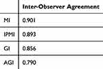
Two trained dental graduates, referred to as Observer 1 and 2, evaluated the images for morphometric parameters. Before the study commenced, 2 observers along with a senior oral radiologist analyzed 10 images to establish consensus on various morphometric parameters. Observer 1 conducted all measurements, while Observer 2 repeated 10% of these on a different day to assess inter-observer reliability. Observer 1 repeated 10% of measurements after 1 month to evaluate intra-observer reliability. The results showed strong inter-observer agreement, with values ranging from 0.75 to 0.90, and intra-observer correlation coefficient of 0.93, indicating excellent reproducibility (Table 1).
 |
Table 1 Showing the Correlation Co-Efficient of Inter-Observer Agreement |
Data and Statistical Analysis
The data was entered in the Microsoft Excel (Redmond, WA, USA) and analysed using IBM-SPSS Version 20. Data was checked for normality using Shapiro–Wilk test and was normally distributed. Mean values of radiomorphometric parameters among the cases and control were analysed using independent t-test and proportions were analysed using chi-square test. A p-value of less than 0.05 was considered statistically significant.
Results
The sample size consisted of panoramic radiographs of 47 patients diagnosed with hypothyroidism and 50 healthy controls. The mean age of hypothyroid cases were 52.06 ± 15.26 years, and 50 healthy controls were 47.30 ± 14.66 years (p value= 0.128). Table 2 shows the age and gender distribution revealing no significant differences in age or sex between the cases and controls.
 |
Table 2 Distribution of Study Participants According to Age and Gender |
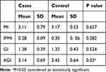
The mean values of MI, PMI and AGI were lower in hypothyroid patients compared to healthy controls. AGI values showed statistically significant differences between cases and controls (p = 0.03) (Table 3).
 |
Table 3 Comparison of Quantitative Radiomorphometric Indices Between Case and Control Group |
The distribution of MCI (mandibular cortical index) at C1, C2 and C3 level in the cases and control groups is shown in Table 4. A significant difference was noted in different mandibular cortical levels between the cases and controls (p=0.001). The significantly higher percentage of cases had MCI at the C1 level as compared to controls.
 |
Table 4 Distribution of the Categorical MCI Data Between Case and Control Groups |
Discussion
Thyroid gland disorders are caused by excessive or insufficient thyroid hormone production.15 Hypothyroidism is one of the most common endocrinopathies, seen 10 times more frequently in females with an overall prevalence of 3.8% to 4.6%.16,17 While some studies have highlighted the adverse effects of hypothyroidism on skeletal structure, others have found no significant increase in fracture risk.9,10,18,19 Study by Bjerkreim et al observed altered bone turnover rate with suppressed TSH levels but concluded that the extent of bone changes varies among patients.20
Radiomorphometric parameters are reliable indicators of BMD. Although bone changes are noted in weight-bearing bones, mandible, especially the posterior region also experiences significant bone resorption. Patients with lower BMD often show reduced MI values, particularly those with osteoporosis and chronic kidney failure.21,22 Our study showed lower values of MI and PMI in hypothyroidism group. In contrast to our results, Yilmaz G et al’s study showed MI and PMI in hypothyroid patients were similar to control group.10 The PMI is also a favourable indicator of BMD. A study by Dagistan and Bilge et al showed significantly lower PMI values in men with osteoporosis.21 Lower PMI was also noted in poorly controlled Type 1 diabetes mellitus patients.23
Several authors have associated osteoporosis with reduction in cortical thickness in mandibular gonial region.24,25 Our study showed higher values for gonial index in hypothyroid group compared to controls. This was not in accordance to results of study by Yilmaz G et al.14 This difference could be due to smaller sample size compared in this study. Several studies have suggested that the AGI is not a reliable measure for identifying undiagnosed low bone mineral density or osteoporosis.26–28 However, Dagistan and Bilge, Abdinian et al, Mahl et al, and Dutra observed AGI was significantly different in patients with osteoporosis than in healthy individuals.21,22,29 We noted a significantly lower values of AGI in patients with hypothyroidism compared to healthy controls.
The MCI developed by Klemetti et al is considered a reliable indicator for BMD determination (16). However, few studies stated that MCI displays negligible changes.30–32 Our study saw majority of hypothyroid patients with C1 cortical index depicting even and sharp endosteal margins without much bone resorption, and the maximum number of patients in the control group had a C2 pattern with minor semilunar defects or lacunar erosions. Overall, in our study, there was evidence of significant difference between the cortical index patterns between case and control groups. Previous study on hypothyroid patients showed no significant changes in MCI between cases and healthy controls.14
Few limitations of the present study were, that we did not consider vitamin D and calcium levels, dietary habits that can alter the mandibular bone density. We could not compare hormonal levels and correlate them with the mandibular bone density. Further studies with larger samples, comparing the dosage of hormone replacement therapy and hormone levels could provide validated data.
Conclusion
This study showed radiomorphometric parameters like MI, PMI, GI to be valuable indicators of bone changes in mandible of patients with systemic diseases like hypothyroidism. Even though lower values of radiomorphometric parameters like mental index (MI), inferior panoramic mandibular index (PMI), gonial index (GI) and antegonial index (AGI) were noted in hypothyroid cases, only AGI showed significant difference between healthy controls and cases. Future multicentric longitudinal studies on a large number of radiographs, applying newer technologies like machine learning, can help us determine whether mandibular morphometric parameters can be a diagnostic tool for predicting changes in bone mineral density.
Institutional Review Board Statement
This research was conducted after obtaining permission from the Kasturba Medical College and Kasturba Hospital Institutional Ethics Committee (IEC: 608/2022, date 01-04-2023). All procedures performed in this study involving human participants were in accordance with the Declaration of Helsinki. Informed consent was taken from all the participants prior to commencement of the study.
Acknowledgment
We are grateful to the Department of Oral and Maxillofacial Radiology, MCODS Manipal for providing us with all the scans and other technical support during the conduct of this study.
Conceptualization, Vathsala Patil, Ananya Nair, Isha Bharadwaj, Komal Smriti and Deepak Singhal; Data curation, Ananya Nair, Isha Bhardwaj, Vathsala Patil, and Komal Smriti; Formal analysis, Deepak Singhal Yogesh Chhaparwal, Shubha Chhaparwal; Investigation, Deepak Singhal; Methodology, Vathsala Patil, Ananya Nair, Isha Bhardwaj, Komal Smriti and Deepak Singhal, Project administration, Vathsala Patil, Ananya Nair, Isha Bhardwaj, Komal Smriti and Deepak Singhal; Software, Supervision, Disha Prabhu, Shubha Chhaparwal; Writing—original draft, Ananya Nair, Isha Bhardwaj, Vathsala Patil, Writing—review and editing, Vathsala Patil, Komal Smriti, Yogesh Chhaparwal and Shubha Chhaparwal.
All authors made a significant contribution to the work reported, whether that is in the conception, study design, execution, acquisition of data, analysis and interpretation, or in all these areas; took part in drafting, revising or critically reviewing the article; gave final approval of the version to be published; have agreed on the journal to which the article has been submitted; and agree to be accountable for all aspects of the work.
Funding
This research received no external funding.
Disclosure
The authors declare that the research was conducted in the absence of any commercial or financial relationships that could be construed as a potential conflict of interest. This paper has been published as a Preprint: https://www.researchsquare.com/article/rs-4696793/v1
References
1. Bassett JD, Williams GR. The molecular actions of thyroid hormone in bone. Trends Endocrinol Metab. 2003;14(8):356–364. doi:10.1016/S1043-2760(03)00144-9
2. Harvey CB, Pj O, Scott AJ, et al. Molecular mechanisms of thyroid hormone effects on bone growth and function. Mol Gene Metabol. 2002;75(1):17–30. doi:10.1006/mgme.2001.3268
3. Stevens DA, Harvey CB, Scott AJ, et al. Thyroid hormone activates fibroblast growth factor receptor-1 in bone. Mol Endocrinol. 2003;17(9):1751–1766. doi:10.1210/me.2003-0137
4. Reddy PA, Harinarayan CV, Sachan A, Suresh V, Rajagopal G. Bone disease in thyrotoxicosis. Indian J Med Res. 2012;135(3):277–286.
5. Lakatos P, Foldes J, Horvath C, et al. Serum interleukin-6 and bone metabolism in patients with thyroid function disorders. J Clin Endocrinol Metab. 1997;82(1):78–81. doi:10.1210/jcem.82.1.3641
6. Vestergaard P, Mosekilde L. Hyperthyroidism, bone mineral, and fracture risk—a meta-analysis. Thyroid. 2003;13(6):585–593. doi:10.1089/10507250332223885
7. Heemstra KA, Hamdy NA, Romijn JA, Smit JW. The effects of thyrotropin-suppressive therapy on bone metabolism in patients with well-differentiated thyroid carcinoma. Thyroid. 2006;16(6):583–591. doi:10.1089/thy.2006.16.5
8. Vestergaard P, Weeke J, Hoeck HC, et al. Fractures in patients with primary idiopathic hypothyroidism. Thyroid. 2000;10(4):335–340. doi:10.1089/thy.2000.10.3
9. Taguchi A. Panoramic radiographs for identifying individuals with undetected osteoporosis. Jpn Dent Sci Rev. 2009;45(2):109–120. doi:10.1016/j.jdsr.2009.05.001
10. Günen-Yılmaz S, Aytekin Z. Evaluation of jaw bone changes in patients with asthma using inhaled corticosteroids with mandibular radiomorphometric indices on dental panoramic radiographs. Med Oral Patologia Oral y Cirugia Bucal. 2023;28(3):e285. doi:10.4317/medoral.25722
11. Nakamoto T, Taguchi A, Ohtsuka M, et al. Dental panoramic radiograph as a tool to detect postmenopausal women with low bone mineral density: untrained general dental practitioners’ diagnostic performance. Osteoporos Int. 2003;14:659–664. doi:10.1007/s00198-003-1419-y
12. Aytekin Z, Yilmaz SG. Evaluation of osseous changes in dental panoramic radiography using radiomorphometric indices in patients with hyperthyroidism. Oral Surg Oral Med Oral Radiol. 2022;133(4):492–499. doi:10.1016/j.oooo.2021.10.011
13. Dhanwal DK. Thyroid disorders and bone mineral metabolism. Indian J Endocrinol Metab. 2011;15(Suppl2):S107–12. doi:10.4103/2230-8210.83339
14. Günen Yilmaz S, Bayrak S. Determination of mandibular bone changes in patients with primary hypothyroidism treated with levothyroxine sodium. Acta Endocrinol. 2023;19(2):201–207. doi:10.4103/2230-8210.83339
15. Ozturk EMA, Artas A. Evaluation of bone mineral changes in panoramic radiographs of Hypothyroid and hyperthyroid patients using fractal dimension analysis. J Clin Densitom. 2024;27(1):101443. doi:10.1016/j.jocd.2023.101443
16. Gulec M, Tassoker M, Erturk M. Evaluation of cortical and trabecular bone structure of the mandible in patients using L-thyroxine. BMC Oral Health. 2023;23(1):886. doi:10.1186/s12903-023-03670-z
17. Zaidi M, Davies TF, Zallone A, et al. Thyroid-stimulating hormone, thyroid hormones, and bone loss. Curr Osteoporos Rep. 2009;7(2):47–52. doi:10.1007/s11914-009-0009-0
18. Gonzalez Rodriguez E, Stuber M, Del Giovane C, et al. Skeletal effects of levothyroxine for subclinical hypothyroidism in older adults: a TRUST randomized trial nested study. J Clin Endocrinol Metab. 2020;105(1):336–343. doi:10.1210/clinem/dgz058
19. Bin-Hong D, Fu-Man D, Yu L, Xu-Ping W, Bing-Feng B. Effects of levothyroxine therapy on bone mineral density and bone turnover markers in premenopausal women with thyroid cancer after thyroidectomy. Endokrynol Pol. 2020;71(1):15–20. doi:10.5603/EP.a2019.0049
20. Bjerkreim BA, Hammerstad SS, Eriksen EF. Bone turnover in relation to thyroid‐stimulating hormone in hypothyroid patients on thyroid hormone substitution therapy. J Thyroid Res. 2022;2022(1):8950546. doi:10.1155/2022/8950546
21. Dagistan S, Bilge OM. Comparison of antegonial index, mental index, panoramic mandibular index and mandibular cortical index values in the panoramic radiographs of normal males and male patients with osteoporosis. Dentomaxillofacial Radiol. 2010;39(5):290–294. doi:10.1259/dmfr/46589325
22. Abdinian M, Salehi MM, Mortazavi M, Salehi H, Kazemi Naeini M. Comparison of dental and skeletal indices between patients under haemodialysis and peritoneal dialysis with healthy individuals in digital panoramic radiography. Dentomaxillofacial Radiol. 2021;50(1):20200108. doi:10.1259/dmfr.20200108
23. Limeira FI, Rebouças PR, Diniz DN, Melo DP, Bento PM. Decrease in mandibular cortical in patients with type 1 diabetes mellitus combined with poor glycemic control. Brazilian Dental j. 2017;28(5):552–558. doi:10.1590/0103-6440201701523
24. Knezović Zlatarić D, Celebić A, Lazić B, et al. Influence of age and gender on radiomorphometric indices of the mandible in removable denture wearers. Coll Antropol. 2002;26:259–266. PMID: 12137308.
25. Knezović-Zlatarić D, Čelebić A. Comparison of mandibular bone density and radiomorphometric indices in wearers of complete or removable partial dentures. Oral Radiology. 2005;21:51–55. doi:10.1007/s11282-005-0030-7
26. Devi BY, Rakesh N, Ravleen N. Diagnostic efficacy of panoramic mandibular index to identify postmenopausal women with low bone mineral densities. J Clin Exp Dent. 2011;3(5):456–461. doi:10.4317/jced.3.e456
27. Padbury AD Jr, TözümTF TM Jr, Taba M, et al. The impact of primary hyperparathyroidism on the oral cavity. J Clin Endocrinol Metab. 2006;91:3439–3445. doi:10.1210/jc.2005-2282
28. Yalcin ED, Avcu N, Uysal S, Arslan U. Evaluation of radiomorphometric indices and bone findings on panoramic images in patients with scleroderma. Oral Surg Oral Med Oral Radiol. 2019;127(1):e23–30. doi:10.1016/j.oooo.2018.08.007
29. Mahl CR, Licks R, Fontanella VR. Comparison of morphometric indices obtained from dental panoramic radiography for identifying individuals with osteoporosis/osteopenia. Radiologia Brasileira. 2008;41:183–187. doi:10.1590/S0100-39842008000300011
30. Gulsahi A, Paksoy CS, Ozden S, Kucuk NO, Cebeci AR, Genc YA. Assessment of bone mineral density in the jaws and its relationship to radiomorphometric indices. Dentomaxillofacial Radiol. 2010;39(5):284–289. doi:10.1259/dmfr/20522657
31. Leite AF, de Souza Figueiredo PT, Guia CM, Melo NS, de Paula AP. Correlations between seven panoramic radiomorphometric indices and bone mineral density in postmenopausal women. Oral Surg Oral Med Oral Pathol Oral Radiol. 2010;109(3):449–456. doi:10.1016/j.tripleo.2009.02.028
32. Unnikrishnan AG, Menon UV. Thyroid disorders in India: an epidemiological perspective. Indian J Endocrinol Metab. 2011;15(Suppl2):S78–81. doi:10.4103/2230-8210.83329
 © 2025 The Author(s). This work is published and licensed by Dove Medical Press Limited. The
full terms of this license are available at https://www.dovepress.com/terms.php
and incorporate the Creative Commons Attribution
- Non Commercial (unported, 3.0) License.
By accessing the work you hereby accept the Terms. Non-commercial uses of the work are permitted
without any further permission from Dove Medical Press Limited, provided the work is properly
attributed. For permission for commercial use of this work, please see paragraphs 4.2 and 5 of our Terms.
Recommended articles
Association Between Hemoglobin Levels and Osteoporosis in Chinese Patients with Type 2 Diabetes Mellitus: A Cross-Sectional Study
Ye T, Lu L, Guo L, Liang M
Diabetes, Metabolic Syndrome and Obesity 2022, 15:2803-2811
Published Date: 14 September 2022
Associations of Obesity Indices with Bone Mineral Densities and Risk of Osteoporosis Stratified Across Diabetic Vascular Disease in T2DM Patients
Zheng S, Zhou J, Wang K, Wang X, Li Z, Chen N
Diabetes, Metabolic Syndrome and Obesity 2022, 15:3459-3468
Published Date: 3 November 2022
Clinical Utility of Romosozumab in the Management of Osteoporosis: Focus on Patient Selection and Perspectives
Lim SY, Bolster MB
International Journal of Women's Health 2022, 14:1733-1747
Published Date: 15 December 2022
The Effects of Switching from Dipeptidyl Peptidase-4 Inhibitors to Glucagon-Like Peptide-1 Receptor Agonists on Bone Mineral Density in Diabetic Patients
Huang CF, Mao TY, Hwang SJ
Diabetes, Metabolic Syndrome and Obesity 2023, 16:31-36
Published Date: 11 January 2023
The Relationship Between Serum 25-Hydroxyvitamin D Levels and Osteoporosis in Postmenopausal Women
Wang D, Yang Y
Clinical Interventions in Aging 2023, 18:619-627
Published Date: 18 April 2023

