")
Back to Journals » International Journal of Nanomedicine » Volume 20
Recent Advances in Aptamers-Based Nanosystems for Diagnosis and Therapy of Cardiovascular Diseases: An Updated Review
Received 21 November 2024
Accepted for publication 7 February 2025
Published 25 February 2025 Volume 2025:20 Pages 2427—2443
DOI https://doi.org/10.2147/IJN.S507715
Checked for plagiarism Yes
Review by Single anonymous peer review
Peer reviewer comments 3
Editor who approved publication: Professor Dong Wang
Hongqin Yu, Jie Yu, Gang Yao
Department of Cardiovascular Medicine, Yantai Mountain Hospital, Yantai, 264000, People’s Republic of China
Correspondence: Gang Yao, Department of Cardiovascular Medicine, Yantai Mountain Hospital, Yantai, 264000, People’s Republic of China, Email [email protected]
Abstract: The increasing global prevalence of cardiovascular diseases highlights the urgent need for innovative diagnostic and therapeutic strategies. Aptamers, small single-stranded nucleic acid molecules with exceptional specificity and affinity for target biomolecules, have emerged as promising tools for precise diagnostics and targeted therapies. Their selective binding capabilities provide valuable insights into the molecular mechanisms underlying cardiovascular conditions. When integrated into nanosystems, aptamers enhance the delivery, bioavailability, and stability of diagnostic and therapeutic agents, addressing challenges of solubility and degradation. This integration enables more targeted drug delivery, advanced imaging techniques, and improved therapeutic interventions, ultimately improving the management of cardiovascular diseases. Recent advancements in aptamer selection methodologies, coupled with their unique three-dimensional structures, have significantly expanded their application potential in cardiovascular health. By combining aptamers with nanosystems, novel approaches to cardiovascular disease diagnosis and treatment are emerging, promising enhanced efficacy, safety, and precision. This review explores recent progress in the development and application of aptamer-based nanosystems in cardiovascular diagnostics and therapies.
Keywords: aptamers, nanosystems, cardiovascular diseases, diagnosis, treatment
Introduction
Cardiovascular diseases are a major global cause of morbidity and mortality, requiring new diagnostic and therapeutic approaches. Traditional diagnostic techniques for cardiovascular diseases (CVDs), biomarker assays and imaging, face sensitivity, specificity, and real-time monitoring challenges. Nanosystems, including nanoparticles, liposomes, quantum dots, and magnetic nanoparticles, offer solutions by enhancing diagnostic accuracy, targeting precision, and enabling real-time monitoring.1 Aptamers, small single-stranded DNA or RNA molecules, offer high specificity and affinity for targets, presenting significant potential for targeted diagnostic and therapeutic strategies in cardiovascular care. When coupled with nanosystems, aptamers enhance detection capabilities, allowing for precise interactions with CVD biomarkers.2
Aptamers are commonly conjugated with gold nanoparticles for optical detection, quantum dots for fluorescence imaging, and magnetic nanoparticles for MRI, providing improved visualization of cardiovascular abnormalities. Additionally, aptamer-functionalized liposomes and nanocarriers improve the delivery and bioavailability of diagnostic agents, facilitating more effective detection of biomarkers like troponins, myoglobins, and C-reactive proteins.3 Integrating aptamers with nanosystems improves diagnostic performance by enhancing sensitivity, lowering detection limits, and enabling real-time monitoring.4 This review explores recent advancements in aptamer-based nanosystems for CVD diagnosis, highlighting their potential to revolutionize diagnostic platforms.
Cardiovascular Diseases (CVDs) Pathophysiology
CVDs represent a major global health concern, with atherosclerosis being a key contributor to their pathophysiology. The accumulation of plaques within arterial walls leads to arterial stiffening and narrowing, significantly influencing conditions like stroke, coronary artery disease, and peripheral vascular disorders. A comprehensive understanding of the molecular mechanisms underlying atherosclerosis is essential for the development of targeted therapeutic strategies aimed at mitigating its detrimental effects on cardiovascular health.2,5 Aptamers, with their high specificity for disease-related biomarkers, offer significant potential in the diagnosis and treatment of CVDs, including atherosclerosis. Integrated into nanosystems, aptamers enable targeted drug delivery and precise diagnostics, facilitating early detection and more effective treatment strategies. This targeted approach enhances personalized care in CVD management, improving therapeutic outcomes while minimizing side effects.6–8
Vascular disorders, characterized by endothelial dysfunction and blood vessel abnormalities, pose a significant challenge to cardiovascular health. Aptamers, as molecular recognition elements, can be engineered to selectively bind to biomarkers associated with vascular pathology. When incorporated into nanosystems, these aptamers enable targeted delivery of therapeutic agents or imaging compounds, facilitating personalized treatment strategies for vascular diseases.9–11
Autoimmune cardiomyopathies, where the immune system targets cardiac tissues, introduce an immunological component to cardiovascular diseases. Aptamers, capable of modulating immune responses, can be integrated into nanosystems to address the immune-driven aspects of these conditions. This approach enables the development of targeted therapies that reduce cardiac tissue damage by specifically targeting the autoimmune components of the disease.12,13 The integration of aptamer-based nanosystems offers a focused, comprehensive approach to cardiovascular health by addressing various aspects of cardiovascular diseases.14 The precision of aptamers, combined with the versatility of nanosystems, provides a platform for early diagnosis and personalized treatment.14–16 As research progresses, this combination is expected to revolutionize cardiovascular medicine, enhancing patient outcomes and quality of life.
Advances in Aptamer-Based Nanosystems
Nanomaterial-based technologies enhance bioanalysis and biomedicine by enabling early disease detection and improving drug and gene delivery. Designed for specific sub-cellular interactions, these technologies have the potential to optimize targeted treatments, improving efficacy and minimizing side effects in clinical applications.17–19
By combining the inherent properties of nanomaterials with the selective recognition capabilities of aptamers, various effective nanomaterial-aptamer complexes have been developed for diverse applications (Figure 1). Aptamers are synthesized using combinatorial oligonucleotide libraries, comprising sequences in the range of 10^12 to 10^15, and are chemically produced through the in vitro systematic evolution of ligands by exponential enrichment (SELEX).20 Following 6 to 18 rounds of selection, populations containing more than 10^13 distinct sequences are meticulously screened, enabling the isolation of nucleic acid species that exhibit high specificity for the intended target.21
 |
Figure 1 Examples of drug delivery systems that employ aptamers as targeting agents. The figure illustrates various strategies, including aptamer-conjugated nanoparticles, aptamer-functionalized liposomes, and aptamer-based delivery complexes, highlighting their potential in improving the precision and efficacy of drug delivery. Reproduced from Gao F, Yin J, Chen Y, Guo C, Hu H, Su J. Recent advances in aptamer-based targeted drug delivery systems for cancer therapy. Front Bioeng Biotechnol. 2022 Aug 16;10:972933. Copyright © 2022 Gao, Yin, Chen, Guo, Hu and Su. Creative Commons Attribution License (CC BY).22 |
Aptamers demonstrate significant versatility, particularly in two key areas. First, their precise binding capabilities position them as promising tools for biomedical applications, such as drug delivery and the development of novel therapeutic systems. A notable example is Macugen, an FDA-approved aptamer targeting vascular endothelial growth factor for the treatment of age-related macular degeneration, which is already in clinical use. Second, aptamers advancement of biosensors. Once a specific aptamer sequence is identified, it can be produced cost-effectively, with high purity. Unlike antibodies, aptamers can be chemically modified with various tags, facilitating their tracking, manipulation, and immobilization on a range of solid substrates.23–25 Aptamer-based nanosystems offer significant advantages in targeted therapies and biosensors, improving precision and efficacy; however, challenges such as stability, delivery efficiency, and potential immunogenicity need to be addressed for broader clinical application.14
Clinical Integration of Aptamer-Based Nanosystems
Nanotechnology has transformative potential across various industries, including electronics, materials science, and healthcare.26 Aptamer-based nanosystems, due to their unique properties and high specificity, offer significant potential in applications as biosensing and targeted drug delivery. Aptamers exhibits strong affinity for specific targets, including proteins, small molecules, or living cells.27,28
Integrating aptamers into nanosystems opens numerous opportunities across various fields like applications in biotechnology, environmental monitoring, and medicine.29 These nanosystems offer significant advancements in biotechnology, enabling the development of research tools and therapeutics30 and are valuable in gene regulation, protein engineering, and the design of targeted therapies due to their ability to selectively target specific molecules.31–34 Targeted drug delivery through aptamer-based nanosystems represents a promising advancement in addressing the limitations of conventional chemotherapy, which often leads to non-specificity and collateral damage to healthy tissues. By encapsulating therapeutic agents in nanoparticles functionalized with aptamers that specifically bind to surface markers on cancer cells, this approach minimizes adverse effects while enhancing the therapeutic efficacy of the treatment.22,34–38
Aptamer-based nanosystems, coupled with signal-amplifying components, enable the development of sensitive biosensors capable of detecting pathogens, contaminants, and disease biomarkers, offering rapid and accurate diagnostics (Figure 2) for early disease detection.39–45 This approach allows for tailored therapeutic interventions, potentially reducing adverse effects and improving treatment outcomes.46–50
 |
Figure 2 Nanotheranostic strategies in cardiovascular diseases. Nanoparticles as liposomes, dendrimers, and metallic nanoparticles, are engineered to simultaneously deliver therapeutic agents and provide real-time diagnostic imaging capabilities (MRI, PET, fluorescence imaging). These systems enable targeted treatment of cardiovascular tissues, improved disease monitoring, and early detection of pathologies, thereby enhancing both therapeutic efficacy and diagnostic accuracy in CVD management. Reproduced from Ramchandani M, Kumari P, Goyal AK. Aptamers as theranostics in cardiovascular diseases. J Nanotheranostics. 2023;4(3):408–428. Copyright: © 2023 by the authors. Licensee MDPI, Basel, Switzerland. This article is an open access article distributed under the terms and conditions of the Creative Commons Attribution (CC BY) license (https://creativecommons.org/licenses/by/4.0/).51 |
Aptamer-based nanosystems have valuable applications in environmental monitoring, enabling sensitive and targeted detection of pathogens, pollutants, and toxins in air and water. These systems enhance the speed and accuracy of toxin detection, contributing to improved environmental health and safety.52–56 Aptamers are a flexible tool with a multitude of applications since researchers may build them to bind to a wide range of targets. To improve their functionality and enable uses like medication delivery, imaging, and theranostics (combined therapy and diagnostics), they can also be functionalized with different nanoparticles.57,58 Aptamer-based nanosystems hold significant potential in medicine and environmental monitoring, offering targeted drug delivery, sensitive biosensing, and personalized therapies.59 However, challenges related to stability, scalability, and delivery optimization must be addressed for broader clinical application.
Diagnostic Applications in Cardiovascular Disease
Aptamer-based nanosystems are increasingly utilized for the diagnosis, prognosis, and risk assessment of CVDs, enabling the identification and detection of specific biomarkers60 associated with these conditions. The flexibility of aptamer design compensates for the drawbacks in their widely used counterparts, antibodies. Typically, antibodies, as a diagnostic tool, do offer greater sensitivity and selectivity towards target molecules. Nevertheless, their high production cost and variations in batches pose a limitation to the experiment.61,62 This has resulted in inaccurate test outcomes, contributing to the widely recognized “reproducibility crisis”.63 Despite efforts to reduce their immunogenicity, the administration of antibodies carries a high risk of triggering immune responses, thereby limiting their clinical application.64 Aptamers, on the other hand, present a useful alternative with low manufacturing cost, and their synthetic nature yields high reproducibility, with reduced batch variations, granting an easy means for large-scale production.65,66 Additionally, aptamers offer high level of customizability, with the help of SELEX, to generate aptamers with high affinity, especially when targeting homologous molecules.67,68 With the advancements in technological fields, recent research suggests a systematic way for isolation and production of aptamers with “programmable binding affinities”,69 which paves way for improved selectivity of aptamers over antibodies. The ability to control aptamer specificity allows for the neutralization of interfering analytes and modulation of inflammatory immune responses, as demonstrated in antibody cross-reactivity with homologous targets.70 This cross-reactivity has been identified to be caused by the complementarity-determining regions intrinsically present in antibodies. Their absence in aptamers automatically makes them safer for use than antibodies.71,72 The small size and low toxicity of aptamers even when conjugated with metallic nanoparticles makes them a more suitable alternative than antibodies for use in experimental and clinical trials.73–76 The development of SOMAmers (slow off-rate modified aptamers) enhances aptamer target diversity by effectively binding hydrophobic analytes.77,78 The nucleic acid aptamers are coupled with hydrophobic moieties allowing SOMAmers to show affinity for such chemical compounds.79
Aptamer-based nanosystems present a promising alternative to antibodies for cardiovascular disease diagnosis, offering cost-effectiveness, high reproducibility, and customizable specificity.80 Their reduced immunogenicity, small size, and ability to bind diverse targets enhance their potential for clinical use, though challenges in delivery optimization persist.81
Biomarker Detection via Aptamers
Biomarkers have gained prominence as critical indicators for early diagnosis and prognostication of CVDs. A diverse array of biomolecular entities, including metabolites, nucleic acids (DNA and RNA), proteins, and hormones, serve as biomarkers. Alterations in the concentration of these molecules, relative to baseline levels, can provide insights into the presence and stage of disease.82 Ongoing research focuses on identifying diverse biomarkers and developing specific aptamers for their detection. The demand for aptamer-based biosensors, or aptasensors, is growing rapidly, as clinicians and researchers seek diagnostic tools that offer broad target specificity. These tools are not only capable of detecting biomolecules like peptides and proteins but also cells and metal ions, addressing the need for accurate and reliable results across various applications.83,84
Aptamers show considerable promise in biomarker identification for CVDs, offering high specificity and reliability for detecting various biomolecules. Their integration into aptasensors enhances early detection and diagnostic accuracy in clinical applications.85,86 This review highlights key CVD biomarkers and their corresponding aptamers or aptasensors, including troponins, myoglobins, and C-reactive proteins (Table 1 and 2).
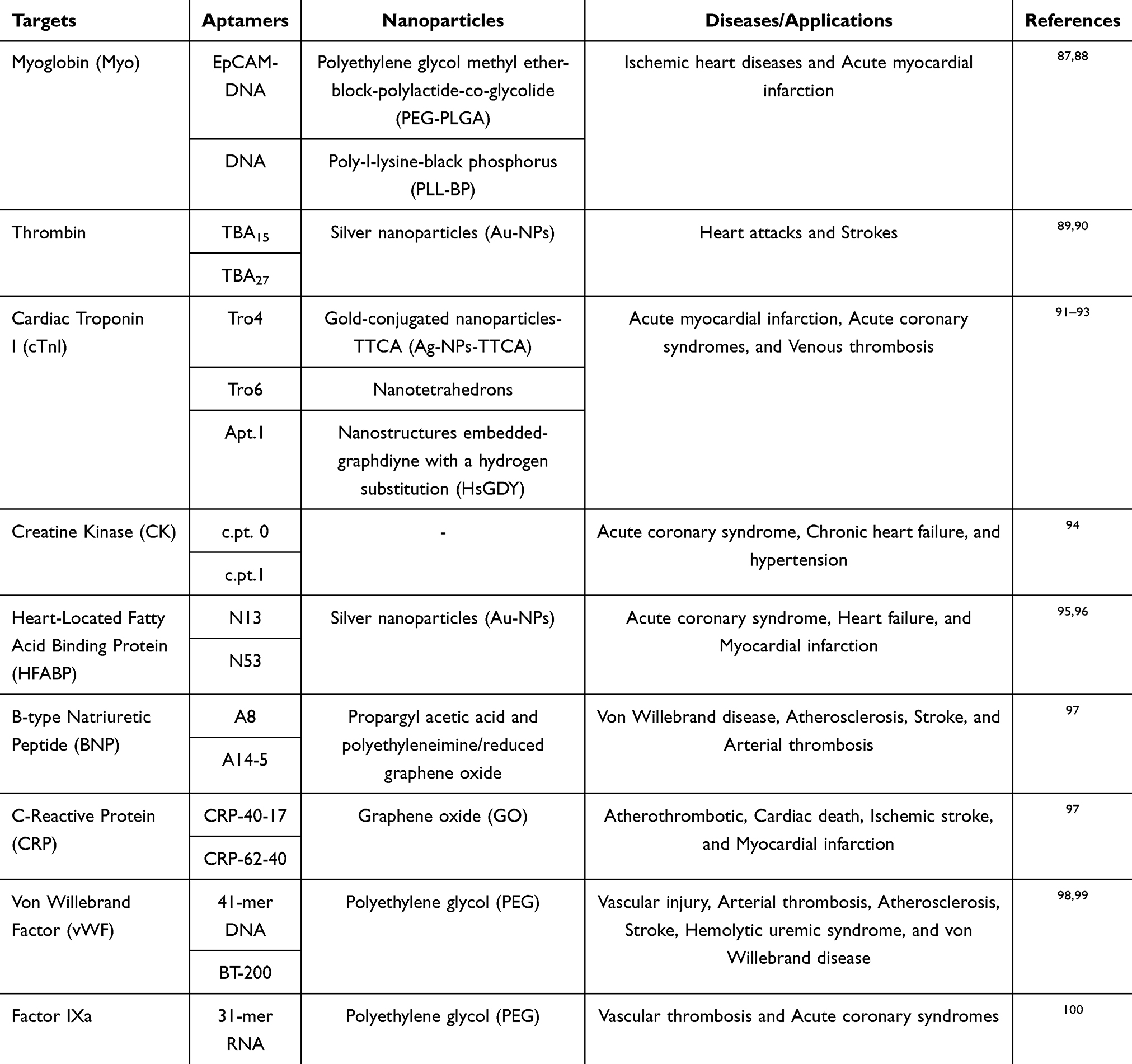 |
Table 1 Applications of Aptamer-Based Nanosystems in the Treatment of CVDs |
 |
Table 2 Applications of Aptamer-Based Nanosystems in the Diagnosis of CVDs |
Troponin as Biomarker
The development of immunological assays for detecting organ-specific protein markers has made cardiac troponin a crucial biomarker for the identification and prediction of acute myocardial infarction.106–109 Troponin T, troponin I, and troponin C, are the three globular proteins that form the Troponin Complex found associated with the regulation of muscle contraction.110 The cardiac troponin I and T (cTnI and cTnT) are the most routinely targeted isoforms due to unique distinction from their complementary skeleton muscle protein isoforms,111,112 thus are referred to as the “gold standard” for AMI detection.113 Damage to myocardial tissues results in the release of troponin into the bloodstream, consequently raising the blood-protein concentration within 4–6 hours, and remaining elevated for the next several days.85
Despite the widespread use of antibody-based ELISA for detecting cardiac troponin I (cTnI), its limit of detection (LOD), typically around 1–10 ng/mL, restricts its ability to accurately detect low concentrations of cTnI.114 The endeavor to produce a more sensitive and responsive detection protocol has yielded an assortment of DNA aptamers that target cTnI. In 2015, 79 bp ssDNA random libraries were screened to find a potential cTnI target. The outcome of this screening produced several favorable aptamer sequences, namely, TnIApt23, TnIApt19, TnIApt18, TnIApt11. Upon further testing, it was found that TnIApt23 had the highest affinity in the nanomolar range (2.69 nM) towards cTnI protein.115 Additionally, a rapid colorimetric assay was developed using new and specific aptamer-AuNanoParticles conjugates, for the detection of Human Cardiac Troponin I. This colorimetric assay was basses on dot blot assay and tolerated an LOD of 5 ng mL−1. Another study selected 2 high-affinity ssDNA aptamers, Apt 3 and Apt 6) and developed a dual-aptamer sandwich Enzyme-Linked Oligonucleotide Assay Method (ELONA) for detection of cTnI. The LOD offered by dual-aptamer ELONAwas 0.05 ng mL−1.116 Current research groups are investigating the possibility developing new versions of aptasensors for cTnI by engineering anti-cTnI aptamers.117 A cTnI aptamer-immobilized screen-printed carbon electrode was developed to achieve highly sensitive cTnI detection, with a LOD of 1.0 pM (24 pg/mL). Further improving sensitivity, flat Au-based nanoplates functionalized with anti-cTnI aptamers enabled detection of cTnI at concentrations as low as 2.4 pg/mL. These advancements demonstrate the potential of cardiac troponin aptamers as a reliable method for early detection of AMI.118
Aptamers targeting cardiac troponin enhance AMI detection sensitivity and precision, offering improved detection limits compared to conventional antibody-based methods.119 However, challenges like limited stability in complex biological environments and potential issues with reproducibility and large-scale production need to be addressed for broader clinical use.119
Myoglobin in Cardiovascular Diagnostics
A monomeric hemoprotein, myoglobin (Mb) is a single chain polypeptide of around 153 amino-acid molecules, widely known for its heam group consist of porphyrin ring with central ferrous iron molecule.120 Myoglobin is low molecular weight protein, and one of the first cardiac biomarkers released into the bloodstream following cardiac tissue damage. Basal blood concentrations of Mb range from 6 to 85 ng/mL, but within two hours of AMI, levels rise to 70–200 ng/mL, establishing Mb as a valuable marker for AMI detection.121,122
Studies to develop a probe for an electrochemical biosensor became fruitful in 2014, when Wang et al were able to successfully generate an anti-Mb aptamer.123 This opened doors for the detection of Mb, as a biomarker of AMI, in a target-induced aptamer displacement assay.124 Continuation of this investigation lead to the creation of an antibody-MB-aptamer sandwich assay where the aptamer/Mb and antibody/Mb possess different binding interactions resulting in different binding affinities. This further lead to the development of a polystyrene microplate which was coated with commercially produced anti-Mb antibodies. The commercially synthesized anti-Mb antibodies were used as capture agents alongside an invertase-aptamer conjugate, used as a secondary probe, to enhance the indicating signals. Hence, an antibody-Mb-aptamer sandwich assay was invented as a means to observe and keep track of AMI.125 In recent studies a 72 nucleotide (nt) DNA-based molecule, which represents Mb, it being synthesized to develop a differential pulse voltammetry-based electrochemical sensor having an LOD of 2.1 pg mL−1.126 Aptamers targeting myoglobin have significant potential for detection of AMI, offering improved sensitivity and specificity compared to conventional methods. However, further optimization of their stability, sensitivity, and integration into practical diagnostic platforms is needed for broader clinical application.127
C-Reactive Protein in Cardiovascular Disease
Produced mainly in the liver, C-reactive protein (CRP) is a 125 kDa pentameric protein synthesized as a result of inflammation, infection or damage to cells of different organs, including the heart.128 Therefore, CRP levels are known to reflect the magnitude of inflammation, despite CRP being a relatively non-specific protein. In normal conditions, CRP levels are noted to be about <10 μg mL−1 in healthy individuals, whereas in the case of AMI, CRP concentration can increase to about 200 μg mL−1.129 CRP is used frequently in clinical diagnosis as a biomarker of AMI, as such, there are a CRP targeting aptamers have been profusely studied and examined.130 One prominent example if the successful design and development of 2 RNA aptamers, CRP-80-17, and a truncated CRP-40-17, by using the graphene oxide-SELEX method.131 OTC Biotech has also played its part in generating aptamers for developing biosensors. One noted example is a CRP DNA aptamer of 20 nt, which was later used in the signal recognition particle-based detection of CRP using an antibody-aptamer sandwich that allows a 5 fg mL−1 LOD.132
Although aptamers have provided a new frontier of clinical diagnosis and their results in trials have been very promising, aptamer-based diagnostic systems currently require more detailed and extensive research before they can be used on a broader level, as in the case of antibody-based ELISA testing kits.
Aptamers present several compelling advantages over conventional diagnostic approaches, such as antibody-based assays (ELISA, Western blot) and molecular probes. Through the SELEX (Systematic Evolution of Ligands by Exponential Enrichment) process, aptamers are selected for their high specificity and affinity for target biomolecules, often exceeding the performance of antibodies in terms of binding strength, with affinities in the picomolar to nanomolar range.133 This high affinity results in enhanced sensitivity and lower detection limits. In contrast to antibodies, which are susceptible to instability and denaturation, aptamers exhibit remarkable thermal and chemical stability, retaining their binding capacity across a broad range of temperatures and pH values. This makes them well-suited for use in demanding conditions and for long-term storage without loss of efficacy.
Moreover, aptamers are cost-effective and scalable due to their ability to be synthesized through automated solid-phase chemical synthesis. This process bypasses the need for animal immunization, offering a more reproducible and economically viable method for large-scale production compared to monoclonal antibodies. The flexibility and versatility of aptamers further enhance their utility, as they can be easily conjugated to various nanoparticles or incorporated into diverse detection platforms, such as surface plasmon resonance (SPR), electrochemical sensors, fluorescence resonance energy transfer (FRET), and lateral flow assays. This versatility facilitates the development of multifunctional biosensors with superior sensitivity and selectivity. Additionally, aptamers are non-toxic and biocompatible, making them ideal for both in vitro and in vivo applications, particularly in diagnostic imaging and targeted therapeutic delivery, where safety is a critical consideration. Finally, aptamers enable rapid, real-time monitoring and label-free detection, offering rapid response times due to their small size and conformational changes upon target binding, which is essential for point-of-care diagnostics.134–136
Therapeutic Applications of Aptamer-Based Nanosystems
Aptamers have shown significant promise in cardiovascular therapeutics, particularly due to their high affinity for specific target molecules. Their potential as antithrombotic and anticoagulant agents is a key area of research in combating CVDs. Ongoing clinical trials are assessing aptamers targeting critical components of the coagulation cascade, including Von Willebrand Factor (vWF), thrombin, and Factor IX, which play essential roles in platelet activation and clot formation. These targets are implicated in conditions like AMI, coronary artery disease (CAD), acute coronary syndrome (ACS), and peripheral vascular disease.137
In order to enable targeted transport, aptamers form conjugates with oligonucleotides, medications, and nanoparticles. The idea that aptamers function as escort molecules was first put forth by Hicke et al in 2000.138 Following this, a great deal of research has been done on their use in cancer treatment, including immunotherapy and chemotherapy. The circulatory system can be exploited to provide therapeutic drugs, a minimally invasive technique that has garnered substantial interest in the cardiovascular realm despite the paucity of research on cardiovascular illnesses. However, the restricted vascular density or permeability hinders this approach’s effectiveness. To maintain physiological homeostasis, immune cells such splenic monocytes are mobilized within the microenvironment.139 Inspired by this phenomenon, a plethora of studies have documented the application of cell-carrying nanoparticles as dynamic targeting techniques for the heart. Lipid nanoparticles (LNPs) containing IOX2, which is an inhibitor of hypoxia-inducible factor (HIF)-1α prolyl hydroxylase-2, have the ability to cling to the outermost layers of monocytes and then dissipate at the sites of injury, reducing the effects of ischemia-reperfusion (IR) injury on the heart.140 A mimetic peptide (MP) was created by imitating an amino acid sequence found in the Cavβ2 cytosolic chaperone’s C-terminal tail. This MP helps restore Cavα1.2 protein density at the plasma membrane by specifically targeting the Cavβ2 globular domain’s Tail Interacting Domain (TID). This is especially important when there are aberrant L-type calcium channel (LTCC) densities in the heart. That being said, it is still unclear how precisely the MP is delivered to heart cells.141 Cu (I)-mediated click chemistry was used to connect the mimetic peptide (MP) with Gint4.T within a novel aptamer-peptide chimera.142 With its resilience to nucleases and single-stranded RNA (ssRNA) structure, gint4.T shows promise in translation by focusing on the platelet-derived growth factor receptor-β (PDGFRβ) and obstructing angiogenesis, migration, and cell proliferation.143 The aptamer-peptide conjugates that were developed enable the mimetic peptide (MP) to be internalized into cardiomyocytes, indicating that the MP is effective in returning the cardiac milieu to normal, which is typified by abnormal intracellular calcium fluxes that rely on L-type calcium channels (LTCC). Within ventricular cardiomyocytes, the sarcoplasmic reticulum Ca2+-ATPase 2a (SERCA2a)-phospholamban (PLN) system plays a major role in regulating intracellular calcium cycling. RNA-Apt30, a phosphorothioate-modified backbone PLN-specific aptamer, was reduced to a 30-nucleotide oligomer after being obtained by SELEX from a random RNA library. The delivery of RNA-Apt30 into adult rat cardiomyocytes is made possible by its conjugation to the cell-penetrating TAT peptide, which is aided by a persistent thiol-maleimide bond. The compound improves cardiac calcium transients and contractility inside these cells.144 Aptamers have been shown to be highly effective in inducing re-endothelialization. They are able to capture endothelial progenitor cells (EPCs), which they have documented and deployed on stent surfaces. This enhancement helps to improve the grafts’ hemocompatibility. Moreover, heparin was designed to stick to the dopamine/polyethyleneimine coating on the stent surface in order to function as an anticoagulant. In turn, this film was electrostatically bound with EPC-capturing aptamers, greatly improving the hemocompatibility of vascular implants.145
Since nanoparticles have unique properties that allow them to be precisely tuned to fulfill therapeutic requirements, nanomedicine has become a highly researched approach to treating cardiovascular disorders.146 The size, shape, composition, surface charge, and flexibility of nanoparticles, when combined with their significant payload and release capabilities, enable them to outperform existing approaches in terms of solubility, diffusivity, half-life, drug release, and biodistribution profiles. As a result, the development of a suitable drug delivery system results in therapies based on nanoparticles that have better and longer-lasting therapeutic efficacy with fewer side effects.147 Aptamers, with their sensor and payload capabilities, are used as in vivo contrast agents in nanoparticle-based systems for diagnostics. Nanoparticles enhance biomaterial functionality in cardiac patches, vascular grafts, and stents, and aid in ischemia-reperfusion treatment. Immune system modulation, targeting specific cell subsets, is a promising approach for CVD therapy, improving efficacy, reducing drug dosage, and minimizing risks of infection and sepsis.148 With regard to aptamer research, the field of cancer has seen greater nanomedicine research than CVDs. There are a number of reasons for this disparity, including funding for research and the excitement and energy created by early clinical trials. This tendency has been notably influenced by the successful regulatory approval of cancer monotherapies like Abraxane and Doxil. It is imperative to acknowledge that the knowledge gained in the field of oncological nanotechnology extends beyond its immediate use. Instead, because the pathophysiology of inflammatory diseases in these two medical domains is similar, the knowledge gained may inspire novel therapeutic approaches in CVDs.148 Additionally, the European Medicines Agency and Food and Drug Administration’s recent approvals indicate the market potential and contemporary significance of nanomedicines in the treatment of cardiovascular illnesses. Regulatory permission has been granted for the first nano-drug, Leqvio (inclisiran), which is intended to treat atherosclerotic cardiovascular diseases. Leqvio works by preventing the liver’s translation of the PCSK9 protein, which lowers levels of low-density lipoprotein cholesterol in the blood and lessens the risk of CVDs.2
Targeting Thrombin in Cardiovascular Therapy
A study demonstrated the effectiveness of thrombin-binding aptamer-conjugated gold nanoparticles (TBA-Au NPs) in regulating thrombin activity related to fibrinogen. Due to their cost-effectiveness and ease of fabrication, TBA29-Au NPs and cTBA29-Au NPs offer promising prospects for treating coagulation disorders. These findings suggest that aptamer-functionalized nanomaterials can effectively control molecular binding, protein recognition, and enzyme activity in biomedical applications.149 Nanoparticle-based delivery of TBA improves its pharmacokinetics, nuclease resistance, and enables multivalency by incorporating multiple TBA copies onto a single scaffold. Fluorescent labeling facilitates tracking and ensures proper folding, enhancing bioactivity and stability. Tris-mTBA, conjugated to Sicastar nanoparticles, effectively and reversibly inhibits thrombin activity toward fibrinogen, demonstrating potential for regulating blood coagulation in biomedical applications.150 The enhanced permeability and retention (EPR) effect, commonly utilized in nanomedicine for anti-tumor therapies, results from the abnormal, permeable vasculature in areas of injury or inflammation, particularly in tumors with disorganized, leaky capillaries. However, in the heart, the EPR effect is less pronounced, even following myocardial infarction, posing challenges for nanoparticle-based drug delivery in cardiac applications.151 Just 0.2% of injected fluorescent-labeled PEG-modified nanoparticles were able to reach the infarcted zone, despite the general consensus that ischemia-reperfusion after myocardial infarction causes the release of cytokines, which in turn increases local vascular permeability and inflammation.152
Nanomedicine provides an innovative approach for targeted nanoparticle accumulation through non-traditional delivery methods. A study using biocompatible and biodegradable calcium phosphate-based nanoparticles to deliver microRNA to the heart demonstrated specific targeting of cardiomyocytes, with minimal accumulation in other cardiac cells or organs.153 The biotech startup NanoPhoria is now investing in the development of technologies that will allow biologics to be delivered directly into cardiomyocytes. The realization of the benefits biomimetic nanoparticles provide over traditional viral vectors is what is driving this research.154 Active targeting enhances drug delivery via nanoparticles in cardiovascular diseases, surpassing passive uptake, with aptamers serving as key ligands for tissue or cell specific absorption. A notable example is the work of Patrick Hsieh’s group, where aptamers were functionalized on monocytes to direct nanocarriers precisely to the infarction site, improving targeted delivery.151
While the proposed formulation requires further refinement for practical application, the chosen aptamer achieved a 5% delivery rate of injected nanoparticles to the infarct site, a significant improvement over EPR-based targeting alone. However, research on using aptamers for drug delivery via nanoparticles remains limited, highlighting the need for more comprehensive studies in this area. Aptamer-conjugated nanoparticles, like TBA-Au NPs, show promise in modulating thrombin activity and advancing drug delivery in cardiovascular applications. However, optimizing delivery mechanisms and improving targeting efficiency remain key challenges, necessitating further research for clinical translation.155
Nanotheranostics in Cardiovascular Treatment
The ability of nanocarriers to aggregate and target at certain locations makes it possible to deliver therapeutic and imaging agents at the same time. These agents can be cardiovascular medications labeled with radionuclides or fluorophores, which provide data on the location of the disease, the kinetics of drug release, and the effectiveness of the treatment (Figure 3).
 |
Figure 3 This figure depicts the application of aptamers as targeted therapeutic and diagnostic agents in CVDs. Aptamers, selected for their high specificity and affinity for biomolecular targets, are engineered to bind to specific molecules involved in cardiovascular pathologies, such as endothelial markers, inflammatory cytokines, or platelet receptors. Reproduced from Di Mauro V, Lauta FC, Modica J, et al. Diagnostic and therapeutic aptamers: a promising pathway to improved cardiovascular disease management. Basic Transl Sci. 2024;9(2):260–277. © 2024 The Authors. Published by Elsevier on behalf of the American College of Cardiology Foundation. Creative Commons CC-BY-NC-ND.2 |
Aptamers exhibit high binding affinity and selectivity for a wide range of targets, including large proteins, ions, small molecules, and living cells. When engineered to target key molecules in the coagulation cascade, factor Xa or thrombin, aptamers provide a more precise and potentially safer alternative to conventional anticoagulants.51 Tailored aptamers targeting specific receptors on endothelial cells can enhance blood flow and induce vasodilation in conditions like hypertension and atherosclerosis. This targeted approach holds significant potential for alleviating symptoms and may reduce the need for invasive procedures such as bypass surgery or angioplasty.156,157
Challenges in Aptamer-Based Nanosystems
Optimizing aptamer selection and functionalization for particular cardiovascular targets is a major challenge. The goal of ongoing research is to optimize the selection procedures in order to increase aptamer specificity and affinity for cardiovascular targets.158 Moreover, stability, pharmacokinetics, and biodistribution present difficulties in the transition of aptamer-based nanosystems from lab to bedside. Many obstacles, including enzymatic degradation and quick clearance, can jeopardize the stability and effectiveness of aptamer-based nanosystems in the in vivo setting. Approaches to improve the stability and extend the half-life of aptamer-functionalized nanoparticles are being researched. The study investigates strategies like surface modifications, encapsulation, and bioconjugation to address these obstacles and enhance the overall pharmacokinetic profile.159
Important factors to take into account for their clinical translation include the immunological response to aptamers and the possibility of off-target effects. Thorough preclinical research is necessary to assess these nanosystems’ safety and biocompatibility. A range of modifications have been investigated in recent research to reduce immunogenicity and improve the overall safety profile of nanosystems based on aptamers.159–161 Nevertheless, there are issues with aptamer production’s scalability and affordability for extensive clinical uses. To fulfill the needs of clinical-scale production, it is imperative to streamline the aptamer synthesis and purification processes. Novel strategies, like cell-based expression systems and enzymatic synthesis, are being investigated to overcome these obstacles.162,163
Conclusion and Future Perspectives
The integration of aptamers and nanosystems is driving significant advancements in the diagnosis and treatment of cardiovascular diseases. These diseases, encompassing conditions such as vascular diseases, autoimmune cardiomyopathies, and atherosclerosis, demand precise and tailored therapeutic approaches. Aptamer-based nanosystems leverage the specificity and affinity of aptamers to target disease-specific biomarkers, troponins, myoglobins, and C-reactive proteins, enabling more accurate diagnostic imaging and enhanced therapeutic targeting. By conjugating aptamers to nanoparticles like gold nanoparticles, quantum dots, and magnetic nanoparticles, these systems improve biomarker detection sensitivity and facilitate targeted drug delivery with minimized off-target effects.
Recent innovations in aptamer-functionalized nanosystems have demonstrated remarkable potential for early disease detection, with improved limits of detection in the range of picomolar to nanomolar levels for cardiovascular biomarkers. Additionally, the development of novel aptamer-based biosensors and the use of aptamers in theranostic platforms integrating both diagnostic and therapeutic functionalities are paving the way for more personalized, efficient treatment modalities. Future research is expected to focus on optimizing the stability and scalability of these systems for clinical application. Additionally, the potential to combine aptamer-based nanosystems with advanced imaging techniques, magnetic resonance imaging (MRI) or positron emission tomography (PET), could further enhance their clinical applicability, offering real-time monitoring of disease progression and treatment response.
The future of aptamer-based nanosystems in cardiovascular care also holds promise for the development of advanced multifunctional platforms that incorporate gene therapy, immune modulation, and personalized medicine. By incorporating the latest advances in nanotechnology and molecular biology, these systems can be fine-tuned to address the complex, heterogeneous nature of cardiovascular diseases, thus providing a path toward more effective and targeted interventions. With ongoing research and development, aptamer-based nanosystems are poised to play a transformative role in precision medicine, improving patient outcomes through earlier detection, better-targeted treatments, and more individualized care strategies.
Data Sharing Statement
Not Applicable. This is a review article and all relevant information is provided in the article.
Ethical Approval and Consent to Participate
Not Applicable. This is a review paper and do not involve direct research on humans or animals.
Consent for Publication
“Not applicable” as this manuscript does not contain data from any individual person.
Author Contributions
All authors made a significant contribution to the work reported, whether that is in the conception, study design, execution, acquisition of data, analysis and interpretation, or in all these areas; took part in drafting, revising or critically reviewing the article; gave final approval of the version to be published; have agreed on the journal to which the article has been submitted; and agree to be accountable for all aspects of the work.
Funding
There is no funding to report.
Disclosure
The Authors declare that they have no competing interests financial or non-financial or any other interests that might be perceived to influence the results and/or discussion reported in this paper.
References
1. Townsend N, Kazakiewicz D, Lucy Wright F, et al. Epidemiology of cardiovascular disease in Europe. Nat Rev Cardiol. 2022;19(2):133–143. doi:10.1038/s41569-021-00607-3
2. Di Mauro V, Lauta FC, Modica J, et al. Diagnostic and therapeutic aptamers: a promising pathway to improved cardiovascular disease management. Basic Transl Sci. 2024;9(2):260–277. doi:10.1016/j.jacbts.2023.06.013
3. Ștefan G, Hosu O, De Wael K, et al. Aptamers in biomedicine: selection strategies and recent advances. Electrochim Acta. 2021;376:137994. doi:10.1016/j.electacta.2021.137994
4. Yang G, Song T, Wang M, et al. Recent advancements in nanosystem-based molecular beacons for RNA detection and imaging. ACS Appl Nano Mater. 2022;5(3):3065–3086. doi:10.1021/acsanm.1c03966
5. Ni S, Zhuo Z, Pan Y, et al. Recent progress in aptamer discoveries and modifications for therapeutic applications. ACS Appl Mater Interfaces. 2020;13(8):9500–9519. doi:10.1021/acsami.0c05750
6. Nimjee SM, Dornbos D, Pitoc GA, et al. Preclinical development of a vWF aptamer to limit thrombosis and engender arterial recanalization of occluded vessels. Mol Ther. 2019;27(7):1228–1241. doi:10.1016/j.ymthe.2019.03.016
7. Soule EE, Bompiani KM, Woodruff RS, Sullenger BA, et al.. Targeting two coagulation cascade proteases with a bivalent aptamer yields a potent and antidote-controllable anticoagulant. nucleic acid therapeutics. Nucleic Acid Ther. 2016;26(1):1–9.
8. Katakami N. Mechanism of development of atherosclerosis and cardiovascular disease in diabetes mellitus. J Atheroscler Thromb. 2018;25(1):27–39. doi:10.5551/jat.RV17014
9. Morelli MB, Gambardella J, Castellanos V, et al. Vitamin C and cardiovascular disease: an update. Antioxidants. 2020;9(12):1227. doi:10.3390/antiox9121227
10. De Stefano A, Mannucci L, Tamburi F, et al. Lp-PLA 2, a new biomarker of vascular disorders in metabolic diseases. Inter j Immuno Pharmacol. 2019;33:2058738419827154. doi:10.1177/2058738419827154
11. Udofot O, Lin L-H, Thiel WH, et al. Delivery of cell-specific aptamers to the arterial wall with an occlusion perfusion catheter. mol Ther Nucleic Acids. 2019;16:360–366. doi:10.1016/j.omtn.2019.03.005
12. Werner S, Wallukat G, Becker N-P, et al. The aptamer BC 007 for treatment of dilated cardiomyopathy: evaluation in doberman pinschers of efficacy and outcomes. ESC Heart Failure. 2020;7(3):844–855. doi:10.1002/ehf2.12628
13. De Bona E, Lidani KCF, Bavia L, et al. Autoimmunity in chronic Chagas disease: a road of multiple pathways to cardiomyopathy? Front Immunol. 2018;9:1842. doi:10.3389/fimmu.2018.01842
14. Urmi R, Banerjee P, Singh M, et al. Revolutionizing biomedicine: aptamer-based nanomaterials and nanodevices for therapeutic applications. Biotechnol Rep. 2024;42:e00843. doi:10.1016/j.btre.2024.e00843
15. Catuogno S, Esposito CL, De Franciscis V. Aptamer-mediated targeted delivery of therapeutics: an update. Pharmaceuticals. 2016;9(4):69. doi:10.3390/ph9040069
16. Sahoo RK, Singh H, Thakur K, et al. Theranostic applications of nanomaterials in the field of cardiovascular diseases. Curr Pharm Des. 2022;28(2):91–103. doi:10.2174/1381612827666210701154305
17. Ravichandran G, Rengan AK. Aptamer-mediated nanotheranostics for cancer treatment: a review. ACS Appl Nano Mater. 2020;3(10):9542–9559. doi:10.1021/acsanm.0c01785
18. Vandghanooni S, Sanaat Z, Barar J, et al. Recent advances in aptamer-based nanosystems and microfluidics devices for the detection of ovarian cancer biomarkers. TrAC Trends in Analytical Chem. 2021;143:116343. doi:10.1016/j.trac.2021.116343
19. Fakruddin M, Hossain Z, Afroz H. Prospects and applications of nanobiotechnology: a medical perspective. J Nanobiotechnol. 2012;10:1–8. doi:10.1186/1477-3155-10-1
20. Wong KY, Liu Y, Wong MS, Liu J, et al.. Cornea‐SELEX for aptamers targeting the surface of eyes and liposomal drug delivery in Exploration. Wiley Online Lib. 2024:
21. P. U A, Raj G, John J, Mohan K M, John F, George J. Aptamers: features, synthesis and applications. Chem Biodivers. 2023;20(10):e202301008. doi:10.1002/cbdv.202301008
22. Gao F, Yin J, Chen Y, et al. Recent advances in aptamer-based targeted drug delivery systems for cancer therapy. Front Bioeng Biotechnol. 2022;10:972933. doi:10.3389/fbioe.2022.972933
23. Wang K, Wang M, Ma T, et al. Review on the selection of aptamers and application in paper-based sensors. Biosensors. 2022;13(1):39. doi:10.3390/bios13010039
24. Mo T, Liu X, Luo Y, et al. Aptamer‐based biosensors and application in tumor theranostics. Cancer Science. 2022;113(1):7–16. doi:10.1111/cas.15194
25. Sheraz M, Sun X-F, Wang Y, et al. Recent developments in aptamer-based sensors for diagnostics. Sensors. 2024;24(23):7432. doi:10.3390/s24237432
26. Shi Y, Zhen X, Zhang Y, et al. Chemically modified platforms for better RNA therapeutics. Chem Rev. 2024;124(3):929–1033. doi:10.1021/acs.chemrev.3c00611
27. Xu C, Tan X, Xie Z, et al. The application of aptamer and research progress in liver disease. Mol Biotechnol. 2024;66:1–19. doi:10.1007/s12033-023-00736-9
28. Chen X, Liao X, Lu G, et al.. Aptamer BT200 is protective against myocardial ischemia-reperfusion injury in mice. J Thromb Haemost. 2024 23:222–34.
29. Podutwar AA, Chandorkar PU, Chabukswar AR, Polshettiwar SA, Jagdale SC, et al.. The intersection of nanotechnology and biotechnology implications for human health nanotechnology in societal development. Springer. 2024;271–305.
30. Goswami A, Garg S, Bhatt E, et al. Nanotechnology-based biosensors for biomedical applications. J Electrochem Soc. 2024;171(9):097508. doi:10.1149/1945-7111/ad7908
31. Omidian H, Babanejad N, Cubeddu LX. Nanosystems in cardiovascular medicine: advancements, applications, and future perspectives. Pharmaceutics. 2023;15(7):1935. doi:10.3390/pharmaceutics15071935
32. Ouyang Q, Wang B, Ahmad W, et al. Development of cobalt oxyhydroxide-aptamer-based upconversion sensing nano-system for the rapid detection of Staphylococcus aureus. Anal BioanalChem. 2022;414(29):8179–8189. doi:10.1007/s00216-022-04352-5
33. Qi S, Duan N, Khan IM, et al. Strategies to manipulate the performance of aptamers in SELEX, post-SELEX and microenvironment. Biotechnol Adv. 2022;55:107902. doi:10.1016/j.biotechadv.2021.107902
34. Ramasamy T, Munusamy S, Ruttala HB, et al. Smart nanocarriers for the delivery of nucleic Acid-Based therapeutics: a comprehensive review. Biotechnol J. 2021;16(2):1900408. doi:10.1002/biot.201900408
35. Moosavian SA, Kesharwani P, Singh V, Sahebkar A, et al.. Aptamer-functionalized liposomes for targeted cancer therapy. Aptamers Engineered Nanocarriers Cancer Ther. 2023;141–172.
36. Yan J, Gao T, Lu Z, et al. Aptamer-targeted photodynamic platforms for tumor therapy. ACS Appl Mater Interfaces. 2021;13(24):27749–27773. doi:10.1021/acsami.1c06818
37. Rangel AE, Hariri AA, Eisenstein M, et al. Engineering aptamer switches for multifunctional stimulus‐responsive nanosystems. Adv Mater. 2020;32(50):2003704. doi:10.1002/adma.202003704
38. Sheikh A, Abourehab MAS, Tulbah AS, et al. Aptamer-grafted, cell membrane-coated dendrimer loaded with doxorubicin as a targeted nanosystem against epithelial cellular adhesion molecule (EpCAM) for triple negative breast cancer therapy. J Drug Delivery Sci Technol. 2023;86:104745. doi:10.1016/j.jddst.2023.104745
39. Zhang Y, Tan J, Zhou L, et al. Synthesis and application of AS1411-functionalized gold nanoparticles for targeted therapy of gastric cancer. ACS omega. 2020;5(48):31227–31233. doi:10.1021/acsomega.0c04605
40. Hianik T. Aptamer-based biosensors. Encyclopedia Interfacial Chemi. 2018;7:11–19.
41. Li D, Liu L, Huang Q, et al. Recent advances on aptamer-based biosensors for detection of pathogenic bacteria. World J Microbiol Biotechnol. 2021;37:1–20. doi:10.1007/s11274-021-03002-9
42. Lou B, Liu Y, Shi M, et al. Aptamer-based biosensors for virus protein detection. TrAC Trends in Analytical Chem. 2022;157:116738. doi:10.1016/j.trac.2022.116738
43. Stanciu LA, Wei Q, Barui AK, et al. Recent advances in aptamer-based biosensors for global health applications. Annu Rev Biomed Eng. 2021;23(1):433–459. doi:10.1146/annurev-bioeng-082020-035644
44. Liu L, Han Z, An F, et al. Aptamer-based biosensors for the diagnosis of sepsis. J nanobiotechnol. 2021;19:1–22. doi:10.1186/s12951-021-00959-5
45. Mehlhorn A, Rahimi P, Joseph Y. Aptamer-based biosensors for antibiotic detection: a review. Biosensors. 2018;8(2):54. doi:10.3390/bios8020054
46. Yoo H, Jo H, Oh SS. Detection and beyond: challenges and advances in aptamer-based biosensors. Mater Adv. 2020;1(8):2663–2687. doi:10.1039/D0MA00639D
47. Yang S, Li H, Xu L, et al. Oligonucleotide aptamer-mediated precision therapy of hematological malignancies. mol Ther Nucleic Acids. 2018;13:164–175. doi:10.1016/j.omtn.2018.08.023
48. Futane A, Narayanamurthy V, Jadhav P, et al. Aptamer-based rapid diagnosis for point-of-care application. Microfluid Nanofluidics. 2023;27(2):15. doi:10.1007/s10404-022-02622-3
49. Nice EC. The status of proteomics as we enter the 2020s: towards personalised/precision medicine. Anal Biochem. 2022;644:113840. doi:10.1016/j.ab.2020.113840
50. Zununi Vahed S, Fathi N, Samiei M, et al. Targeted cancer drug delivery with aptamer-functionalized polymeric nanoparticles. J Drug Targeting. 2019;27(3):292–299. doi:10.1080/1061186X.2018.1491978
51. Ramchandani M, Kumari P, Goyal AK. Aptamers as theranostics in cardiovascular diseases. J Nanotheranostics. 2023;4(3):408–428. doi:10.3390/jnt4030018
52. Wu L, Wang Y, Xu X, et al. Aptamer-based detection of circulating targets for precision medicine. Chem Rev. 2021;121(19):12035–12105. doi:10.1021/acs.chemrev.0c01140
53. Shaban SM, Kim D-H. Recent advances in aptamer sensors. Sensors. 2021;21(3):979. doi:10.3390/s21030979
54. Kim SH, Thoa TTT, Gu MB. Aptasensors for environmental monitoring of contaminants in water and soil. Curr Opin Environ Sci Health. 2019;10:9–21. doi:10.1016/j.coesh.2019.09.003
55. Kudłak B, Wieczerzak M. Aptamer based tools for environmental and therapeutic monitoring: a review of developments, applications, future perspectives. Crit Rev Environ Sci Technol. 2020;50(8):816–867. doi:10.1080/10643389.2019.1634457
56. McConnell EM, Nguyen J, Li Y. Aptamer-based biosensors for environmental monitoring. Front Chem. 2020;8:434. doi:10.3389/fchem.2020.00434
57. Guan B, Zhang X. Aptamers as versatile ligands for biomedical and pharmaceutical applications. Int j Nanomed. 2020;15:1059–1071. doi:10.2147/IJN.S237544
58. Lúcio M, Fernandes E, Gonçalves H, Machado S, Gomes AC, Oliveira ME, et al.. Graphene-based nanosystems: versatile nanotools for theranostics and bioremediation Theranostics-an old concept in new clothing. 2021.
59. Vandghanooni S, Eskandani M, Barar J, et al. Recent advances in aptamer-armed multimodal theranostic nanosystems for imaging and targeted therapy of cancer. Eur J Pharm Sci. 2018;117:301–312. doi:10.1016/j.ejps.2018.02.027
60. Tuerk C, Gold L. Systematic evolution of ligands by exponential enrichment: RNA ligands to bacteriophage T4 DNA polymerase. science. Science. 1990;249(4968):505–510. doi:10.1126/science.2200121
61. Bradbury A, Plückthun A. Reproducibility: standardize antibodies used in research. Nature. 2015;518(7537):27–29. doi:10.1038/518027a
62. Hong X, Tian G, Zhu Y, et al. Exogeneous metal ions as therapeutic agents in cardiovascular disease and their delivery strategies. Regenerative Bio. 2024;11:103. doi:10.1093/rb/rbad103
63. Weller MG. Quality issues of research antibodies. Anal Chem Insights. 2016;11:
64. Ramsland PA, Farrugia W. Crystal structures of human antibodies: a detailed and unfinished tapestry of immunoglobulin gene products. J Mol Recog. 2002;15(5):248–259. doi:10.1002/jmr.585
65. Groff K, Brown J, Clippinger AJ. Modern affinity reagents: recombinant antibodies and aptamers. Biotechnol Adv. 2015;33(8):1787–1798. doi:10.1016/j.biotechadv.2015.10.004
66. McKeague M, Bradley CR, Girolamo AD, et al. Screening and initial binding assessment of fumonisin B1 aptamers. Int J mol Sci. 2010;11(12):4864–4881. doi:10.3390/ijms11124864
67. White R, Rusconi C, Scardino E, et al. Generation of species cross-reactive aptamers using “toggle. SELEX Molecular Therapy. 2001;4(6):567–573. doi:10.1006/mthe.2001.0495
68. Geiger A, Burgstaller P, Von der Eltz H, Roeder A, Famulok M., et al.. RNA aptamers that bind L-arginine with sub-micromolar dissociation constants and high enantioselectivity. Nucleic Acids Res. 1996;24(6):1029–1036. doi:10.1093/nar/24.6.1029
69. Song P, Fan C. Selecting aptamers with programmed affinities. Nature Chemistry. 2023;15(6):747–748. doi:10.1038/s41557-023-01215-z
70. Predki PF, Mattoon D, Bangham R, et al. Protein microarrays: a new tool for profiling antibody cross-reactivity. Human Antibodies. 2005;14(1–2):7–15. doi:10.3233/HAB-2005-141-202
71. Sotnikov DV, Zherdev AV, Zvereva EA, et al. Changing cross-reactivity for different immunoassays using the same antibodies: theoretical description and experimental confirmation. Appl Sci. 2021;11(14):6581. doi:10.3390/app11146581
72. Trier NH, Houen G. Antibody cross-reactivity in auto-immune diseases. Int J mol Sci. 2023;24(17):13609. doi:10.3390/ijms241713609
73. Arshavsky-Graham S, Urmann K, Salama R, et al. Aptamers Antibodies as Capture Probes in Optical Porous Silicon Biosensors. Analyst.2020;145(14):4991–5003.
74. Liu Q, Jin C, Wang Y, et al. Aptamer-conjugated nanomaterials for specific cancer cell recognition and targeted cancer therapy. NPG Asia Materials. 2014;6(4):e95–e95. doi:10.1038/am.2014.12
75. McKeague M, DeRosa MC. Challenges and opportunities for small molecule aptamer development. J Nucleic Acids. 2012;2012(1):748913. doi:10.1155/2012/748913
76. Dam DHM, Culver KSB, Kandela I, et al. Biodistribution and in vivo toxicity of aptamer-loaded gold nanostars. Nanomed Nanotechnol Biol Med. 2015;11(3):671–679. doi:10.1016/j.nano.2014.10.005
77. Eid C, Palko JW, Katilius E, et al. Rapid slow off-rate modified aptamer (SOMAmer)-based detection of C-reactive protein using isotachophoresis and an ionic spacer. Anal Chem. 2015;87(13):6736–6743. doi:10.1021/acs.analchem.5b00886
78. Duo J, Chiriac C, Huang RY-C, et al. Slow off-rate modified aptamer (SOMAmer) as a novel reagent in immunoassay development for accurate soluble glypican-3 quantification in clinical samples. Anal Chem. 2018;90(8):5162–5170. doi:10.1021/acs.analchem.7b05277
79. Elskens JP, Elskens JM, Madder A. Chemical modification of aptamers for increased binding affinity in diagnostic applications: current status and future prospects. Int J mol Sci. 2020;21(12):4522. doi:10.3390/ijms21124522
80. Ahmad A, Imran M, Ahsan H. Biomarkers as biomedical bioindicators: approaches and techniques for the detection, analysis, and validation of novel Biomarkers of diseases. Pharmaceutics. 2023;15(6):1630. doi:10.3390/pharmaceutics15061630
81. Brown A, Brill J, Amini R, et al. Development of better aptamers: structured library approaches, selection methods, and chemical modifications. Angew Chem Int Ed. 2024;63(16):e202318665. doi:10.1002/anie.202318665
82. Wang J, Tan G-J, Han L-N, et al. Novel biomarkers for cardiovascular risk prediction. JGC. 2017;14(2):135. doi:10.11909/j.issn.1671-5411.2017.02.008
83. Hong P, Li W, Li J. Applications of aptasensors in clinical diagnostics. Sensors. 2012;12(2):1181–1193. doi:10.3390/s120201181
84. Song S, Wang L, Li J, et al. Aptamer-based biosensors. TrAC Trends in Analytical Chem. 2008;27(2):108–117. doi:10.1016/j.trac.2007.12.004
85. Sakthivel K, L in WC, Lee YY, et al.. Advancements in electrochemical biosensing of cardiovascular disease biomarkers. J Mat Chem B. 2024.
86. Ghosh G, Sharma R. Affinity-based clinical biomarkers for early disease detection. In: Biosensors for Personalized Healthcare. Springer; 2024:49–68.
87. Yu T, Xu Q, Chen X, et al. Biomimetic nanomaterials in myocardial infarction treatment: harnessing bionic strategies for advanced therapeutics. Mater Today Bio. 2024;25:100957. doi:10.1016/j.mtbio.2024.100957
88. Banerjee S, Mukherjee R. Two-dimensional material-based novel drug delivery system. In: Two-Dimensional Hybrid Composites: Synthesis, Properties and Applications. Springer; 2024:259–278.
89. Virgilio A, Benigno D, Aliberti C, et al. Probing the effects of chemical modifications on anticoagulant and antiproliferative activity of thrombin binding aptamer. Int J mol Sci. 2024;26(1):134. doi:10.3390/ijms26010134
90. Quazi MZ, Choi JH, Kim M, et al. DNA and nanomaterials: a functional combination for DNA sensing. ACS Appl Bio Mater. 2024;7(2):778–786. doi:10.1021/acsabm.3c01190
91. Cheremiskina A, Krasitskaya VV, Generalov VM, et al. Novel SOI-biosensor topology for the detection of an acute myocardial infarction marker—troponin I. Mod Technolo Med. 2024;16(1):37. doi:10.17691/stm2024.16.1.04
92. Zhang Y, Yu W, Zhang L, Li P, et al.. Nanozyme‐based visual diagnosis and therapeutics for myocardial infarction: the application and strategy. J Adv Res. 2024.
93. Qiao X, Li K, Xu J, et al. Novel electrochemical sensing platform for ultrasensitive detection of cardiac troponin I based on aptamer-MoS2 nanoconjugates. Biosens Bioelectron. 2018;113:142–147. doi:10.1016/j.bios.2018.05.003
94. Zhang J, Lv X, Feng W, et al. Aptamer-based fluorometric lateral flow assay for creatine kinase MB. Mikrochim Acta. 2018;185:1–8. doi:10.1007/s00604-018-2905-4
95. Rezar R, Jirak P, Gschwandtner M, et al. Heart-type fatty acid-binding protein (H-FABP) and its role as a biomarker in heart failure: what do we know so far? J Clin Med. 2020;9(1):164. doi:10.3390/jcm9010164
96. Kakoti A, Goswami P. Multifaceted analyses of the interactions between human heart type fatty acid binding protein and its specific aptamers. Biochimica Et Biophysica Acta (BBA)-General Subjects. 2017;1861(1):3289–3299. doi:10.1016/j.bbagen.2016.08.011
97. Grabowska I, Sharma N, Vasilescu A, et al. Electrochemical aptamer-based biosensors for the detection of cardiac biomarkers. ACS omega. 2018;3(9):12010–12018. doi:10.1021/acsomega.8b01558
98. Kovacevic KD, Greisenegger S, Langer A, et al. The aptamer BT200 blocks von Willebrand factor and platelet function in blood of stroke patients. Sci Rep. 2021;11(1):3092. doi:10.1038/s41598-021-82747-7
99. Huang R-H, Fremont DH, Diener JL, et al. A structural explanation for the antithrombotic activity of ARC1172, a DNA aptamer that binds von Willebrand factor domain A1. Structure. 2009;17(11):1476–1484. doi:10.1016/j.str.2009.09.011
100. Zhou J, Rossi J. Aptamers as targeted therapeutics: current potential and challenges. Nat Rev Drug Discov. 2017;16(3):181–202. doi:10.1038/nrd.2016.199
101. Daraee H, Eatemadi A, Abbasi E, et al. Application of gold nanoparticles in biomedical and drug delivery. Artif Cells Nanomed Biotechnol. 2016;44(1):410–422. doi:10.3109/21691401.2014.955107
102. Kulabhusan PK, Jeevanandam J, Acquah C, Danquah MK, et al.. Aptamer-mediated drug delivery system for cardiovascular diseases, in combination drug delivery approach as an effective therapy for various diseases. Elsevier. 2022;107–127.
103. Li D, Shi Y, Liu G. Aptamer-based probes for molecular imaging. Aptamers Med Appl. 2021;31–52.
104. Hemdan M, Ali MA, Doghish AS, et al. Innovations in biosensor technologies for healthcare diagnostics and therapeutic drug monitoring: applications, recent progress, and future research challenges. Sensors. 2024;24(16):5143. doi:10.3390/s24165143
105. Kaur A, Rohilla R, Rana S, et al. Recent advances in protein biomarkers based enzymatic biosensors for non-communicable diseases. TrAC Trends in Analytical Chem. 2024;174:117683. doi:10.1016/j.trac.2024.117683
106. Antman EM, Grudzien C, Mitchell RN, et al. Detection of unsuspected myocardial necrosis by rapid bedside assay for cardiac troponin T. Am Heart J. 1997;133(5):596–598. doi:10.1016/S0002-8703(97)70156-X
107. McPherson RA, Pincus MR. Henry’s clinical diagnosis and management by laboratory methods E-book. Elsevier Health Sci. 2021.
108. Vasan RS. Biomarkers of cardiovascular disease: molecular basis and practical considerations. Circulation. 2006;113(19):2335–2362. doi:10.1161/CIRCULATIONAHA.104.482570
109. Khan SS, Coresh J, Pencina MJ, et al. Novel prediction equations for absolute risk assessment of total cardiovascular disease incorporating cardiovascular-kidney-metabolic health: a scientific statement from the American Heart Association. Circulation. 2023;148(24):1982–2004. doi:10.1161/CIR.0000000000001191
110. van de Locht M, Borsboom TC, Winter JM, et al. Troponin variants in congenital myopathies: how they affect skeletal muscle mechanics. Int J mol Sci. 2021;22(17):9187. doi:10.3390/ijms22179187
111. Celeski M, Segreti A, Crisci F, et al. The role of cardiac troponin and other emerging biomarkers among athletes and beyond: underlying mechanisms, differential diagnosis, and guide for interpretation. Biomolecules. 2024;14(12):1630. doi:10.3390/biom14121630
112. Ma H, Cassedy A, O’Kennedy R. The role of antibody-based troponin detection in cardiovascular disease: a critical assessment. J Immunol Methods. 2021;497:113108. doi:10.1016/j.jim.2021.113108
113. Komarova N, Panova O, Titov A, et al. Aptamers targeting cardiac biomarkers as an analytical tool for the diagnostics of cardiovascular diseases: a review. Biomedicines. 2022;10(5):1085. doi:10.3390/biomedicines10051085
114. Qin X, Li D, Qin X, et al. Electrochemical detection of the cardiac biomarker cardiac troponi. I View. 2024;5(5):20240025. doi:10.1002/VIW.20240025
115. Dorraj GS, Rassaee MJ, Latifi AM, et al. Selection of DNA aptamers against human cardiac troponin I for colorimetric sensor based dot blot application. J Bio. 2015;208:80–86. doi:10.1016/j.jbiotec.2015.05.002
116. Cen Y, Wang Z, Ke P, et al. Development of a novel ssDNA aptamer targeting cardiac troponin I and its clinical applications. Anal BioanalChem. 2021;413:7043–7053. doi:10.1007/s00216-021-03667-z
117. Asl SK, Rahimzadegan M, Ostadrahimi R. The recent advancement in the chitosan hybrid-based scaffolds for cardiac regeneration after myocardial infarction. Carbohydr Polym. 2023;300:120266. doi:10.1016/j.carbpol.2022.120266
118. Lee H, Youn H, Hwang A, et al. Troponin aptamer on an atomically flat Au nanoplate platform for detection of cardiac troponin I. Nanomaterials. 2020;10(7):1402. doi:10.3390/nano10071402
119. Kim SY, Lee J-P, Shin W-R, et al. Cardiac biomarkers and detection methods for myocardial infarction. Mol Cellular Toxicol. 2022;18(4):443–455. doi:10.1007/s13273-022-00287-1
120. Kendrew J, Dickerson R, Strandberg B, Hart R, et al.. Structure of myoglobin. Sci Not Quiet Life. 1997;4:169–175.
121. Negahdary M. Aptamers in nanostructure-based electrochemical biosensors for cardiac biomarkers and cancer biomarkers: a review. Biosens Bioelectron. 2020;152:112018. doi:10.1016/j.bios.2020.112018
122. Asl SK, Rahimzadegan M. The recent progress in the early diagnosis of acute myocardial infarction based on myoglobin biomarker: nano-aptasensors approaches. J Pharm Biomed Anal. 2022;211:114624. doi:10.1016/j.jpba.2022.114624
123. Wang Q, Liu W, Xing Y, et al. Screening of DNA aptamers against myoglobin using a positive and negative selection units integrated microfluidic chip and its biosensing application. Anal Chem. 2014;86(13):6572–6579. doi:10.1021/ac501088q
124. Wang Q, Liu F, Yang X, et al. Sensitive point-of-care monitoring of cardiac biomarker myoglobin using aptamer and ubiquitous personal glucose meter. Biosens Bioelectron. 2015;64:161–164. doi:10.1016/j.bios.2014.08.079
125. Wang Q, Liu L, Yang X, et al. Evaluation of medicine effects on the interaction of myoglobin and its aptamer or antibody using atomic force microscopy. Anal Chem. 2015;87(4):2242–2248. doi:10.1021/ac503885e
126. Sharma A, Bhardwaj J, Jang J. Label-free, highly sensitive electrochemical aptasensors using polymer-modified reduced graphene oxide for cardiac biomarker detection. ACS omega. 2020;5(8):3924–3931. doi:10.1021/acsomega.9b03368
127. Kazemi Asl S, Rahimzadegan M. Recent advances in the fabrication of nano-aptasensors for the detection of troponin as a main biomarker of acute myocardial infarction. Crit Rev Anal Chem. 2023;53(3):594–613. doi:10.1080/10408347.2021.1967721
128. Sonawane MD, Nimse SB. C-Reactive protein: a major inflammatory biomarker. Anal Methods. 2017;9(23):3400–3413. doi:10.1039/C7AY00711F
129. Castro AR, Silva SO, Soares SC. The use of high sensitivity C-reactive protein in cardiovascular disease detection. J Pharm Pharm Sci. 2018;21:496–503. doi:10.18433/jpps29872
130. Tang M-Q, et al. Advances in aptamer-based sensing assays for C-reactive protein. Anal BioanalChem. 2022;414:1–18. doi:10.1007/s00216-021-03780-z
131. Yang X, Wang Y, Wang K, et al. DNA aptamer-based surface plasmon resonance sensing of human C-reactive protein. RSC Adv. 2014;4(58):30934–30937. doi:10.1039/C4RA05011H
132. Wu B, Jiang R, Wang Q, et al. Detection of C-reactive protein using nanoparticle-enhanced surface plasmon resonance using an aptamer-antibody sandwich assay. Chem. Commun. 2016;52(17):3568–3571. doi:10.1039/C5CC10486F
133. Ospina-Villa JD, Cisneros-Sarabia A, Sánchez-Jiménez MM, et al. Current advances in the development of diagnostic tests based on aptamers in parasitology: a systematic review. Pharmaceutics. 2020;12(11):1046. doi:10.3390/pharmaceutics12111046
134. Chang -C-C. Recent advancements in aptamer-based surface plasmon resonance biosensing strategies. Biosensors. 2021;11(7):233. doi:10.3390/bios11070233
135. Mahmoudpour M, Ding S, Lyu Z, et al. Aptamer functionalized nanomaterials for biomedical applications: recent advances and new horizons. Nano Today. 2021;39:101177.
136. Kim YS, Raston NHA, Gu MB. Aptamer-based nanobiosensors. Biosens Bioelectron. 2016;76:2–19. doi:10.1016/j.bios.2015.06.040
137. Liu M, Zaman K, Fortenberry YM. Overview of the therapeutic potential of aptamers targeting coagulation factors. Int J mol Sci. 2021;22(8):3897. doi:10.3390/ijms22083897
138. Hicke BJ, Stephens AW. Escort aptamers: a delivery service for diagnosis and therapy. J Clin Invest. 2000;106(8):923–928. doi:10.1172/JCI11324
139. Mentkowski KI, Euscher LM, Patel A, et al. Monocyte recruitment and fate specification after myocardial infarction. Am J Physiol Cell Physiol. 2020;319(5):C797–C806. doi:10.1152/ajpcell.00330.2020
140. Huang -S-S, Lee K-J, Chen H-C, et al. Immune cell shuttle for precise delivery of nanotherapeutics for heart disease and cancer. Sci Adv. 2021;7(17):eabf2400. doi:10.1126/sciadv.abf2400
141. Rusconi F, Ceriotti P, Miragoli M, et al. Peptidomimetic targeting of Ca v β2 overcomes dysregulation of the L-type calcium channel density and recovers cardiac function. Circulation. 2016;134(7):534–546. doi:10.1161/CIRCULATIONAHA.116.021347
142. Romanelli A, Affinito A, Avitabile C, et al. An anti-PDGFRβ aptamer for selective delivery of small therapeutic peptide to cardiac cells. PLoS One. 2018;13(3):e0193392. doi:10.1371/journal.pone.0193392
143. Camorani S, Esposito CL, Rienzo A, et al. Inhibition of receptor signaling and of glioblastoma-derived tumor growth by a novel PDGFRβ aptamer. Mol Ther. 2014;22(4):828–841. doi:10.1038/mt.2013.300
144. Sakai H, Ikeda Y, Honda T, et al. A cell-penetrating phospholamban-specific RNA aptamer enhances Ca2+ transients and contractile function in cardiomyocytes. J mol Cell Cardiol. 2014;76:177–185. doi:10.1016/j.yjmcc.2014.09.006
145. Deng J, Yuan S, Li X, et al. Heparin/DNA aptamer co-assembled multifunctional catecholamine coating for EPC capture and improved hemocompatibility of vascular devices. Mater Sci Eng C. 2017;79:305–314. doi:10.1016/j.msec.2017.05.057
146. Iafisco M, Alogna A, Miragoli M, Catalucci D, et al.. Cardiovascular Nanomedicine: The Route Ahead. Nanomedicine; 2019:2391–2394.
147. Pala R, Anju VT, Dyavaiah M, et al. Nanoparticle-mediated drug delivery for the treatment of cardiovascular diseases. Int j Nanomed. 2020;15:3741–3769. doi:10.2147/IJN.S250872
148. Smith BR, Edelman ER. Nanomedicines for cardiovascular disease. Nat Cardiovasc Res. 2023;2(4):351–367. doi:10.1038/s44161-023-00232-y
149. Shiang YC, Huang -C-C, Wang T-H, et al. Aptamer‐conjugated nanoparticles efficiently control the activity of thrombin. Adv Funct Mater. 2010;20(18):3175–3182. doi:10.1002/adfm.201000642
150. Riccardi C, Russo Krauss I, Musumeci D, et al. Fluorescent thrombin binding aptamer-tagged nanoparticles for an efficient and reversible control of thrombin activity. ACS Appl Mater Interfaces. 2017;9(41):35574–35587. doi:10.1021/acsami.7b11195
151. George TA, Hsu -C-C, Meeson A, et al. Nanocarrier-based targeted therapies for myocardial infarction. Pharmaceutics. 2022;14(5):930. doi:10.3390/pharmaceutics14050930
152. Lundy DJ, Chen K-H, Toh EK-W, et al. Distribution of systemically administered nanoparticles reveals a size-dependent effect immediately following cardiac ischaemia-reperfusion injury. Sci Rep. 2016;6(1):25613. doi:10.1038/srep25613
153. Modica J, Di Mauro V, Barandalla-Sobrados M, et al. Nano-miR-133a replacement therapy blunts pressure overload–induced heart failure. Circulation. 2021;144(24):1973–1976. doi:10.1161/CIRCULATIONAHA.121.055866
154. Shen W, Liu S, Ou L. rAAV immunogenicity, toxicity, and durability in 255 clinical trials: a meta-analysis. Front Immunol. 2022;13:1001263. doi:10.3389/fimmu.2022.1001263
155. Huang -S-S, Wei S-C, Chang H-T, et al. Gold nanoparticles modified with self-assembled hybrid monolayer of triblock aptamers as a photoreversible anticoagulant. J Control Release. 2016;221:9–17. doi:10.1016/j.jconrel.2015.11.028
156. MacRitchie N, Di Francesco V, Ferreira MF, Guzik TJ, Decuzzi P, Maffia P, et al.. Nanoparticle theranostics in cardiovascular inflammation. Seminars in Immunology2021 56,
157. Wu X, Chen J, Wu M, et al. Aptamers: active targeting ligands for cancer diagnosis and therapy. Theranostics. 2015;5(4):322. doi:10.7150/thno.10257
158. Shariati L, Esmaeili Y, Rahimmanesh I, et al. Nanobased platform advances in cardiovascular diseases: early diagnosis, imaging, treatment, and tissue engineering. Environ Res. 2023;238:116933. doi:10.1016/j.envres.2023.116933
159. Sabir F, Barani M, Mukhtar M, et al. Nanotreatment of cardiovascular diseases: an overview. Chemosensors. 2021;9:67. doi:10.3390/chemosensors9040067
160. Sheikh A, Md S, Kesharwani P. Aptamer grafted nanoparticle as targeted therapeutic tool for the treatment of breast cancer. Biomed Pharmacother. 2022;146:112530. doi:10.1016/j.biopha.2021.112530
161. Odeh F, Nsairat H, Alshaer W, et al. Aptamers chemistry: chemical modifications and conjugation strategies. Molecules. 2019;25(1):3. doi:10.3390/molecules25010003
162. Chandler M, Panigaj M, Rolband LA, et al. Challenges in optimizing RNA nanostructures for large-scale production and controlled therapeutic properties. Nanomedicine. 2020;15(13):1331–1340. doi:10.2217/nnm-2020-0034
163. Agrahari V, Agrahari V. Facilitating the translation of nanomedicines to a clinical product: challenges and opportunities. Drug Discovery Tod. 2018;23(5):974–991. doi:10.1016/j.drudis.2018.01.047
 © 2025 The Author(s). This work is published and licensed by Dove Medical Press Limited. The
full terms of this license are available at https://www.dovepress.com/terms.php
and incorporate the Creative Commons Attribution
- Non Commercial (unported, 3.0) License.
By accessing the work you hereby accept the Terms. Non-commercial uses of the work are permitted
without any further permission from Dove Medical Press Limited, provided the work is properly
attributed. For permission for commercial use of this work, please see paragraphs 4.2 and 5 of our Terms.
© 2025 The Author(s). This work is published and licensed by Dove Medical Press Limited. The
full terms of this license are available at https://www.dovepress.com/terms.php
and incorporate the Creative Commons Attribution
- Non Commercial (unported, 3.0) License.
By accessing the work you hereby accept the Terms. Non-commercial uses of the work are permitted
without any further permission from Dove Medical Press Limited, provided the work is properly
attributed. For permission for commercial use of this work, please see paragraphs 4.2 and 5 of our Terms.
Recommended articles
Nanotechnology: A Promising Approach for Cancer Diagnosis, Therapeutics and Theragnosis
Dessale M, Mengistu G, Mengist HM
International Journal of Nanomedicine 2022, 17:3735-3749
Published Date: 26 August 2022
Updated Perspectives on the Diagnosis and Management of Onychomycosis
Falotico JM, Lipner SR
Clinical, Cosmetic and Investigational Dermatology 2022, 15:1933-1957
Published Date: 15 September 2022
Nontuberculous Mycobacteria Lung Disease (NTM-LD): Current Recommendations on Diagnosis, Treatment, and Patient Management
Pathak K, Hart S, Lande L
International Journal of General Medicine 2022, 15:7619-7629
Published Date: 1 October 2022
Leiomyoma with Bizarre Nuclei: A Current Update
Guo E, Li C, Hu Y, Zhao K, Zheng Q, Wang L
International Journal of Women's Health 2022, 14:1641-1656
Published Date: 25 November 2022
Challenges in the Early Diagnosis and Treatment of Chronic Inflammatory Demyelinating Polyradiculoneuropathy in Adults: Current Perspectives
van Doorn IN, Eftimov F, Wieske L, van Schaik IN, Verhamme C
Therapeutics and Clinical Risk Management 2024, 20:111-126
Published Date: 14 February 2024