")
Back to Journals » International Journal of Nanomedicine » Volume 20
Research Progress of Multifunctional Hydrogels in Promoting Wound Healing of Diabetes
Authors He J , Chen J, Liu T , Qin F, Wei W
Received 13 February 2025
Accepted for publication 30 May 2025
Published 16 June 2025 Volume 2025:20 Pages 7549—7578
DOI https://doi.org/10.2147/IJN.S519100
Checked for plagiarism Yes
Review by Single anonymous peer review
Peer reviewer comments 3
Editor who approved publication: Professor Lijie Grace Zhang
Jiansong He, Jiemei Chen, Taotao Liu, Fuli Qin, Weipeng Wei
Department of Pharmacy, The First Affiliated Hospital of Guangxi Medical University, Nanning, People’s Republic of China
Correspondence: Weipeng Wei, Department of Pharmacy, The First Affiliated Hospital of Guangxi Medical University, No. 6 Shuangyong Road, Nanning, Guangxi, 530021, People’s Republic of China, Email [email protected]
Abstract: Diabetic wound healing represents a crucial and complex subject in clinical medicine, because of its physiological mechanism and pathological state, the conventional treatment methods are often limited. In recent years, multifunctional hydrogels have emerged as a focal point in the research field regarding the healing of diabetic wounds. This is attributed to their outstanding biocompatibility, the capacity for controlling drug release, and the traits of facilitating cell migration and proliferation. This paper reviews the fundamental materials, modification strategies for functionality, the principles underlying drug release, and the latest application advancements of multifunctional hydrogels in the context of facilitating the healing process of diabetic wounds. By introducing bioactive molecules and utilizing 3D bioprinting technology, researchers continue to optimize the properties of hydrogels to adapt to various wound conditions, which demonstrates great promise in the use of wound dressings. Taking the microenvironment of diabetic wounds into consideration, strategies for antibacterial, anti-inflammatory, immunomodulatory, antioxidant, and pro-angiogenic effects are integrated with multifunctional hydrogels. This paper systematically analyzes the existing challenges and explores the future research directions, and emphasizes the potential of multifunctional hydrogels in improving wound healing of diabetes and their clinical application prospects.
Keywords: hydrogels, diabetic wound, multi-functional, wound dressing
Introduction
The process of wound healing is intricate and dynamic, characterized by the careful coordination of multiple repair cells, immune cells, cytokines, and signaling pathways. Disruptions in the regulation of these elements can result in prolonged healing times or complete failure of the wound to heal, potentially leading to the development of chronic wounds.1 Diabetic wounds are highly prevalent among chronic wounds. Diabetes, a chronic disease that severely impacts public health globally, has seen an increasing prevalence because of lifestyle alterations and the aging trend of the population. According to the 10th edition of the International Diabetes Federation (IDF) Diabetes Atlas, approximately 537 million individuals were estimated to be living with diabetes in 2021. This number is anticipated to rise to 643 million by 2030 and reach 783 million by 2045. Additionally, it is believed that around 541 million people had impaired glucose tolerance in 2021. Furthermore, projections suggest that more than 6.7 million individuals aged 20 to 79 will die from diabetes-related complications in 2021.2 Even more worryingly, the occurrence rate of diabetic wounds in the diabetic patient group reaches as high as 25%. Notably, within five years after wound healing, 66% of the patients suffer from ulcer recurrence. Moreover, following ulcer recurrence, 12% of the patients will have to have an amputation. This situation has become the primary factor leading to hospital admissions and non-traumatic amputations among diabetic patients.3 Additionally, the US government alone spends approximately $30 billion annually on chronic wound care.4 The high incidence, low healing rate, and strong recurrence tendency of diabetic wounds not only increase patient suffering but also degrade their life quality and bring about a substantial economic burden, representing a pressing clinical challenge that requires urgent attention.
Normal wound healing chiefly includes four intertwined and successive procedures: hemostasis, inflammatory, proliferative, and remodeling stage.5 In the early stages of injury, blood exudate and fibrinogen coagulate to form a clot. Subsequently, neutrophils exude and migrate to the site, clearing extracellular debris and inactivated matrix, thus achieving initial wound cleanliness. Macrophages also produce and release various cytokines and growth factors that assist in disintegrating cellular debris and promoting new blood vessel formation, reinstating the blood vessel structure of the tissue and establishing the groundwork for the development of granulation tissue. The proliferation stage is principally typified by the generation of granulation tissue and the buildup of extracellular matrix (ECM). During the remodeling phase, highly vascularized granulation tissue transforms into a scar with sparse blood vessels.6,7 In contrast, the wound healing process in individuals with diabetes is impacted by a range of factors, including persistent hyperglycemia, concurrent bacterial infections, excessive oxidative stress and inflammation, impaired angiogenesis, neuropathy, compromised cellular repair functions, and ECM metabolic imbalance.8 The interplay of these various factors can result in delayed healing or even non-healing of wounds in diabetic patients, thereby augmenting the likelihood of infection and gravely influencing the patient’s living standard. As a result, the development of novel and effective wound healing strategies has emerged as a crucial emphasis of present-day research.
With the rapid development of biomaterials science, multifunctional hydrogels have garnered extensive interest as an innovative biomaterial within the wound-healing domain. Owing to their distinctive physical and chemical characteristics, hydrogels possess considerable potential for use in wound healing applications.9 Hydrogels, with their high-water content, good biocompatibility, biodegradability, and superior controlled release characteristics, are considered an ideal choice for promoting wound healing.10 Hydrogels have a three-dimensional network structure and remarkable water absorption abilities, which enable them to absorb exudates from wounds. This effectively maintains a moist environment that supports the healing process and assists in the removal of necrotic tissue. The translucency of their material permits unobstructed inspection of the wound, and they can carry active molecules, making them a comfortable and easily replaceable material. Hydrogels can effectively relieve wound pain, lower wound temperature, reduce the infection rate, and thus accelerate wound healing. Additionally, hydrogels feature high porosity and flexibility, and by integrating functional polymers or bioactive agents into their distinct reticular structure, they are capable of accurately regulating the micro-environment within chronic wounds, providing analgesic effects and promoting cell proliferation and tissue regeneration.11,12 Furthermore, researchers have functionalized hydrogels by incorporating antibacterial agents and growth factors, endowing them with antibacterial, healing-promoting, and drug release-controlling capabilities. These modifications make hydrogels even more effective in promoting diabetic wound healing, further enhancing their potential in this area.8,13 In diabetic wound healing applications, the mechanical properties of hydrogels are closely related to their crosslinking methods, both of which collectively influence material stability, drug-loading capacity, and mechanical compatibility with tissues. Physically crosslinked hydrogels (eg, via hydrogen bonds or hydrophobic interactions) typically exhibit dynamic reversibility, enabling responsiveness to changes in the wound microenvironment (eg, pH, enzymes), but often suffer from low mechanical strength.14 In contrast, chemically crosslinked hydrogels (eg, via covalent bonds or photopolymerization) significantly enhance tensile/compressive resistance (eg, achieving elastic moduli of 10–100 kPa), though potentially at the expense of biocompatibility.15 Recent studies have employed dual-crosslinking strategies (eg, combining Schiff base reactions in gelatin-oxidized dextran with Ca²+ ionic crosslinking) to balance mechanical strength and self-healing properties, or introduced nanocomposite networks (eg, silicate nanosheets) to tailor hydrogel moduli closer to those of skin tissue (~20 kPa), thereby minimizing secondary damage from mechanical stress on wounds.16,17 Furthermore, the incorporation of dynamic covalent bonds (eg, boronate esters) has enabled better adaptation of mechanical properties to the dynamic healing process of wounds, offering new insights for long-term management of diabetic chronic wounds.14 Figure 1 illustrates the mechanistic role of hydrogel in diabetic wound repair. Panel A delineates the localized drug delivery process of the hydrogel at the diabetic wound site. By sustaining the release of active components (eg, antibacterial, antioxidant, and anti-inflammatory agents), the hydrogel modulates the wound microenvironment, promotes angiogenesis, and regulates the activity of immune cells (eg, neutrophils, T lymphocytes, and B lymphocytes), thereby establishing favorable conditions for wound regeneration. Panel B elaborates the therapeutic mechanism of the hydrogel in diabetic infected wounds. Through sustained release of active components, the hydrogel orchestrates the functional regulation of macrophages (M1 phenotype), fibroblasts, and endothelial cells, thereby facilitating critical wound-healing processes such as myofibroblast differentiation and keratinocyte migration. Concurrently, its inherent antibacterial, anti-inflammatory, and antioxidant properties synergistically suppress infection and accelerate tissue repair, ultimately enabling effective healing of diabetic wounds. Collectively, Figure 1 underscores the hydrogel’s capacity to improve the diabetic wound microenvironment and enhance tissue repair through multi-target, multi-pathway synergistic mechanisms.
 |
Figure 1 Illustrates the mechanistic role of hydrogel in diabetic wound repair. (A) delineates the localized drug delivery process of the hydrogel at the diabetic wound site. By sustaining the release of active components (eg, antibacterial, antioxidant, and anti-inflammatory agents), the hydrogel modulates the wound microenvironment, promotes angiogenesis, and regulates the activity of immune cells (eg, neutrophils, T lymphocytes, and B lymphocytes), thereby establishing favorable conditions for wound regeneration. (B) elaborates the therapeutic mechanism of the hydrogel in diabetic infected wounds. Through sustained release of active components, the hydrogel orchestrates the functional regulation of macrophages (M1 phenotype), fibroblasts, and endothelial cells, thereby facilitating critical wound-healing processes such as myofibroblast differentiation and keratinocyte migration. Concurrently, its inherent antibacterial, anti-inflammatory, and antioxidant properties synergistically suppress infection and accelerate tissue repair, ultimately enabling effective healing of diabetic wounds. |
In recent years, hydrogel dressings have achieved remarkable progress in diabetic wound management. Smart-responsive hydrogels (eg, temperature-, pH-, or enzyme-activated systems) have garnered significant attention due to their dynamic adaptability to the wound microenvironment.8,18 Chitosan-based self-healing hydrogels, in particular, demonstrate tremendous potential owing to their excellent biocompatibility, inherent antibacterial properties, and tissue-regenerative capabilities.10 Extensive studies confirm that functional hydrogel dressings accelerate wound healing by maintaining an optimal moist environment, enhancing angiogenesis and modulating inflammatory responses.18,19 A systematic evaluation further substantiates that hydrogel dressings outperform conventional wound dressings in reducing healing time and infection rates in diabetic foot ulcers.20 The integration of nanotechnology—such as drug-loaded nanoparticles and growth factor delivery systems—has further augmented their therapeutic efficacy.21 While pharmacological interventions (eg, growth factors and anti-inflammatory drugs) remain important adjunctive therapies,22 hydrogels have emerged as one of the most promising strategies due to their ability to precisely regulate pathological microenvironments characterized by hyperglycemia, hypoxia, and bacterial infection.23
Distinct from previous reviews, this paper innovatively explores the topic from dual dimensions of mechanistic elucidation and technological innovation: 1) Mechanistic dimension: We systematically elucidate the key factors contributing to the delayed healing of diabetic wounds, including hyperglycemia-induced microvascular damage, immune dysfunction, collagen metabolic disorders, and persistent inflammatory responses. 2) Technological dimension: We focus on the therapeutic mechanisms of multifunctional hydrogel dressings, particularly the synergistic effects of microenvironment-responsive strategies (eg, antibacterial, immunomodulatory, and antioxidant properties), while critically reviewing recent advances in research and clinical applications. Furthermore, this paper analyzes the mechanical properties, functional modifications, biodegradability, and controlled drug-release mechanisms of hydrogels, along with the challenges in clinical translation. The findings aim to provide novel insights for addressing diabetic wound healing and facilitate the clinical application of hydrogel-based technologies.
The literature search for this review was conducted using PubMed and Web of Science databases. A combination of keywords (eg, “hydrogel”, “diabetic wound”, “healing” and their synonyms) was employed, along with boolean operators (AND/OR) to refine the search query. The publication date was restricted to 2013–2025. The inclusion criteria comprised: original research articles, reviews, and clinical trials published in English that explicitly addressed the mechanisms of multifunctional hydrogels in diabetic wound applications. Studies were excluded if they focused on single-function hydrogels or were published in non-English languages. Title/abstract screening, full-text review, and data extraction were performed to ensure high relevance to the topic.
Mechanisms of Delayed Healing in Diabetic Wounds
In diabetic wounds, the presence of hyperglycemia is associated with prolonged inflammation, oxidative stress damage, impaired angiogenesis, and an increased risk of bacterial infection. These factors hinder the growth and multiplication of dermal and epidermal cells and result in the compromised renewal of skin in the context of diabetic wounds.24,25 Additionally, diabetes is a chronic condition, and patients often lack the ability to manage their wounds effectively, which increases the probability of pathogenic bacteria infiltrating into the wound, disrupting the wound microenvironment, and ultimately leading to delayed healing or even the risk of amputation.26 The mechanisms underlying the occurrence and delayed healing of diabetic wounds can be categorized as follows:
Hyperglycemia-Induced Microvascular Damage
Hyperglycemia can damage endothelial cells, leading to microvascular changes. These microvascular alterations reduce blood supply, which in turn limits the delivery of oxygen and nutrients to the wound area, causing local hypoxia. This impedes cell proliferation and regeneration, as insufficient oxygen impairs the function of fibroblasts and keratinocytes, ultimately affecting the healing process.27 Prolonged hyperglycemia can also induce arteriosclerosis, further compromising blood flow.
Immune Dysfunction
The immune system in diabetic patients is impaired, especially the functions of neutrophils and macrophages.28 A hyperglycemic environment can impair the chemotactic capacity and phagocytic function of white blood cells, leading to a decreased resistance to infections and a delay in the resolution of inflammatory responses. This makes diabetic patients more susceptible to infections and hampers wound healing.29
Neuropathy
Diabetes can lead to sensory neuropathy, which results in a diminished ability to perceive pain and injury. This may prevent patients from recognizing trauma in a timely manner, thereby delaying medical attention and treatment.30 Additionally, diabetes can cause autonomic neuropathy, affecting vascular regulation and sweat gland function, which further exacerbates local ischemia and dryness. Furthermore, neuropathy may contribute to the development of foot ulcers, as patients may no longer experience pain, leading to neglect of proper care for minor wounds.31
Impaired Collagen Synthesis and Remodeling
Diabetes affects the synthesis and remodeling of collagen, an essential component in wound healing. Elevated blood glucose levels result in a rise in advanced glycation end-products (AGEs), which affect cellular functions and tissue repair. This inhibits collagen synthesis and interferes with its crosslinking and remodeling, resulting in delayed wound healing.32
Impaired Extracellular Matrix Remodeling
Diabetes alters the composition and structure of the ECM, which impacts its supporting and guiding functions in wound healing. Additionally, the imbalance between matrix metalloproteinases (MMPs) and their inhibitors (TIMPs) in diabetic patients disrupts the degradation and remodeling of the ECM.33 Within a diabetic environment, fibroblasts exhibit diminished proliferation and migration abilities, resulting in inadequate collagen production.34 The weakened migratory ability of epidermal cells at the wound edges delays re-epithelialization and wound healing.
Inflammatory Response
Diabetic patients often experience chronic low-grade inflammation, which affects the normal healing process. Inflammatory cells exert a vital influence during the initial phases of healing; however, over-inflammation can cause damage to the surrounding tissues and impede healing.35 The wound environment in diabetic patients is typically conducive to bacterial growth, making infections more likely and further delaying healing.36 Due to impaired immune function, diabetic patients have a diminished response to infections, leading to more frequent and severe infections.
Other Factors
Diabetic patients often suffer from inadequate or imbalanced nutritional intake, with deficiencies in essential nutrients such as vitamin C and zinc, which can impair wound healing. Additionally, a lack of physical activity may lead to poor circulation, further hindering wound healing. Long-term diabetes and its complications can also contribute to increased psychological stress, negatively affecting treatment adherence and the healing process.37,38
Given that impaired healing in diabetic wounds involves multidimensional pathophysiological interactions, conventional single-target therapies often fail to achieve effective intervention. The vicious cycle formed by microvascular damage, chronic inflammation, and extracellular matrix metabolic dysregulation not only exacerbates tissue hypoxia and oxidative stress but also creates a microenvironment conducive to microbial colonization and aberrant protease activation. In recent years, multifunctional hydrogel dressings have emerged as an innovative solution to disrupt this pathological loop. By leveraging their unique physicochemical properties and bioactive functions, these dressings enable spatiotemporal modulation of the wound microenvironment—simultaneously targeting hyperglycemia-induced cascade damage and activating tissue regeneration programs through biomimetic microenvironment engineering.
Multifunctional Hydrogel Dressings in Diabetic Wound Healing Mechanisms
Regulation of Local Inflammatory Response
Compared to normal wound healing, chronic wounds in diabetes exhibit varying degrees of dysregulated inflammation during the healing process, with the abnormal transformation of M1 macrophages towards M2 macrophages being a key factor.39 At various phases of the wound-healing process, M1 macrophages act in the inflammatory phase to eliminate foreign pathogens and clear necrotic tissue. In the proliferative phase, M1 macrophages polarize to M2 macrophages, which stimulate the generation of blood vessels and the development of granulation tissue. M2 macrophages also activate fibroblasts, facilitating collagen deposition, thereby maintaining the wound site’s integrity in the maturation stage.39,40 However, in diabetic wounds, hyperglycemic microenvironments stimulate macrophages to secrete pro-inflammatory cytokines, disrupting M1 macrophage polarization and inducing chronic inflammation, which results in delayed wound healing.41 Therefore, regulating the shift from M1 to M2 macrophages offers great promise for improving diabetic wound healing.42
To address this mechanism, Yang et al39 developed a hydrogel system based on hyaluronic acid (HA) that incorporates paeoniflorin (PF) (HA-PF) to modify the phenotype and functionality of macrophages in wounds associated with diabetes. The results indicated that HA-PF effectively aided in the PF-induced transition of macrophages from the M1 (pro-inflammatory) state to the M2 (anti-inflammatory and pro-healing) state, thereby enhancing the healing process of diabetic wounds. Zhao et al43 copolymerized green tea derivatives, 3-acrylamido phenylboronic acid, and acrylamide to create a smart hydrogel dressing (EACPA hydrogel) with adequate mechanical properties, self-healing abilities, and tissue adhesion. Experimental studies showed that this hydrogel was able to influence the recruitment of macrophages by facilitating the shift from M1 to M2 macrophage phenotypes. In a dorsal wound model of diabetic mice, the EACPA hydrogel demonstrated a considerably greater wound-healing rate when compared with the non-treated group. Chen et al44 incorporated adipose-derived mesenchymal stem cells into a thermosensitive hydrogel composed of iso-propyl acrylamide and found that the experimental group exhibited a reduction in the expression of pro-inflammatory M1 macrophage expression and an increase in M2 macrophage expression, along with improved granulation tissue formation and ECM secretion. Fu et al45 developed a hydrogel made entirely from natural components, namely small molecule catechol and collagen. This hydrogel was designed to enhance the healing of diabetic wounds by controlling the heterogeneity of macrophages. The hydrogel exhibited strong bio-adhesive qualities, antibacterial characteristics, and the ability to scavenge reactive oxygen species (ROS). Both in vitro and in vivo studies showed that the hydrogel facilitated the transformation of pro-inflammatory (M1) macrophages into anti-inflammatory (M2) macrophages while increasing the expression of anti-inflammatory factors, thereby aiding in the healing of diabetic wounds.
Reducing the Risk of Bacterial Infections
Diabetic patients often experience a series of immune system and microcirculation dysfunctions due to prolonged hyperglycemia, which impedes the process of wound healing and substantially raises the likelihood of bacterial and fungal infections. This risk is even more common in elderly patients and ranks as the primary factor contributing to mortality in elderly diabetic individuals.46 Common pathogenic bacteria include staphylococcus aureus and gram-negative bacteria. Superficial infections are primarily caused by staphylococcus aureus, while deep tissue infections are more commonly associated with pseudomonas aeruginosa, a gram-negative bacillus.47 Traditional topical and oral antibiotics often require repeated administration, which increases the burden on the body and may even result in the appearance of multi-drug resistant.48
Research has indicated that the porous reticular structure within hydrogel materials has the capacity to regulate the release rate of antibiotics, resulting in sustained antibacterial effects. Some hydrogel materials can also achieve controlled drug release triggered by alterations in the surrounding microenvironment, demonstrating excellent drug-loading properties.49 To prevent bacterial resistance, there has been growing attention to the antibacterial capabilities of inorganic metals and materials that are non-metallic. He et al50 created a dual-stage hydrogel material that possessed both antibacterial and drug-release functions, which was founded on a single antimicrobial hydrogel dressing. This material was created using substances such as silver nanoparticles, carboxymethyl chitosan (CS), and sodium polyacrylate. In the early stages of dressing application, silver nanoparticles exert their antibacterial effects to prevent wound infection, while later stages involve the gradual release of cytokines encapsulated within the hydrogel material, which work synergistically to promote better wound healing. Bao et al51 developed an iron-heme-mimetic photosensitizer (MnO2@Fe-TCPP(Zn)), which is predicated on the iron requirements of bacteria. This compound can be absorbed and retained by various bacterial strains, including Escherichia coli, Staphylococcus aureus, and methicillin-resistant Staphylococcus aureus, demonstrating significant antibacterial efficacy through photodynamic therapy (PDT). To enhance wound healing following antibacterial treatment, MnO2@Fe-TCPP(Zn) was incorporated into a zwitterionic hydrogel noted for its biocompatibility and anti-fouling characteristics, resulting in a nanocomposite antibacterial hydrogel (PSB-MnO2@Fe-TCPP(Zn)). In a diabetic mouse model simulating multi-bacterial infections, the PSB-MnO2@Fe-TCPP(Zn) hydrogel dressing significantly facilitated skin regeneration by effectively combating bacterial infections, reducing inflammation, and enhancing angiogenesis.
In addition to some metal materials exhibiting antibiotic-like capabilities, certain inorganic non-metallic materials also show excellent antibacterial properties. Demirci et al52 incorporated boric acid and sodium borate into hydrogel materials to create boron-containing wound dressings, which were applied in the repair of diabetic rat wounds, demonstrating significant antibacterial properties against both bacteria and fungi. Cheng et al53 designed a sprayable antimicrobial hydrogel using antimicrobial macromolecules (quaternized CS), photothermal antimicrobial nanoparticles (ε-poly-L-lysine grafted graphene quantum dots), and compatible macromolecules (benzaldehyde-terminated four-armed polyethylene glycol). This hydrogel responds to acidic environments triggered by bacterial activity. Under the condition of visible light (such as xenon light), it brings about a combined action of chemical-based treatment and photothermal therapy, leading to bacterial membrane disruption and bacterial inactivation. This process promotes fibroblast migration and proliferation, improves the adhesion between platelets and endothelial cells, and eventually speeds up the recovery of diabetic wounds with infections.
Promotion of Neovascularization
Neovascularization plays a vital role in enhancing the microenvironment associated with diabetes, supplying essential nutrients to wounds, and facilitating the process of wound closure. This is not only because blood flow provides essential nutrients to newly forming tissues, but also because it is crucial in aiding the remodeling and reorganization of the wound tissue. In diabetic wounds, conditions including hyperglycemia, oxidative stress-induced injury, and an imbalance in inflammatory responses contribute to the downregulation of various angiogenesis-related cytokines (eg, VEGF) and the impairment of the hypoxia-inducible factor-1α/vascular endothelial growth factor (HIF-1α/VEGF) signaling pathway, which ultimately leads to delayed wound healing in diabetes.54
Yang et al55 have created a thermosensitive hydrogel using Poloxamer 407, which incorporates keratinocyte growth factor-2 (KGF-2) and fibroblast growth factor-21 (FGF-21). This hydrogel accelerated epithelialization, granulation tissue formation, collagen synthesis, and angiogenesis by inhibiting inflammation and enhancing the levels of alpha-smooth muscle actin (α-SMA), collagen type III, pan-cytokeratin, and transforming growth factor-beta (TGF-β).
Additionally, it upregulated the expression of VEGF and CD31, accelerating wound healing in burn-induced skin wounds of type 2 diabetic rats. Shao et al56 developed a self-adaptive multifunctional hydrogel that exhibits both self-healing properties and injectability through a reaction involving boronic esters. This reaction occurs between the phenylboronic acid groups of 3-carboxyl-4-fluorophenylboronic acid-grafted quaternized CS and the hydroxyl groups present in polyvinyl alcohol. The hydrogel is designed to encapsulate the pro-angiogenic drug desferrioxamine (DFO) in the form of gelatin microspheres (DFO@G). The boronic ester bonds within the hydrogel are capable of responding adaptively to conditions such as hyperglycemia and the presence of hydrogen peroxide, thereby reducing oxidative stress and enabling the release of DFO@G during the initial stages of wound healing. Furthermore, a sustained release of DFO is achieved by interacting with overexpressed MMPs. In studies involving a full-thickness diabetic wound model, the DFO@G-loaded hydrogel significantly promotes angiogenesis by increasing the expression of HIF-1 and various angiogenic growth factors, ultimately leading to enhanced collagen deposition and accelerated wound closure. Thangavel et al57 crosslinked L-glutamic acid into CS hydrogels, and the results indicated that, compared to pure CS hydrogels, the L-glutamic acid-CS composite hydrogel-treated diabetic wounds exhibited a higher density of freshly-sprouted blood vessels, greater collagen deposition, and a more rapid rate of wound-healing. Endothelial dysfunction is a key factor leading to impaired angiogenesis in diabetic patients, which delays wound healing. Exosomes or extracellular vesicle (EV) have emerged as potential therapeutic carriers for delivering drug cargo to diseased cells. Huang et al58 designed an all-peptide printable hydrogel platform that utilizes highly efficient and precise one-step click chemistry. This platform is composed of thiolated γ-polyglutamic acid, glycidyl methacrylate-conjugated γ-polyglutamic acid, and thiolated arginine-glycine-aspartate sequences. The innovative use of click chemistry in this context enhances the hydrogel’s functionality and potential applications in wound healing and tissue engineering. Using this all-peptide printable hydrogel platform, VEGF 165-overexpressed human umbilical vein endothelial cells are printed, resulting in the creation of a living material characterized by high cell viability and precise control over cell spatial distribution. This cell-laden hydrogel platform significantly accelerates diabetic wound healing in rats through sustained release of VEGF 165, Which promotes angiogenesis, mitigates damage to vascular endothelial mitochondria, alleviates tissue hypoxia, downregulates inflammation, and facilitates ECM remodeling.
Anti-Oxidant Stress Damage
Under normal circumstances, when the body experiences damage or injury, an imbalance occurs between the oxidative and antioxidant systems, leading to the production of various oxidative stress products such as superoxide anions, nitric oxide, and hydroxyl radicals. In a typical situation, an appropriate quantity of free radicals is advantageous for the organism as it aids in safeguarding against external damaging factors.59 However, for diabetic patients, the presence of hyperglycemia limits the expression of the antioxidant system, resulting in a sustained increase in oxidative stress products. This leads to the accumulation of advanced glycation end products (AGEs), which further impairs the function of cells around the wound area and contributes to the prolonged healing of diabetic wounds.60 The main mechanisms in which oxidative stress hampers the wound-healing process in diabetic patients61 are as follows: 1) the production of ROS directly oxidizes macromolecules such as membrane lipids, structural proteins, enzymes, and nucleic acids, which in turn leads to cellular dysfunction and ultimately cell death; 2) abnormal redox signaling. In oxidative stress, ROS (such as H2O2) that are produced in a non-physiological manner can lead to abnormal redox signaling.
Currently, research on antioxidative hydrogel dressings for challenging diabetic wounds remain in the preliminary stages. Li et al62 introduced a hydrogel enhanced with an engineered nanenzyme, which consists of natural polymers, specifically hydrazide-modified HA and aldehyde-modified HA, along with a peroxidase-mimicking nanenzyme obtained from metal-organic frameworks, namely mesoporous manganese-cobalt oxide that is coated with ε-poly-lysine. This hydrogel is able to both scavenge the elevated endogenous ROS in diabetic wounds and jointly generate oxygen via ROS-induced oxygen-production mechanisms. It can also safeguard skin cells, including keratinocytes, fibroblasts, and endothelial cells, from cell death and proliferation suppression induced by ROS and hypoxic conditions. Furthermore, it triggers the polarization of macrophages from the pro-inflammatory (M1) phenotype to the anti-inflammatory (M2). This action effectively promotes cell multiplication, the process of re-epithelialization, the deposition of collagen, and angiogenesis. Consequently, it remarkably expedites the healing course of diabetic wounds. Xu et al63 developed a multifunctional hydrogel, termed Regesi-CS, by incorporating regenerative silica (Regesi) into a non-crosslinked CS solution. This process involved freeze-drying the mixture and subsequently rehydrating it to form the hydrogel. The results showed that the hydrogel exhibited robust antimicrobial activity against Escherichia coli, Staphylococcus aureus, and methicillin-resistant Staphylococcus aureus (MRSA). It also facilitated fibroblast proliferation and migration while effectively scavenging intracellular ROS, providing protection to fibroblasts under oxidative stress conditions. In a diabetic wound model infected with MRSA, the hydrogel demonstrated its effectiveness in promoting wound healing by eliminating the bacterial infection, enhancing the formation of granulation tissue, fostering collagen deposition, and improving angiogenesis. Zhu et al64 introduced stromal-derived factor-1 (SDF-1), a molecule that promotes angiogenesis and cell migration, into a citrate-based material undergoing continuous polycondensation, thereby preparing a thermally expandable hydrogel capable of sustained release of SDF-1. This hydrogel not only provides antioxidative benefits but also speeds up granulation tissue development and promotes rapid wound healing.
Improving Local Tissue Hypoxia
Oxygen is of utmost importance in the generation of human energy and has the capacity to produce adenosine triphosphate by means of aerobic glycolysis.65 When it comes to wound healing, an ample supply of oxygen proves to be of great significance. The reason lies in that repair activities such as the synthesis of collagen, the resistance against bacteria, and the multiplication of cells necessitate a large quantity of energy.66 The supply of oxygen in chronic wounds associated with diabetes is often severely compromised due to impaired blood vessels, excessive permeation, and heightened metabolic requirements. In necrotic regions, tissue oxygen levels frequently drop below 10 mmHg, in contrast to normal tissue, which has transcutaneous oxygen levels of around 40 mmHg. This deficiency hinders critical healing processes, including collagen synthesis and angiogenesis, leading to prolonged wound healing times.54 Current treatments, including hyperbaric oxygen therapy and topical oxygen therapy, often struggle to effectively reduce wound hypoxia. In some cases, these methods may result in localized hyperoxia, which can trigger epigenetic alterations and disrupt the cell cycle.67 Impaired blood vessels restrict the diffusion of oxygen through the bloodstream, while tight tissue also hinders the permeation of gaseous oxygen into wound sites.68
After diabetes induces injury, it promptly restricts the oxygen supply to the wound due to compromised vasculature, leading to a state of hypoxia in the surrounding area. This hypoxia worsens due to the influx of inflammatory cells, which have a substantial demand for oxygen. Although acute hypoxia can stimulate cell growth and trigger tissue repair, persistent hypoxia in chronic wounds hinders angiogenesis, re-epithelialization, and the synthesis of ECM, ultimately disrupting the healing process.69 As a result, improving oxygen levels in the tissue surrounding the wound is essential for the healing of chronic wounds. To address the problem of localized hypoxia around the wound. Yang et al65 enhanced the traditional gel-based wound dressing by integrating lyophilized nanoparticles that encapsulate oxygen, which release dissolved oxygen directly to the wound surface. Their findings indicate that nano-oxygenated hydrogels facilitate cell migration, promote angiogenesis, and enhance cellular vitality by alleviating the hypoxic conditions experienced by epithelial cells, endothelial cells, and fibroblasts. Patil’s team70 prepared an oxygen-releasing hydrogel dressing using fluorinated methyl acrylamide CS, which was pre-saturated with medical-grade oxygen. The dressing was applied to diabetic mouse dorsal wounds, and by assessing wound vascular regeneration, collagen formation, healing degree, and oxygen levels around the wound, the findings demonstrated that the hydrogel releasing oxygen remarkably expedited the wound healing process. Rehman et al71 utilized graphene oxide and employed UV-crosslinking to fabricate GelMA hydrogel materials, which demonstrated a notable ability to promote keratinocyte migration and enhance wound healing in scratch assays. Furthermore, Lee et al72 created a micro-network system embedded within a gelatin hydrogel matrix to address the limitations of three-dimensional diffusion concerning nutrients and oxygen, thereby stimulating the generation of microvessels in ischemic and hypoxic areas, which has potential applications for diabetic wounds.
Regulation of Blood Glucose Levels
Chronic hyperglycemia can lead to insulin (INS) resistance, which in turn leads to the buildup and circulation of leftover glucose within the bloodstream. Metabolic disorders resulting from diabetes also contribute to the overproduction of mitochondrial superoxide, which first functions as an oxygen free radical and later evolves into various ROS.73 The surplus glucose present in bodily tissues and the bloodstream undergoes non-enzymatic reactions with the amino groups found in proteins, lipids, and nucleic acids within an oxidative environment, resulting in the creation of intricate structures referred to as AGEs.74 In cases of chronic hyperglycemia, AGEs bind to their receptor (RAGE), which triggers oxidative stress, leads to the formation of dysfunctional macrophages, and extends inflammatory responses. This disrupts the intracellular physiological activities of endothelial cells, monocytes, and fibroblasts, preventing their normal participation in the healing process and impairing angiogenesis and re-epithelialization.75 Extracellularly, the accumulating AGEs cause abnormal cross-linking, disrupting the normal structure of the ECM. The interaction between AGEs and RAGE triggers the production of pro-inflammatory cytokines, adhesion molecules, and chemokines, which attract additional inflammatory cells into the tissues, resulting in a persistent inflammatory response.76
The topical administration of INS has also been demonstrated to remarkably enhance the process of wound healing. However, due to INS’s short half-life and its susceptibility to inactivation in wounds rich in peptide enzymes, particularly in diabetic wounds, the local administration of INS still faces substantial challenges.77 Studies have reported that hydrogels can serve as carriers for INS sustained release, thereby extending its action time.78 Hua et al79 integrated INS into an injectable multifunctional hydrogel to meet the complex and varying wound environments encountered by diabetic patients. This approach facilitates the controlled release of the drug as needed. The results demonstrated that the hydrogel could release INS in response to the specific wound conditions characterized by elevated glucose levels typical of chronic diabetic wounds, thereby promoting wound healing more effectively. Chen et al80 created a multifunctional hydrogel dressing that utilizes carbon monoxide gas therapy, demonstrating a continuous and controlled release of INS. This mechanism significantly enhances the healing process for MRSA-infected diabetic wounds induced by streptozotocin.
Inhibition of Protease Activation
The failure of chronic wounds to heal effectively is partly linked to the imbalance between proteases and their inhibitors. In cases of chronic wounds, there is a significant rise in MMPs, including collagenase and gelatinases A and B, accompanied by a notable increase in serine proteases. These serine proteases degrade matrix components, such as fibronectin, along with numerous essential growth factors that play a crucial role in ECM remodeling and cellular proliferation.75 Regulated degradation of ECM is essential for eliminating damaged components and facilitating cell migration and angiogenesis.81 Numerous proteases contribute to ECM degradation, with members of the MMP family being the most significant.82 MMPs are zinc-containing endopeptidases in the body that exist in dynamic equilibrium with their respective tissue inhibitors of metalloproteinases (TIMPs) in normal wounds, playing a vital role in wound repair.83 In the persistent inflammation associated with diabetic chronic wounds, pro-inflammatory cytokines enhance the production of MMPs while simultaneously reducing the expression of tissue inhibitors of metalloproteinases (TIMPs), disrupting the dynamic balance between MMPs and TIMPs. This imbalance results in the breakdown of associated growth factors and the newly formed ECM.84 Moreover, ECM degradation products themselves further induce inflammation. The stimulation by pro-inflammatory cytokines and ECM degradation contributes to the movement of additional inflammation-related cells toward the wound site, further reinforcing and perpetuating the chronic inflammatory response, thereby greatly limiting the formation and the development of granulation tissue while hindering the shift of chronic wounds from the inflammatory phase to the proliferative phase.85 Consequently, effectively regulating MMP activity in the wound area has the ability to drive ECM formation, thereby facilitating wound healing.
In response to this challenge, Lin et al86 developed an injectable double-strand HA hydrogel (HA-Gel) that is responsive to MMP-9, primarily made from oxidized hyaluronic acid (HA-CHO) and aldehyde-functionalized gelatin (Gel-ADH). The hydrogel was combined with the drug cinnamaldehyde (CA) and locally injected into the site of injury, where it formed a gel that adapted to the contours of the wound. As a result of MMP-9 activity, the gelatin underwent degradation and steadily released CA, which played a critical role in inhibiting ferroptosis in endothelial cells present at the site of the wound, thereby accelerating the restoration of diabetic wounds. Lan et al87 developed a thermosensitive hydrogel material, based on methylcellulose, to load siRNA targeting human MMP-9 for local and sustained delivery. The results indicated that in diabetic wound rats, the hydrogel released encapsulated siMMP-9 into the wound tissue through temperature-sensitive control, allowing for localized and sustained delivery. This led to the silencing of the MMP-9 gene and significantly promoted the closure of diabetic wounds. Castleberry et al88 demonstrated that the use of self-assembling wound dressings to silence MMP-9 expression enhanced the healing process of diabetic wounds. Consequently, an overabundance of MMP-9 may play a significant role in the impairment of diabetic wound healing.89 Banerjee et al73 delivered a combination of RAGE and MMP-9 inhibitors into a hydrogel to achieve sustained release and protect pro-healing signals from the protease-rich diabetic wound microenvironment. The findings suggest that the immune-hydrogel can influence the microenvironment at the wound-hydrogel interface by targeting AGE/RAGE signaling pathways and reducing MMP-9 expression. This modulation helps to improve macrophage function and promotes faster healing of diabetic wounds.
Drug Release Mechanisms of Hydrogels
The diabetic wound microenvironment is characterized by complex features such as hyperglycemia, acidity, reactive oxygen species (ROS) overload, and dysregulated protease activity, which impose dynamic adaptability requirements on the drug release behavior of hydrogels. Moreover, in wound healing, the drug release mechanism of multifunctional hydrogels serves as the cornerstone of precision therapy. Its design must account for both the dynamic properties of the wound microenvironment (eg, pH fluctuations, oxidative stress, bacterial infection) and the spatiotemporal controllability of drug release. Currently, the primary drug release mechanisms of hydrogels include: diffusion-controlled release, swelling-controlled release, and enzyme degradation-controlled release. The release kinetics of these mechanisms are closely associated with the pathological features of diabetic wounds.
Diffusion-Controlled Release
Diffusion-controlled release serves as the fundamental mechanism for drug release from hydrogels, wherein drug molecules migrate through the hydrogel network pores driven by concentration gradients, with the release kinetics governed by Fick’s law. The release rate is strongly influenced by crosslinker concentration and porous structure.90 However, in diabetic wounds, the hyperglycemic microenvironment increases tissue fluid viscosity, reducing the drug diffusion coefficient. Additionally, excessive inflammatory factors (eg, TNF-α, IL-6) in wound exudate may adsorb onto the hydrogel surface, forming a biofilm barrier that further impedes diffusion. Moreover, the burst release effect inherent in conventional diffusion-controlled systems can lead to drug wastage or localized toxicity. To address these challenges, researchers have optimized diffusion pathways by modulating crosslinking density or incorporating nanochannels (eg, mesoporous silica loading). For instance, Cherri et al91 developed a redox-degradable hydrogel loaded with an antibacterial peptide (vancomycin) in a straightforward gram-scale synthesis, where adjusting disulfide bond density reduced vancomycin diffusion rate by 40%, achieving sustained antibacterial efficacy.
Swelling-Controlled Release
Swelling-controlled release modulates drug release kinetics through hydration-induced expansion of the hydrogel network, which increases porosity and facilitates drug diffusion. Upon absorbing wound exudate, the hydrogel swells, enlarging its mesh size and accelerating drug release. However, diabetic wounds often exhibit “dry-wet alternation” due to vascular dysfunction, and conventional pH-responsive hydrogels may suffer from burst release under fluctuating wound pH (4.5–7.4). To address this, glucose oxidase (GOx)-integrated hydrogels have been developed. GOx catalyzes the oxidation of wound glucose into gluconic acid, which protonates carboxyl-containing hydrogels, triggering on-demand swelling and release. For example, Scheja et al92 engineered a GOx/Fe³+-alginate hydrogel that demonstrated a 2.3-fold increase in swelling ratio under hyperglycemic conditions, with synchronized insulin release. Furthermore, photothermal-responsive swelling (eg, Pd@Au nanoframe-embedded hydrogels) enables dynamic modulation of swelling behavior under near-infrared (NIR) laser irradiation, allowing spatiotemporally precise release of antibacterial agents (eg, mupirocin) while simultaneously enabling real-time infection monitoring.93
Enzyme-Responsive Degradation-Controlled Release
Enzyme-responsive degradation represents an advanced drug release mechanism wherein hydrogel networks undergo progressive cleavage by specific enzymes, making it particularly suitable for diabetic wound environments characterized by elevated protease activity. In this context, overexpressed proteases in diabetic wounds - including MMP-9 and elastase - serve as natural triggering signals for controlled drug release. The strategic incorporation of enzyme-cleavable motifs (eg, MMP-sensitive peptide sequences such as GPLGIAGQ) into hydrogel networks enables precise, microenvironment-responsive drug delivery. Experimental studies demonstrated that the VEGF-loaded gelatin-hyaluronic acid enzyme-responsive hydrogel exhibited a strong linear correlation (R²=0.91) between drug release kinetics and wound MMP-9 concentration, and significantly enhanced angiogenic efficacy in diabetic rat models.94
Release Kinetics Tailored to Wound Healing Phases
The drug release kinetics model provides a theoretical basis for mechanism optimization. Based on Korsmeyer-Peppas model analysis, rapid antimicrobial release is required during the early inflammatory phase of diabetic wound healing, where diffusion-dominated Higuchi kinetics demonstrate high fitting accuracy (n ≈ 0.45). In contrast, sustained growth factor release is optimal during the proliferative phase, with swelling/degradation-synergized zero-order kinetics (n > 0.89) proving more effective. Recent strategies employ multilayered core-shell structured hydrogels for spatiotemporal controlled release. For instance, Lee et al95 developed a chitosan/collagen composite hydrogel, where the outer layer rapidly releases Ag+ via swelling for antibacterial action, while the core undergoes MMP-2-mediated degradation to sustain bFGF release, reducing diabetic ulcer healing time to 14 days (vs 21 days in controls). Furthermore, smart hydrogels (eg, MXene@PDA composites) integrate photothermal stimulation with oxyhemoglobin catalysis, enabling NIR-modulated oxygen release kinetics to simultaneously scavenge ROS and promote angiogenesis, significantly enhancing diabetic wound healing efficiency.96
The above findings demonstrate that diabetic wound healing requires multi-mechanism coordination: pH/enzyme-responsive swelling and degradation can adapt to dynamic wound microenvironmental changes; Photothermal/electrical stimulation-modulated diffusion enables precise on-demand drug release; Kinetic modeling guides material design to balance the conflict between burst release and sustained delivery. Future research should focus on: Multi-stimuli-responsive systems (eg, pH/temperature/enzyme triple-responsive hydrogels); Bioinspired dynamic networks (eg, life-like self-healing hydrogels with autonomous adaptation); Machine learning -driven release prediction to tailor drug delivery to distinct pathological phases, addressing the multifactorial complexity of diabetic wound healing.
Types of Hydrogels Currently Being Investigated for the Treatment of Diabetic Wounds
In recent years, researchers have enhanced the structure and functionality of hydrogels, imparting them with properties such as antibacterial, anti-inflammatory, cell proliferation-promoting, and wound-healing acceleration abilities (Table 1). For instance, hydrogels prepared from natural polymers (eg, gelatin, HA) or synthetic polymers (eg, polyethylene glycol, polyurethane) not only maintain a moist wound environment to prevent drying but also facilitate controlled drug release for sustained local treatment of the wound. Furthermore, the design of smart hydrogels, such as those that react to variations in pH, temperature, or glucose levels, has demonstrated considerable promise in the targeted treatment of diabetic wounds. These advancements provide innovative strategies for managing diabetic wounds and establish a basis for clinical application. The following are several representative hydrogel dressings from recent studies:
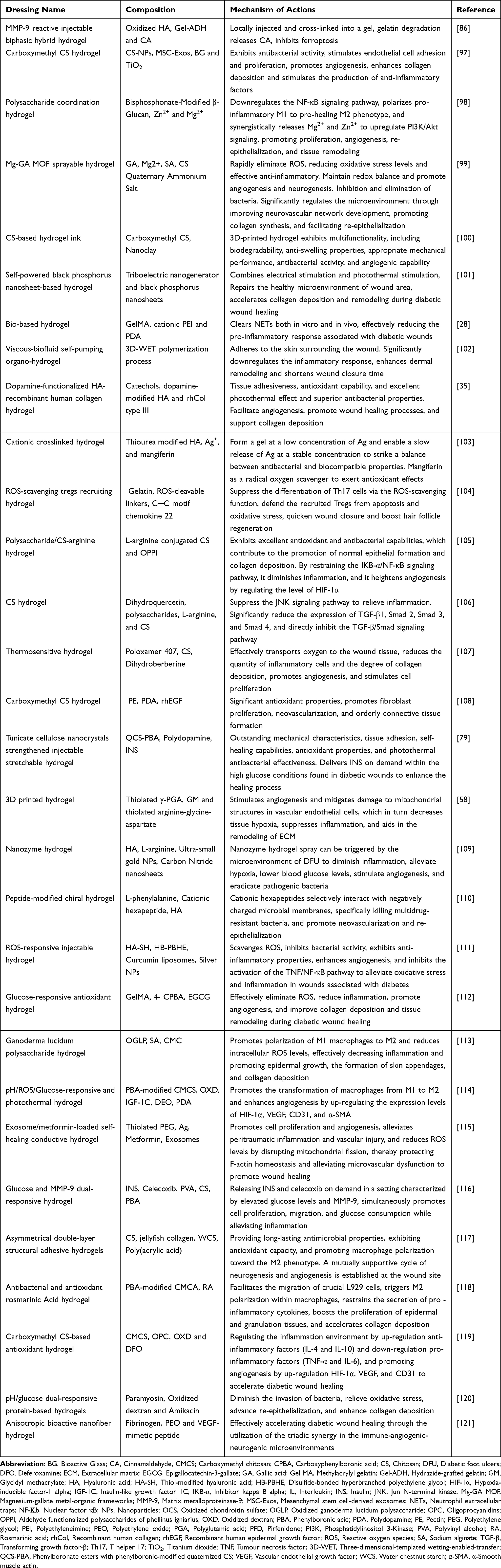 |
Table 1 Recent Studies on Hydrogel Dressings for the Treatment of Diabetic Wounds |
Antibacterial Hydrogels
Antibacterial hydrogels are vital in the repair of diabetic wounds, especially in their function of inhibiting infections and enhancing the healing process. Diabetic patients, due to weakened immune function, are far more likely to be afflicted by bacterial infections. These infections can decelerate the wound-healing process and lead to complications. Antibacterial hydrogels, by embedding antimicrobial agents (eg, silver ions, ethylene oxide, or natural antibacterial substances), can continuously release these agents, effectively inhibiting bacterial growth and minimizing the likelihood of infection.19 Concurrently, these hydrogels provide a moist healing environment that promotes cell migration, tissue repair, and alleviates inflammation, thus accelerating wound healing. The attributes of their biocompatibility and localized drug release qualities render them highly suitable for treating diabetic wounds.122 Currently, antibacterial hydrogels are primarily categorized into: self-antibacterial hydrogels, antimicrobial agent-containing hydrogels, and hydrogels containing agents that activate antibacterial functions.
Self-Antibacterial Hydrogels – Chitosan (CS) Series
Self-antibacterial hydrogels have shown distinctive benefits in the healing of diabetic wounds. Unlike conventional antibacterial hydrogels, self-antibacterial hydrogels utilize their inherent antibacterial properties (such as those imparted by surface modifications with natural polymers or synthetic materials) to form a stable protective layer at the wound site, successfully suppressing bacterial development and lowering the likelihood of infection.123 These hydrogels not only preserve moisture at the wound site and facilitate the healing process, but they also demonstrate low cytotoxicity and favorable biocompatibility. This ensures that the surrounding healthy tissue remains unharmed throughout the wound healing process.10 Additionally, by slowly and continuously releasing antibacterial agents, self-antibacterial hydrogels help accelerate the healing of diabetic wounds. The benefits are particularly significant for diabetic individuals with impaired immune functions, as it notably decreases the rate of chronic wound infections and enhances the effectiveness of treatments.124
With the exception of CS, most hydrogels derived from natural sources do not possess significant antibacterial properties. Research has indicated that CS-based hydrogels can be effectively utilized for wound healing due to their natural antibacterial traits.125,126 CS hydrogels are capable of absorbing cellular secretions and creating a thin protective layer over the cells, which helps to block microbial intrusion from the outside environment.127 The antibacterial action of CS is primarily linked to its positively charged polysaccharide structure that includes free amino and hydroxyl groups, which facilitates ease of chemical modification and cross-linking.128 Owing to its positive charge, CS has a heightened affinity for negatively charged bacteria, engaging with them through both electrostatic and hydrophobic interactions. This interaction facilitates the accumulation of CS on the surface of bacterial cell walls, where it disrupts the integrity of the bacterial membrane. Consequently, this disruption leads to cytoplasmic leakage and the release of proteins and other vital cellular constituents, ultimately culminating in the lysis and death of the bacteria.129,130 Lin et al131 created a hydrogel composed of CS and HA that is both drug-free and non-crosslinked, which combines the multiple functions of CS and HA to synergistically heal diabetic wounds infected with MRSA. The findings indicated that CS/HA hydrogels possess broad-spectrum antibacterial properties, effectively promoting fibroblast proliferation and migration. Additionally, these hydrogels demonstrate excellent ROS scavenging abilities, providing significant protection to cells under oxidative stress conditions. In diabetic mouse wounds infected with MRSA, CS/HA hydrogels significantly advanced wound healing by eradicating MRSA infection and enhancing epidermal regeneration, collagen deposition, and angiogenesis. Furthermore, quaternary ammonium salts, which have bacterial membrane-disrupting activity, which are frequently utilized in conjunction with polymers to improve their antibacterial effectiveness. Using CS and konjac glucomannan as raw materials, a hydrogel for wound injection was prepared through Schiff base linkage, which showed significant antibacterial efficacy against both Gram-positive Staphylococcus aureus and Gram-negative Escherichia coli, while also demonstrating promising performance in promoting wound healing.132 Shao et al56 created an injectable, adaptive multifunctional hydrogel with self-healing properties by grafting 3-carboxy-4-fluorophenylboronic acid onto quaternized CS. The phenylboronic acid groups of this hydrogel reacted with the hydroxyl groups of polyethylene glycol via boronate ester bonds. This hydrogel adapts to high glucose and hydrogen peroxide levels, mitigating oxidative stress. It promotes angiogenesis by enhancing the expression of HIF-1 and VEGF, resulting in collagen deposition and expedited wound healing. Wang et al133 formulated a multifunctional hydrogel dressing based on CS, which incorporated quaternary ammonium salt and catechol-modified CS, thioctic acid-functionalized poly(ethylene glycol)s, along with polydopamine-coated honeycomb manganese dioxide nanoparticles. This hydrogel demonstrated superoxide dismutase and catalase activities, allowing it to scavenge ROS while producing oxygen to reduce oxidative stress and improve the hypoxic conditions in wounds. Additionally, it facilitated the modulation of macrophage polarization to lessen inflammation. This hydrogel effectively managed diabetic wound infections associated with Staphylococcus aureus, reduced oxidative stress, inhibited inflammation, encouraged neovascularization, and promoted the synthesis of dermal collagen, thus expediting the wound healing process.
Hydrogels Loaded with Antibacterial Agents
Since most hydrogels do not inherently possess antibacterial properties, they need to be loaded with antibacterial agents (such as inorganic ions, inorganic particles, organic natural products, antibiotics, antimicrobial peptides, etc). that can continuously release drugs at the wound site. This local drug delivery helps control infection and achieve antibacterial or bacteriostatic effects, thereby promoting rapid wound healing.134 Antibiotics represent the most frequently utilized and efficient agents for combating bacterial infections, but the overuse of antibiotics in recent years has severely impaired their efficacy. Therefore, how to precisely control the use of antibiotics and maximize their effectiveness has become a research hotspot. Hydrogels loaded with antibiotics can ensure that antibiotics reach bactericidal concentrations directly at the local site, avoiding the toxic side effects of systemic application. Common antibiotics loaded into hydrogels include ciprofloxacin, gentamicin, and vancomycin. Mendes et al135 introduced a spongy-like hydrogel based on gellan gum (GG), designed as a topical and controlled drug delivery system (DDS) for vancomycin and clindamycin, aimed at enhancing dual antibiotic treatment for MRSA in diabetic foot infections (DFI). The developed DDS possesses properties suitable for local application and facilitates controlled release of both antibiotics, notably decreasing in vitro cytotoxic effects associated with antibiotics while preserving their antibacterial efficacy. The hydrogel treatment for MRSA-infected wounds significantly reduced bacterial load, minimized persistent infiltration of inflammatory cells, and promoted wound healing in diabetic patients. Wang et al136 developed a drug-delivery hydrogel that responds to inflammation by linking silver nanoclusters with vancomycin and incorporating pH-sensitive micelles containing nimesulide. The hydrogel demonstrated outstanding biocompatibility and hemostatic characteristics, showcasing notable antibacterial and anti-inflammatory effects. It facilitated wound healing in diabetic infected wounds by promoting continuous hemostasis along with antimicrobial and anti-inflammatory mechanisms. Xu et al134 developed a highly stretchable, adhesive, transparent, and antibacterial hydrogel using a one-pot radical polymerization process involving N-[tris(hydroxymethyl)] acrylamide (THMA) and acrylic acid (AA). CS and the antibiotic tobramycin were utilized as dynamic physical crosslinkers in the formulation. In vivo studies conducted on a Staphylococcus aureus-infected full-thickness diabetic skin wound model demonstrated that the tobramycin-loaded hydrogel exhibited prolonged antibacterial activity and significantly accelerated wound healing.
Antibacterial metal ions such as Ag+, Cu2+, and Zn2+ have been widely used in hydrogels to enhance their antibacterial effects.137 Alven et al138 prepared a hydrogel loaded with silver nanoparticles, which exhibited good antibacterial efficacy against Staphylococcus aureus, Bacillus subtilis, Pseudomonas aeruginosa, and Escherichia coli, significantly improving diabetes-induced wound healing. Lian et al99 introduced the application of magnesium-gallate metal-organic frameworks (Mg-GA MOF) to adjust the oxidative stress microenvironment and created a hydrogel dressing composed of sodium alginate/CS quaternary ammonium salts. The CS quaternary ammonium salts are antimicrobial, while GA provides sustained release to eliminate ROS, reduce oxidative stress, promote macrophage polarization, and maintain redox balance. Magnesium ions assist in promoting angiogenesis and neurogenesis. This hydrogel dressing effectively facilitates the rapid healing of diabetic wounds by enhancing the regeneration of the neurovascular network, speeding up re-epithelialization, and promoting collagen deposition. Zhang et al139 developed an intelligent stimuli-responsive multifunctional hydrogel that enables pH-triggered sequential delivery of silver-curcumin polydopamine nanoparticles (PDA@Ag&Cur NPs) and vascular endothelial growth factor (VEGF) by targeting the acidic wound microenvironment. Both in vitro and in vivo studies demonstrated that this hydrogel effectively eradicated bacterial infections, suppressed excessive inflammation, and promoted angiogenesis, leading to significantly accelerated diabetic wound healing. This study highlights a time-dependent precision drug delivery strategy based on dynamic wound healing stages, establishing a novel paradigm for multi-phase synergistic intervention in chronic wound therapy. Long et al140 designed a novel micro-fibrillated cellulose (MFC)-reinforced natural polymer composite sponge using carboxymethyl chitosan (CMC) and oxidized starch (OS) as matrix materials. Through functionalization with silver nanoparticles (AgNPs) and recombinant human type III collagen (rhCol III), this system exhibited broad-spectrum antimicrobial activity while promoting the proliferation and migration of L929 fibroblasts. It significantly accelerated wound healing by suppressing inflammation, promoting angiogenesis, and enhancing cell proliferation.
Hydrogel Capable of Activate Antibacterial Function
Hydrogels Loaded with Photothermal Materials
Hydrogels loaded with photothermal materials have shown unique advantages in diabetic wound healing, particularly in promoting healing and combating infection. By embedding photothermal materials (such as gold nanoparticles, carbon nanotubes, etc). into hydrogels, these materials can generate localized heat under near-infrared (NIR) light exposure and increase in local temperature enhances blood circulation, accelerates metabolism, and stimulates cell proliferation, contributing to wound healing.141,142 Moreover, the photothermal effect generates substantial heat, which disrupts bacterial proteins and effectively inhibits bacterial growth, thereby reducing the risk of infection. This is particularly important for diabetic patients, who often have weakened immune systems, as preventing wound infection is crucial.143
Jiang et al144 constructed an intelligent black phosphorus hydrogel with sprayable and NIR responsive properties. When irradiated by NIR laser, it produces local heat, which speeds up the blood flow in the microcirculation. It mediates lidocaine release in an “on-demand” manner, providing pain relief for diabetic wounds, eliminating bacteria, and reducing inflammation. Furthermore, the hydrogel promotes endothelial cell proliferation and angiogenesis to protect tissues from external influences and accelerate chronic wound healing. Huang et al145 prepared an NIR-responsive, ROS-generating, antibacterial nanohydrogel incorporating black phosphorus quantum dots. Under NIR irradiation, the hydrogel produced ROS, lipid peroxidation, glutathione, adenosine triphosphate accumulation and bacterial membrane destruction, which had synergistic effect on inhibiting MRSA, and promoted wound healing of MRSA infection in diabetic rats by reducing inflammatory response and regulating the expression of VEGF and basic fibroblast growth factor (bFGF). Ding et al146 prepared novel gold cage (AuNCs) modified with epigallocateching allate (EGCG), which produced a large amount of ROS under NIR irradiation to promote bacterial membrane rupture, induce bacterial lysis and apoptosis, facilitate the migration and proliferation of human umbilical endothelial cells, mitigate mitochondrial damage and oxidative stress under conditions of infection. It also significantly increased the expression of bFGF and VEGF, promoted cell migration and angiogenesis in diabetic rats, inhibited Staphylococcus aureus activity, reduced cell oxidative stress, restored damaged mitochondrial function, and promoted wound healing.
Hydrogels with ROS-Responsive Materials
Hydrogels incorporating ROS-responsive materials hold significant potential in the repair of diabetic wounds, particularly in promoting wound healing and regulating the local redox environment. Diabetic wounds are often associated with chronic inflammation and oxidative stress, contributing to a delay in the healing process. By embedding ROS-responsive materials (such as catalase, metal oxide nanoparticles, etc). into hydrogels, these materials can respond to the local oxidative stress environment at the wound site, automatically releasing substances with antioxidant properties, reduces excessive ROS accumulation, suppress the inflammatory response and enhance the wound healing process.147
Chen et al148 developed an injectable thermosensitive hydrogel based on niobium carbide (Nb2C), which exhibits both antioxidant and antibacterial properties. This hydrogel effectively eliminates ROS, reduces oxidative damage, eradicates bacterial infections, and fosters angiogenesis to support the healing of diabetic wounds. Zhang et al149 developed a flexible conductive hydrogel dressing that possesses intrinsic ROS scavenging properties and electroactivity, aimed at wound combination therapy and monitoring. The antioxidant hydrogel, when used in conjunction with electrical stimulation, synergistically enhances the healing of chronic diabetic wounds. It achieves this by regulating oxidative stress, reducing inflammation, and promoting key processes such as re-epithelialization, angiogenesis, and collagen deposition. Wei et al150 created an innovative spongy macroporous hydrogel with a stable macroporous structure and anti-swelling properties. This hydrogel exhibits excellent anti-swelling ability, strong antibacterial performance, and ROS-scavenging ability (96% scavenging rate at 120 minutes), which together facilitate diabetic wound healing.
Environment-Specific Responsive Hydrogels
Environmentally responsive hydrogels possess significant potential for applications in the healing of diabetic wounds. These hydrogels are capable of responding to particular local stimuli present at the wound site, including factors like pH, glucose concentration, and temperature. This responsiveness facilitates accurate drug release, sustains a moist environment for the wound, encourages cellular growth and repair, and effectively modulates local inflammatory responses.151 Through these functionalities, environmentally responsive hydrogels enhance the healing process of diabetic wounds while simultaneously decreasing the risk of infections and complications. This ultimately leads to improved treatment outcomes for patients.8 With the continuous innovation of technology, these intelligent hydrogels are projected to assume a more significant role in the repair of diabetic wounds.
Zhao et al152 created injectable gels that are responsive to both pH and glucose by crosslinking phenylboronic acid with a schiff base. The raw materials used in this process included phenylboronic acid modified CS, polyvinyl alcohol, and polyethylene glycol capped with benzaldehyde. During the in-situ crosslinking process, protein drugs and living cells can be encapsulated in the hydrogel, indicating that the hydrogel has a sustained or glucose-induced stimulus response to release drugs. Consequently, the hydrogel combined with INS and fibroblasts can function as a bioactive dressing for the healing of diabetic wounds. The addition of INS and L929 cells to the hydrogel facilitates angiogenesis and collagen accumulation, thereby improving the wound healing process. Huang et al153 introduced a temperature-responsive self-contractile nanofiber/hydrogel composite dressing, which consists of electrospun poly (lactic acid-trimethyl carbonate) nanofibers integrated with antimicrobial peptide Epinecidin-1@CSnanoparticle-loaded methacrylated gelatin hydrogel. This composite dressing effectively alters the microenvironment of diabetic wounds, encourages collagen accumulation and angiogenesis, alleviates inflammation, and enhances wound closure through forces triggered by temperature-induced contraction. Chen et al154 developed a copper-based MOF nanozyme and incorporated glucose oxidase (GOX), resulting in the creation of Cu-MOF/GOX. This was subsequently combined with CS-Arg (CS modified by L-Arginine) and Pluronic (F127) to formulate a multifunctional Cu-MOF/GOX-Gel thermosensitive hydrogel. At the wound site, the GOX utilized elevated blood glucose levels to produce H2O2 (hydrogen peroxide) and gluconic acid, thereby triggering an effective antibacterial self-cascade catalytic reaction during the early phases of wound healing. As the catalysis of the in situ produced H2O2 continued, nitric oxide (NO) was gradually released from the hydrogel, fostering angiogenesis and collagen production, which in turn expedited the overall healing process. Liang et al151 developed a hydrogel dressing that releases metformin in response to both pH and glucose levels. This dressing boasts the ability to release metformin in a stimulus-responsive manner while enhancing adhesion and self-healing capabilities. It features a dual dynamic adhesion mechanism that allows for easy removal and effective adhesion, making it suitable for treating wounds associated with diabetic foot conditions. Hua et al79 formulated an injectable multifunctional hydrogel (QPTx) that demonstrates outstanding mechanical properties along with triple responsiveness to pH, temperature, and glucose. This responsiveness is attributed to dynamic covalent cross-linking involving dynamic Schiff base bonds and phenylboronate esters, utilizing phenylboronic-modified quaternized CS (QCS-PBA), polydopamine-coated tunicate cellulose crystals (PDAn@TCNCs), and polyvinyl alcohol (PVA). Additionally, these hydrogels have the capacity to encapsulate INS drugs, enabling them to adjust to the intricate and fluctuating wound environment of diabetic patients for on-demand drug release, ultimately facilitating diabetic wound healing. Pan et al155 developed a hydrogel dressing (HPC) that responds to both pH and glucose through a dual dynamic crosslinking process involving Schiff base and phenylborate ester. This was achieved by combining m-aminophenylboronic acid with adiponed-hydrazide dual-functionalized HA (AHP) and oxychondroitin sulfate (OCS). Subsequently, polydopamine reduced graphene oxide complex glycine modified fullerene (GPC) having photothermal/photodynamic synergistic antibacterial properties and pirfenidone (PFD), a drug having angiogenesis promoting and inflammation inhibiting effects, were loaded into HPC hydrogel, which exhibited significant wound healing promoting effects by reducing infection and inflammation, accelerating wound healing, and promoting the regeneration of the epidermis, deposition of collagen, and formation of new blood vessels.
In recent years, green synthesis strategies have emerged as an environmentally friendly and efficient approach for diabetic wound therapy by integrating natural antibacterial components (eg, plant extracts, biocompatible polysaccharides) with smart responsive materials. For instance, antibacterial active compounds derived from mint extracts can be incorporated into thermosensitive hydrogels via green extraction techniques (eg, supercritical CO2 extraction). This approach not only circumvents the antibiotic resistance issues associated with traditional synthetic antimicrobials but also enables controlled drug release through temperature-responsive behavior, thereby enhancing diabetic wound healing efficacy.156 Studies demonstrate that thermosensitive hydrogels based on poloxamer/chitosan systems (eg, Poloxamer 407/chitosan) can undergo phase transitions in response to wound microenvironment temperature changes, enabling precise release of natural antibacterial components (eg, mint-derived phenolic compounds) and pro-healing factors. This dual functionality synergistically suppresses drug-resistant bacterial infections while promoting angiogenesis.107
High Adhesive Hydrogels
High adhesive hydrogels offer significant advantages in diabetic wound healing. Because of their remarkable adhesive qualities, these hydrogels can securely attach to the surface of wounds, reducing the risk of exposure, effectively safeguarding the area from outside contaminants, and preserving a moist environment to enhance the healing process.157 Moreover, the strong adhesive capability allows these hydrogel dressings to release medication (such as antimicrobial agents and growth factors) in a direct and gradual manner at the site of injury, thereby facilitating tissue regeneration, inhibiting infection, and enhancing local microcirculation as well as nutrient delivery during the wound healing process.8 Their excellent biocompatibility and stability render them an ideal repair material for treating chronic diabetic wounds.
Liu et al158 developed a modified polyacrylamide hydrogel with strong adhesive capability by grafting nitrogen-containing bases (A, G, C, T, U) onto acrylamide and then conducting either single or paired polymerization to obtain the base-grafted polyacrylamide hydrogel. The adhesion of hydrogels polymerized with A-U, A-T, or G-C pairs was considerably improved as a result of the hydrogen bonds formed between the base pairs. The hydrogel was used in adhesion and tensile experiments with various materials, and the findings demonstrated that this hydrogel demonstrated outstanding adhesive characteristics on surfaces such as glass, rubber, plastic, iron products, wooden materials, and animal tissues. Xu et al134 developed an innovative hydrogel characterized by high stretchability, excellent adhesion, transparency, and antibacterial properties. This hydrogel was synthesized through the free radical polymerization of THMA and AA. CS and the antibiotic tobramycin served as dynamic physical crosslinkers in its formulation. The resulting hydrogel demonstrated significant adhesion to diverse surfaces and exhibited prolonged antibacterial efficacy against Staphylococcus aureus in full-thickness diabetic skin wound models, thereby significantly enhancing the wound healing process. Zhang et al159 developed an adhesive HA hydrogel that forms in situ. Upon exposure to light irradiation, the nitrosoformaldehyde group generated by this hydrogel can covalently bond with amino groups present on tissue surfaces, resulting in enhanced tissue adhesion. This mechanism significantly promotes diabetic wound healing by modulating macrophage polarization and facilitating angiogenesis. Liu et al160 developed a conductive hydrogel patch characterized by exceptional transparency, strong adhesion, and hemostatic properties. This innovative patch allows for visual monitoring of wound healing progress and effectively enhances the healing of diabetic wounds. It achieves this through several mechanisms, including accelerating hemostasis, enhancing intercellular communication, preventing wound infections, facilitating collagen deposition, and promoting angiogenesis. Wang et al161 developed a hydrogel that responds to both pH and ROS, which displayed remarkable mechanical properties as well as robust adhesion attributed to Schiff base and phenylboronic ester linkages. This hydrogel accelerated diabetic wound healing by modulating macrophage phenotype through the NF-κB/JAK-STAT signaling pathway, alleviating oxidative stress, and enhancing both cell migration and angiogenesis. Liu et al162 developed a multifunctional hydrogel dressing characterized by temperature resistance, durability, strong adhesion, and inherent antimicrobial properties. The hydrogel demonstrated super-stretchability (>1400%), long-lasting moisture retention and adhesion, which effectively promoting diabetic wound healing by hastening collagen deposition, spurring angiogenesis, and curbing bacterial proliferation. Yin et al163 developed a multifunctional bio-adhesive hydrogel through dynamic covalent and light-triggered covalent bonds. The adhesive strength of hydrogel measured at 180 kPa is 4.35 times that of fibrin glue, which can significantly reduce bacterial load, accelerate polarization of M2 macrophages, mitigate inflammation, and enhance the healing process of wounds.
Diabetes Wound Dressing Currently Used in Clinic
Currently, the treatment of diabetic wounds relies on comprehensive management strategies, including controlling blood glucose, preventing infection, promoting wound healing and alleviating pain and discomfort. In clinical practice, the choice of diabetic wound dressings is particularly important, because diabetic patients are often accompanied by microangiopathy and neuropathy, resulting in slow wound healing process and prone to infection. In recent years, many innovative dressings have been introduced and achieved remarkable results in clinical practice. Currently Commonly used diabetes wound dressings include the following types:
Moisture-retentive dressings: Such as hydrocolloid dressings, alginate dressings, and polyurethane dressings, which have excellent absorbency and gel-forming properties. These dressings can quickly absorb exudate from the wound, maintaining a moist environment that promotes cell migration and proliferation, encourages epithelialization and angiogenesis, reduces dryness and crusting, and significantly accelerates the healing process.20,164
Silver ion dressings: Silver has strong antibacterial properties, which can effectively prevent wound infections and maintain a sterile wound environment. Silver ions penetrate bacterial cell membranes, disrupt their structure, and inhibit bacterial growth. Silver ion dressings are effective in inhibiting the growth of common microorganisms, including staphylococcus aureus and pseudomonas aeruginosa, thereby lowering infection susceptibility and promoting wound healing to a certain degree.165,166
Biomaterial-based dressings: Such as collagen dressings, skin substitutes and growth factor dressings. These dressings through hydrogel-based sustained-release systems to continuously release high concentrations of bioactive factors over extended periods, stimulating skin cell proliferation, migration and differentiation. This facilitates the healing process of wounds and improves the restorative capability of skin structures. Biomaterial-based dressings have demonstrated effective outcomes, particularly in managing chronic diabetic foot ulcers and other difficult-to-treat wounds.167,168
Negative pressure wound therapy (NPWT): NPWT is a technique that accelerates wound healing by applying external negative pressure to draw exudate from the wound and promote blood flow. It is especially effective for extensive and profound diabetic wounds, as it can notably lower the likelihood of infection, minimize swelling, and encourage the development of granulation tissue.169,170
In conclusion, various types of dressings are available for the management of diabetic wounds. When choosing the suitable dressing in clinical settings, it is important to take into account elements like the wound classification, the risk of infection, and the individual patient’s circumstances. Thoughtful selection of different dressing types can greatly enhance the rate of healing for diabetic wounds, minimize complications, and improve the patient’s overall quality of life. Table 2 includes some of the currently used adjunctive materials for the treatment of diabetic wounds.
 |
Table 2 Some Clinically Used Dressings for Treating Diabetic Wounds |
Critical Comparison of Different Hydrogel Types
Hydrogels, as hydrophilic materials with three-dimensional network structures, have demonstrated broad application potential in biomedical engineering, flexible electronics, environmental science, and related fields. In the context of diabetic wound healing, significant progress has been made in the design and application of multifunctional hydrogels. However, the advantages and limitations of different hydrogel types directly influence their clinical applicability. Table 3 provides a critical comparative analysis of existing hydrogel systems, evaluating them based on material composition, functional properties, and clinical performance.
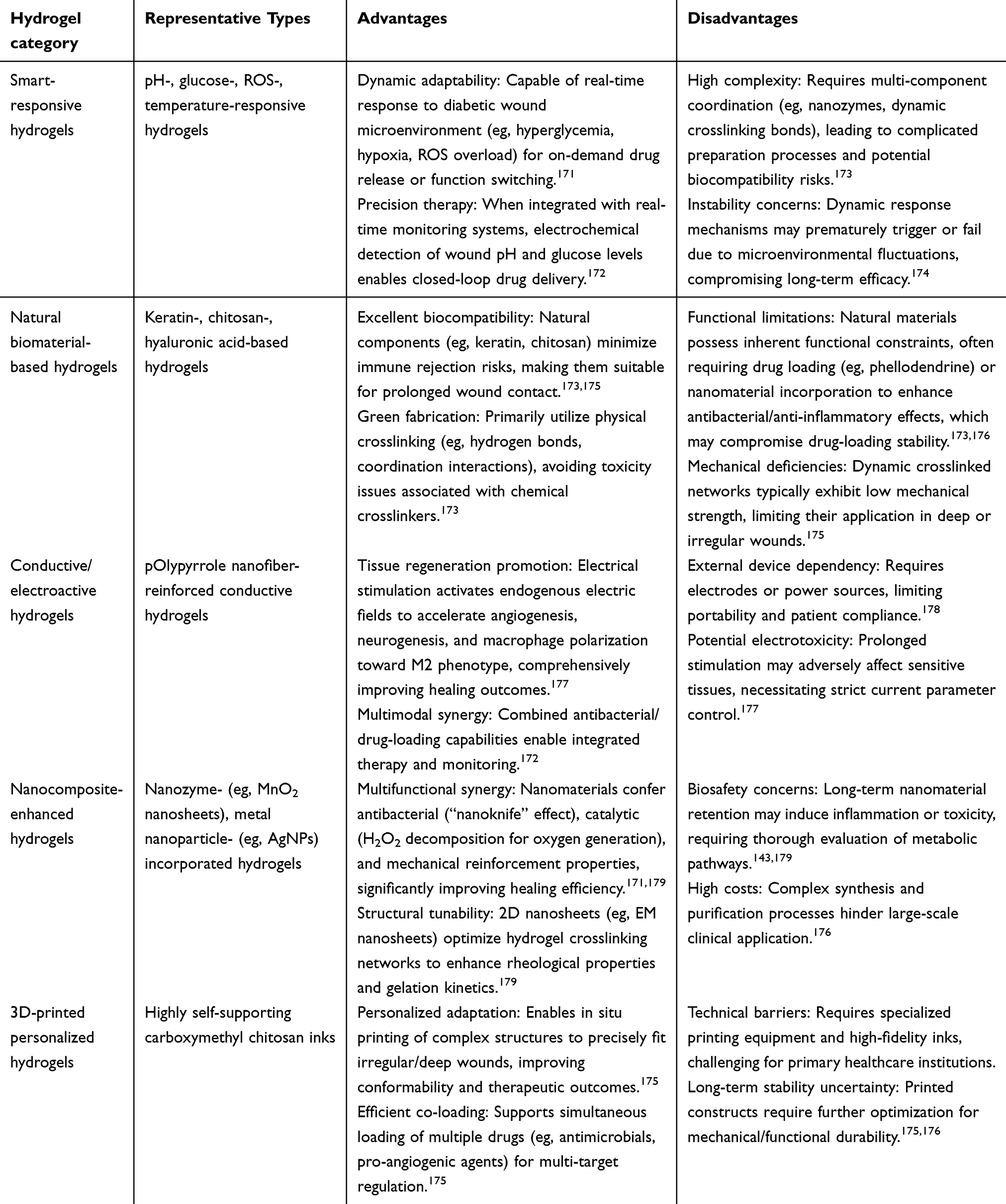 |
Table 3 Comparative Analysis of Hydrogel Types for Diabetic Wound Healing |
Based on the above analysis, the advantages and disadvantages of different hydrogel types are intrinsically linked to their design objectives. Among them, smart-responsive hydrogels are suitable for dynamic microenvironment regulation but require a balance between response speed and stability; natural material-based hydrogels prioritize biocompatibility but often necessitate functional enhancement; conductive hydrogels excel in promoting tissue regeneration but rely on external devices; nanocomposite hydrogels offer comprehensive functionality but face bottlenecks in safety and cost; while 3D-printed hydrogels demonstrate significant advantages in personalization but are limited in widespread adoption due to technical barriers. Future research should focus on simplifying multifunctional integration (eg, reducing component complexity), validating long-term biosafety, and optimizing clinical translation feasibility (eg, through low-cost fabrication techniques).
Conclusion and Perspectives
In hydrogel-based research for diabetic wound healing, the most promising future directions include: 1) Smart responsive hydrogels: Developing materials capable of dynamically responding to the wound microenvironment (eg, pH, enzymes, ROS, or glucose levels) to achieve on-demand drug release and real-time monitoring.114 2) Multifunctional synergistic therapy: Integrating antibacterial, anti-inflammatory, angiogenic, and cell-recruiting properties through biomimetic designs that mimic the complex functions of the extracellular matrix (ECM).180 3) 3D bioprinting technology: Combining patient-specific cells and growth factors to fabricate personalized hydrogel dressings tailored to complex wound geometries.58 4) Long-term sustained-release systems: Optimizing the degradation kinetics of drug-loaded hydrogels to ensure prolonged delivery of therapeutic agents (eg, exosomes or siRNA).103 5) Integration with emerging technologies: Combining approaches such as photothermal therapy, electrical stimulation, or gene editing to enhance tissue regeneration.179,181
Although hydrogel technology demonstrates significant therapeutic potential, critical challenges remain in its long-term stability and scalability—two pivotal factors for successful clinical translation and industrial application. Regarding long-term stability, hydrogels must maintain structural integrity and functional persistence in complex wound microenvironments, while resisting premature degradation caused by enzymatic hydrolysis, hydrolysis, or mechanical stress.171 Strategies such as advanced crosslinking (eg, dynamic covalent bonds, nanocomposite reinforcement) or enzyme-resistant polymers (eg, PEGylation) can enhance stability, though biocompatibility must be carefully balanced. Furthermore, prolonged hydrogel retention may trigger foreign body reactions, necessitating optimization between degradability and functional duration.182 For scalability, synthesis protocols, material costs, and sterilization conditions must meet industrial-scale production requirements. For instance, photo-crosslinked or thermosensitive hydrogels are more amenable to mass production due to their rapid gelation properties, whereas natural polymer-based hydrogels require solutions for batch-to-batch variability.179,183 Future research should integrate automated manufacturing technologies with standardized evaluation systems to accelerate the transition of hydrogels from laboratory benches to clinical and commercial markets.
However, significant challenges remain in translating hydrogel-based therapies from the lab to clinical applications: 1) Biocompatibility and long-term safety: Systematic evaluation is needed for hydrogel degradation byproducts, potential nanomaterial accumulation toxicity, and immunomodulatory effects—particularly regarding long-term tissue responses post-implantation.184,185 2) Scalable manufacturing and stability: The mechanical properties of existing hydrogels (eg, adhesiveness, self-healing capacity) may degrade in complex wound environments. Optimizing synthesis techniques (eg, spray-coating, dynamic crosslinking strategies) is critical to meet industrial-scale production requirements.186 3) Standardized evaluation systems: Unified animal models and clinical assessment criteria must be established, including metrics such as wound closure rates, angiogenesis indicators, and dynamic monitoring of microbial load, to improve cross-study comparability.171,172 4) Personalized treatment adaptation: Hydrogels should be tailored to individual patient variations (eg, glucose fluctuations, infection types) through adaptive designs, with machine learning-assisted optimization of dynamic drug release profiles to achieve precision medicine.172,181 Thus, future research must bridge the gap between material innovation and clinical needs by leveraging interdisciplinary approaches (eg, bioelectronics, nanomedicine, AI) to accelerate translation, ultimately delivering effective and safe solutions for diabetic wound management.
Meanwhile, regulatory oversight still faces certain challenges, such as: 1) Ambiguous classification criteria: As a “drug-device combination product”, hydrogels require clear regulatory pathways. For example, hydrogels loaded with growth factors (eg, VEGF) may need to meet both pharmaceutical and medical device approval standards.187 2) Lack of standardized testing: Unified testing protocols must be established for hydrogel biodegradability, long-term toxicity, and immunogenicity. Additionally, there are ethical concerns, including: 1) Animal testing ethics: Compliance with the 3R principles (replacement, reduction, refinement) is required. For instance, the use of porcine models must undergo ethical review.188 2) Patient privacy and data security: Smart diagnostic/therapeutic hydrogels (eg, the system developed by Sun Yat-sen University’s team) involve collecting patient physiological data, necessitating encrypted storage and compliant usage.172 3) Equity and accessibility: Advanced hydrogel technologies (eg, nanozyme composite systems) may exacerbate healthcare disparities due to high costs. Strategies like insurance coverage or public funding should be explored.189,190
Although the current clinical translation of hydrogel technology faces multiple bottlenecks, including material performance, production processes, and regulatory/ethical challenges. Its progression from lab to bedside can be accelerated through dynamic functional design, cost-effective process optimization, and interdisciplinary collaboration, all supported by rigorous regulatory frameworks and ethical guidelines.
Author Contributions
All authors made a significant contribution to the work reported, whether that is in the conception, study design, execution, acquisition of data, analysis and interpretation, or in all these areas; took part in drafting, revising or critically reviewing the article; gave final approval of the version to be published; have agreed on the journal to which the article has been submitted; and agree to be accountable for all aspects of the work.
Funding
This work was supported by the National Natural Science Foundation of China (82460735).
Disclosure
The authors report no conflicts of interest in this work.
References
1. Jia B, Li G, Cao E, et al. Recent progress of antibacterial hydrogels in wound dressings. Mater Today Bio. 2023;19:100582. doi:10.1016/j.mtbio.2023.100582
2. Sun H, Saeedi P, Karuranga S, et al. IDF diabetes atlas: global, regional and country-level diabetes prevalence estimates for 2021 and projections for 2045. Diabetes Res Clin Pract. 2023;204:110945. doi:10.1016/j.diabres.2023.110945
3. Liu J, Chen Z, Liu H, et al. Nickel-based metal-organic frameworks promote diabetic wound healing via scavenging reactive oxygen species and enhancing angiogenesis. Small. 2024;20(10):e2305076. doi:10.1002/smll.202305076
4. Saeedi P, Petersohn I, Salpea P, et al. Global and regional diabetes prevalence estimates for 2019 and projections for 2030 and 2045: results from the International Diabetes Federation Diabetes Atlas, 9th edition. Diabetes. Res Clin Pract. 2019;157:107843. doi:10.1016/j.diabres.2019.107843
5. Chin JS, Madden L, Chew SY, et al. Drug therapies and delivery mechanisms to treat perturbed skin wound healing. Adv. Drug Deliv Rev. 2019;149-150:2–18. doi:10.1016/j.addr.2019.03.006
6. Freedman BR, Hwang C, Talbot S, et al. Breakthrough treatments for accelerated wound healing. Sci Adv. 2023;9(20):eade7007. doi:10.1126/sciadv.ade7007
7. Peña OA, Martin P. Cellular and molecular mechanisms of skin wound healing. Nat. Rev Mol Cell Biol. 2024;25(8):599–616. doi:10.1038/s41580-024-00715-1
8. Chen Y, Wang X, Tao S, et al. Research advances in smart responsive-hydrogel dressings with potential clinical diabetic wound healing properties. Mil Med Res. 2023;10(1):37. doi:10.1186/s40779-023-00473-9
9. Xiong Y, Chen L, Liu P, et al. All-in-One: multifunctional hydrogel accelerates oxidative diabetic wound healing through timed-release of exosome and fibroblast growth factor. Small. 2023;19(21):e2303158. doi:10.1002/smll.202303158
10. Zhang X, Liang Y, Huang S, et al. Chitosan-based self-healing hydrogel dressing for wound healing. Adv Colloid Interface Sci. 2024;332:103267. doi:10.1016/j.cis.2024.103267
11. Zheng Y, Cheng Y, Chen J, et al. Injectable hydrogel-microsphere construct with sequential degradation for locally synergistic chemotherapy. ACS Appl Mater Interfaces. 2017;9(4):3487–3496. doi:10.1021/acsami.6b15245
12. Liu L, Feng X, Pei Y, et al. α-Cyclodextrin concentration-controlled thermo-sensitive supramolecular hydrogels. Mater Sci Eng C Mater Biol Appl. 2018;82:25–28. doi:10.1016/j.msec.2017.08.045
13. Qian Y, Zheng Y, Jin J, et al. Immunoregulation in diabetic wound repair with a photoenhanced glycyrrhizic acid hydrogel scaffold. Adv Mater. 2022;34(29):e2200521. doi:10.1002/adma.202200521
14. Zhang Z, Yao A, Raffa P. Transparent, highly stretchable, self-healing, adhesive, freezing-tolerant, and swelling-resistant multifunctional hydrogels for underwater motion detection and information transmission. Adv Funct Mater. 2024;34(49):2470292. doi:10.1002/adfm.202470292
15. Zeng H, Tang L, Huang L, et al. A novel multi-functional PVA-alginate hydrogel with dynamic bond crosslinking for infected wound repair. Carbohydr Polym. 2025;362:123636. doi:10.1016/j.carbpol.2025.123636
16. Guo B, Liang Y, Dong R. Physical dynamic double-network hydrogels as dressings to facilitate tissue repair. Nat Protoc. 2023;18(11):3322–3354. doi:10.1038/s41596-023-00878-9
17. Fan L, He Z, Peng X, et al. Injectable, intrinsically antibacterial conductive hydrogels with self-healing and pH stimulus responsiveness for epidermal sensors and wound healing. ACS Appl Mater Interfaces. 2021;13(45):53541–53552. doi:10.1021/acsami.1c14216
18. Wang H, Xu Z, Zhao M, et al. Advances of hydrogel dressings in diabetic wounds. Biomater Sci. 2021;9(5):1530–1546. doi:10.1039/D0BM01747G
19. Liang Y, He J, Guo B. Functional hydrogels as wound dressing to enhance wound healing. ACS Nano. 2021;15(8):12687–12722. doi:10.1021/acsnano.1c04206
20. Zhao H, Wu Y, Xie Y, et al. Hydrogel dressings for diabetic foot ulcer: a systematic review and meta-analysis. Diabetes Obes Metab. 2024;26(6):2305–2317. doi:10.1111/dom.15544
21. Bai Q, Han K, Dong K, et al. Potential applications of nanomaterials and technology for diabetic wound healing. Int J Nanomed;2020. 9717–9743. doi:10.2147/IJN.S276001
22. Dixon D, Edmonds M. Managing diabetic foot ulcers: pharmacotherapy for wound healing. Drugs. 2021;81(1):29–56. doi:10.1007/s40265-020-01415-8
23. Zhao Y, Zhao Y, Xu B, et al. Microenvironmental dynamics of diabetic wounds and insights for hydrogel-based therapeutics. J Tissue Eng. 2024;15:20417314241253290. doi:10.1177/20417314241253290
24. Qi X, Cai E, Xiang Y, et al. An immunomodulatory hydrogel by hyperthermia-assisted self-cascade glucose depletion and ROS scavenging for diabetic foot ulcer wound therapeutics. Adv Mater. 2023;35(48):e2306632. doi:10.1002/adma.202306632
25. Cao J, Wu B, Yuan P, et al. Rational Design of multifunctional hydrogels for wound repair. J Funct Biomater. 2023;14(11):553. doi:10.3390/jfb14110553
26. Peng Y, Guo Y, Ge X, et al. Construction of programmed time-released multifunctional hydrogel with antibacterial and anti-inflammatory properties for impaired wound healing. J Nanobiotechnology. 2024;22(1):126. doi:10.1186/s12951-024-02390-y
27. Xiang P, Jiang M, Chen X, et al. Targeting grancalcin accelerates wound healing by improving angiogenesis in diabetes. Adv Sci (Weinh). 2024;11(14):e2305856.
28. Xiao Y, Ding T, Fang H, et al. Innovative bio-based hydrogel microspheres micro-cage for neutrophil extracellular traps scavenging in diabetic wound healing. Adv Sci (Weinh). 2024;11(21):e2401195. doi:10.1002/advs.202401195
29. Huang C, Dong L, Zhao B, et al. Anti-inflammatory hydrogel dressings and skin wound healing. Clin Transl Med. 2022;12(11):e1094. doi:10.1002/ctm2.1094
30. Yu FX, Lee PSY, Yang L, et al. The impact of sensory neuropathy and inflammation on epithelial wound healing in diabetic corneas. Prog Retin Eye Res. 2022;89:101039. doi:10.1016/j.preteyeres.2021.101039
31. Bi S, He C, Zhou Y, et al. Versatile conductive hydrogel orchestrating neuro-immune microenvironment for rapid diabetic wound healing through peripheral nerve regeneration. Biomaterials. 2025;314:122841. doi:10.1016/j.biomaterials.2024.122841
32. Cheng F, Wang S, Zheng H, et al. Ceria nanoenzyme-based hydrogel with antiglycative and antioxidative performance for infected diabetic wound healing. Small Methods. 2022;6(11):e2200949. doi:10.1002/smtd.202200949
33. Liu J, Chen Z, Wang J, et al. Encapsulation of curcumin nanoparticles with MMP 9-responsive and thermos-sensitive hydrogel improves diabetic wound healing. ACS Appl Mater Interfaces. 2018;10(19):16315–16326. doi:10.1021/acsami.8b03868
34. Jere SW, Houreld NN, Abrahamse H. Effect of photobiomodulation on cellular migration and survival in diabetic and hypoxic diabetic wounded fibroblast cells. Lasers Med Sci. 2021;36(2):365–374. doi:10.1007/s10103-020-03041-y
35. Wang Y, Zhang Y, Yang YP, et al. Versatile dopamine-functionalized hyaluronic acid-recombinant human collagen hydrogel promoting diabetic wound healing via inflammation control and vascularization tissue regeneration. Bioact Mater. 2024;35:330–345. doi:10.1016/j.bioactmat.2024.02.010
36. Liu W, Gao R, Yang C, et al. ECM-mimetic immunomodulatory hydrogel for methicillin-resistant Staphylococcus aureus-infected chronic skin wound healing. Sci Adv. 2022;8(27):eabn7006. doi:10.1126/sciadv.abn7006
37. Summer M, Ali S, Fiaz U, et al. Revealing the molecular mechanisms in wound healing and the effects of different physiological factors including diabetes, age, and stress. J Mol Histol. 2024;55(5):637–654. doi:10.1007/s10735-024-10223-3
38. Burgess JL, Wyant WA, Abdo Abujamra B, et al. Diabetic wound-healing science. Medicina (Kaunas). 2021;57(10):1072. doi:10.3390/medicina57101072
39. Yang H, Song L, Sun B, et al. Modulation of macrophages by a paeoniflorin-loaded hyaluronic acid-based hydrogel promotes diabetic wound healing. Mater Today Bio. 2021;12:100139. doi:10.1016/j.mtbio.2021.100139
40. Mallik SB, Jayashree BS, Shenoy RR. Epigenetic modulation of macrophage polarization- perspectives in diabetic wounds. J Diabetes Complications. 2018;32(5):524–530. doi:10.1016/j.jdiacomp.2018.01.015
41. Xu F, Zhang C, Graves DT. Abnormal cell responses and role of TNF-α in impaired diabetic wound healing. Biomed Res Int. 2013;2013:754802. doi:10.1155/2013/754802
42. Boniakowski AE, Kimball AS, Jacobs BN, et al. Macrophage-mediated inflammation in normal and diabetic wound healing. J Immunol. 2017;199(1):17–24. doi:10.4049/jimmunol.1700223
43. Zhao X, Pei D, Yang Y, et al. Green tea derivative driven smart hydrogels with desired functions for chronic diabetic wound treatment. Adv Funct Mater. 2021;31(18):2009442. doi:10.1002/adfm.202009442
44. Chen S, Shi J, Zhang M, et al. Mesenchymal stem cell-laden anti-inflammatory hydrogel enhances diabetic wound healing. Sci Rep. 2015;5(1):18104. doi:10.1038/srep18104
45. Fu YJ, Shi YF, Wang LY, et al. All-natural immunomodulatory bioadhesive hydrogel promotes angiogenesis and diabetic wound healing by regulating macrophage heterogeneity. Adv Sci (Weinh). 2024;11(33):e2404890.
46. Armstrong DG, Tan TW, Boulton AJM, et al. Diabetic foot ulcers: a review. JAMA. 2023;330(1):62–75. doi:10.1001/jama.2023.10578
47. Pilipenko I, Murzova A, Savin A, et al. Dual-function hydrogel dressings with a dynamic exchange of iron ions and an antibiotic drug for treatment of infected wounds. ACS Appl Bio Mater. 2023;6(5):1896–1905. doi:10.1021/acsabm.3c00124
48. Chai C, Zhang P, Ma L, et al. Regenerative antibacterial hydrogels from medicinal molecule for diabetic wound repair. Bioact Mater. 2022;25:541–554. doi:10.1016/j.bioactmat.2022.07.020
49. Zhang D, Zhou W, Wei B, et al. Carboxyl-modified poly(vinyl alcohol)-crosslinked chitosan hydrogel films for potential wound dressing. Carbohydr Polym. 2015;125:189–199. doi:10.1016/j.carbpol.2015.02.034
50. He H, Xia DL, Chen YP, et al. Evaluation of a two-stage antibacterial hydrogel dressing for healing in an infected diabetic wound. J Biomed Mater Res B Appl Biomater. 2017;105(7):1808–1817. doi:10.1002/jbm.b.33543
51. Bao J, Mi J, Xia Y, et al. Heme-mimetic photosensitizer with iron-targeting and internalizing properties for enhancing PDT activity and promoting infected diabetic wound healing. ACS Appl Bio Mater. 2024;7(6):4116–4132. doi:10.1021/acsabm.4c00427
52. Demirci S, Doğan A, Aydın S, et al. Boron promotes streptozotocin-induced diabetic wound healing: roles in cell proliferation and migration, growth factor expression, and inflammation. Mol Cell Biochem. 2016;417(1–2):119–133. doi:10.1007/s11010-016-2719-9
53. Cheng C, Zhong H, Zhang Y, et al. Bacterial responsive hydrogels based on quaternized chitosan and GQDs-ε-PL for chemo-photothermal synergistic anti-infection in diabetic wounds. Int J Biol Macromol. 2022;210:377–393. doi:10.1016/j.ijbiomac.2022.05.008
54. Zhu Y, Wang Y, Jia Y, et al. Roxadustat promotes angiogenesis through HIF-1α/VEGF/VEGFR2 signaling and accelerates cutaneous wound healing in diabetic rats. Wound Repair Regen. 2019;27(4):324–334. doi:10.1111/wrr.12708
55. Yang X, Yang R, Chen M, et al. KGF-2 and FGF-21 poloxamer 407 hydrogel coordinates inflammation and proliferation homeostasis to enhance wound repair of scalded skin in diabetic rats. BMJ Open Diabetes Res Care. 2020;8(1):e001009. doi:10.1136/bmjdrc-2019-001009
56. Shao Z, Yin T, Jiang J, et al. Wound microenvironment self-adaptive hydrogel with efficient angiogenesis for promoting diabetic wound healing. Bioact Mater. 2022;20:561–573. doi:10.1016/j.bioactmat.2022.06.018
57. Thangavel P, Ramachandran B, Chakraborty S, et al. Accelerated healing of diabetic wounds treated with L-glutamic acid loaded hydrogels through enhanced collagen deposition and angiogenesis: an in vivo study. Sci Rep. 2017;7(1):10701. doi:10.1038/s41598-017-10882-1
58. Huang J, Yang R, Jiao J, et al. A click chemistry-mediated all-peptide cell printing hydrogel platform for diabetic wound healing. Nat Commun. 2023;14(1):7856. doi:10.1038/s41467-023-43364-2
59. Li X, Zou J, Lin A, et al. Oxidative stress, endothelial dysfunction, and n-acetylcysteine in type 2 diabetes mellitus. Antioxid Redox Signal. 2024;40(16–18):968–989. doi:10.1089/ars.2023.0524
60. Forman HJ, Zhang H. Targeting oxidative stress in disease: promise and limitations of antioxidant therapy. Nat Rev Drug Discov. 2021;20(8):652. doi:10.1038/s41573-021-00267-5
61. Zhang W, Zha K, Xiong Y, et al. Glucose-responsive, antioxidative HA-PBA-FA/EN106 hydrogel enhanced diabetic wound healing through modulation of FEM1b-FNIP1 axis and promoting angiogenesis. Bioact Mater. 2023;30:29–45. doi:10.1016/j.bioactmat.2023.07.006
62. Li Z, Zhao Y, Huang H, et al. A Nanozyme-immobilized hydrogel with endogenous ROS-scavenging and oxygen generation abilities for significantly promoting oxidative diabetic wound healing. Adv Healthc Mater. 2022;11(22):e2201524. doi:10.1002/adhm.202201524
63. Xu J, Lin Y, Wang Y, et al. Multifunctional regeneration silicon-loaded chitosan hydrogels for MRSA-infected diabetic wound healing. Adv Healthc Mater. 2024;13(10):e2303501. doi:10.1002/adhm.202303501
64. Zhu Y, Hoshi R, Chen S, et al. Sustained release of stromal cell derived factor-1 from an antioxidant thermoresponsive hydrogel enhances dermal wound healing in diabetes. J Control Release. 2016;238:114–122. doi:10.1016/j.jconrel.2016.07.043
65. Yang Z, Chen H, Yang P, et al. Nano-oxygenated hydrogels for locally and permeably hypoxia relieving to heal chronic wounds. Biomaterials. 2022;282:121401. doi:10.1016/j.biomaterials.2022.121401
66. Yang Z, Ren K, Chen Y, et al. Oxygen-generating hydrogels as oxygenation therapy for accelerated chronic wound healing. Adv Healthc Mater. 2024;13(3):e2302391. doi:10.1002/adhm.202302391
67. Mutluoglu M, Cakkalkurt A, Uzun G, et al. Topical oxygen for chronic wounds: a PRO/CON debate. J Am Coll Clin Wound Spec. 2014;5(3):61–65. doi:10.1016/j.jccw.2014.12.003
68. Golledge J, Singh TP. Systematic review and meta-analysis of clinical trials examining the effect of hyperbaric oxygen therapy in people with diabetes-related lower limb ulcers. Diabet Med. 2019;36(7):813–826. doi:10.1111/dme.13975
69. Guan Y, Niu H, Liu Z, et al. Sustained oxygenation accelerates diabetic wound healing by promoting epithelialization and angiogenesis and decreasing inflammation. Sci Adv. 2021;7(35):eabj0153. doi:10.1126/sciadv.abj0153
70. Patil PS, Fathollahipour S, Inmann A, et al. Fluorinated methacrylamide chitosan hydrogel dressings improve regenerated wound tissue quality in diabetic wound healing. Adv Wound Care (New Rochelle). 2019;8(8):374–385. doi:10.1089/wound.2018.0887
71. Ur Rehman SR, Augustine R, Zahid AA, et al. Graphene oxide loaded hydrogel for enhanced wound healing in diabetic patients. Annu Int Conf IEEE Eng Med Biol Soc. 2019;2019:3943–3946. doi:10.1109/EMBC.2019.8857341
72. Lee JB, Kim DH, Yoon JK, et al. Microchannel network hydrogel induced ischemic blood perfusion connection. Nat Commun. 2020;11(1):615. doi:10.1038/s41467-020-14480-0
73. Banerjee A, Singh P, Sheikh PA, et al. Simultaneous regulation of AGE/RAGE signaling and MMP-9 expression by an immunomodulating hydrogel accelerates healing in diabetic wounds. Biomater Adv. 2024;163:213937. doi:10.1016/j.bioadv.2024.213937
74. Ahmad S, Khan H, Siddiqui Z, et al. AGEs, RAGEs and s-RAGE; friend or foe for cancer. Semin Cancer Biol. 2018;49:44–55. doi:10.1016/j.semcancer.2017.07.001
75. Eming SA, Martin P, Tomic-Canic M. Wound repair and regeneration: mechanisms, signaling, and translation. Sci Transl Med. 2014;6(265):265sr6. doi:10.1126/scitranslmed.3009337
76. Hudson BI, Lippman ME. Targeting RAGE signaling in inflammatory disease. Annu Rev Med. 2018;69(1):349–364. doi:10.1146/annurev-med-041316-085215
77. Moura LI, Dias AM, Leal EC, et al. Chitosan-based dressings loaded with neurotensin--an efficient strategy to improve early diabetic wound healing. Acta Biomater. 2014;10(2):843–857. doi:10.1016/j.actbio.2013.09.040
78. Rajalekshmy GP, Ramesan RM, Geetha CS, et al. In Vivo Wound-Healing Efficacy of Insulin-Loaded Strontium-Cross-Linked Alginate-Based Hydrogels in Diabetic Rats. ACS Biomater Sci Eng. 2024;10(4):2552–2566. doi:10.1021/acsbiomaterials.3c01692
79. Hua S, Zhang Y, Zhu Y, et al. Tunicate cellulose nanocrystals strengthened injectable stretchable hydrogel as multi-responsive enhanced antibacterial wound dressing for promoting diabetic wound healing. Carbohydr Polym. 2024;343:122426. doi:10.1016/j.carbpol.2024.122426
80. Chen J, Chen D, Chen J, et al. An all-in-one CO gas therapy-based hydrogel dressing with sustained insulin release, anti-oxidative stress, antibacterial, and anti-inflammatory capabilities for infected diabetic wounds. Acta Biomater. 2022;146:49–65. doi:10.1016/j.actbio.2022.04.043
81. Rousselle P, Montmasson M, Garnier C. Extracellular matrix contribution to skin wound re-epithelialization. Matrix Biol. 2019;75-76:12–26. doi:10.1016/j.matbio.2018.01.002
82. Rohani MG, Parks WC. Matrix remodeling by MMPs during wound repair. Matrix Biol. 2015;44-46:113–121. doi:10.1016/j.matbio.2015.03.002
83. Cabral-Pacheco GA, Garza-Veloz I, Castruita-De la Rosa C, et al. The roles of matrix metalloproteinases and their inhibitors in human diseases. Int J Mol Sci. 2020;21(24):9739. doi:10.3390/ijms21249739
84. Kuhad A, Singh P, Chopra K. Matrix metalloproteinases: potential therapeutic target for diabetic neuropathic pain. Expert Opin Ther Targets. 2015;19(2):177–185. doi:10.1517/14728222.2014.960844
85. Jia Y, Zhang X, Yang W, et al. A pH-responsive hyaluronic acid hydrogel for regulating the inflammation and remodeling of the ECM in diabetic wounds. J Mater Chem B. 2022;10(15):2875–2888. doi:10.1039/D2TB00064D
86. Lin C, Hu Y, Lin Z, et al. MMP-9 responsive hydrogel promotes diabetic wound healing by suppressing ferroptosis of endothelial cells. Bioact Mater. 2024;43:240–254. doi:10.1016/j.bioactmat.2024.09.006
87. Lan B, Zhang L, Yang L, et al. Sustained delivery of MMP-9 siRNA via thermosensitive hydrogel accelerates diabetic wound healing. J Nanobiotechnology. 2021;19(1):130. doi:10.1186/s12951-021-00869-6
88. Castleberry SA, Almquist BD, Li W, et al. Self-assembled wound dressings silence MMP-9 and improve diabetic wound healing in vivo. Adv Mater. 2016;28(9):1809–1817.
89. Tardáguila-García A, García-Morales E, García-Alamino JM, et al. Metalloproteinases in chronic and acute wounds: a systematic review and meta-analysis. Wound Repair Regen. 2019;27(4):415–420.
90. Song Z, Jiang J, Gao H, et al. Drug release and kinetics properties of rosin-based polyacrylamide hydrogels. Chem Ind for Prod. 2018;38(3):25–32.
91. Cherri M, Stergiou PS, Ahmadian Z, et al. Redox-responsive hydrogels loaded with an antibacterial peptide as controlled drug delivery for healing infectious wounds. Adv Healthc Mater. 2024;13(27):e2401289. doi:10.1002/adhm.202401289
92. Scheja S, Domanskyi S, Gamella M, et al. Glucose-triggered insulin release from Fe -cross-linked alginate hydrogel: experimental study and theoretical modeling. Chemphyschem. 2017;18(12):1541–1551. doi:10.1002/cphc.201700195
93. Wang F, Deng S, Song C, et al. Pd@Au nanoframe hydrogels for closed-loop wound therapy. ACS Nano. 2025;19(15):15069–15080. doi:10.1021/acsnano.5c01864
94. Bang FS, Leeberg V, Ding M, et al. The effect of VEG stimulation in diabetic foot ulcers: a systematic review. Wound Repair Regen. 2024;32(4):384–392. doi:10.1111/wrr.13171
95. Lee YH, Hong YL, Wu TL. Novel silver and nanoparticle-encapsulated growth factor co-loaded chitosan composite hydrogel with sustained antimicrobility and promoted biological properties for diabetic wound healing. Mater Sci Eng C Mater Biol Appl. 2021;118:111385. doi:10.1016/j.msec.2020.111385
96. Li Y, Fu R, Duan Z, et al. Artificial nonenzymatic antioxidant MXene nanosheet-anchored injectable hydrogel as a mild photothermal-controlled oxygen release platform for diabetic wound healing. ACS Nano. 2022;16(5):7486–7502. doi:10.1021/acsnano.1c10575
97. Shang S, Zhuang K, Chen J, et al. A bioactive composite hydrogel dressing that promotes healing of both acute and chronic diabetic skin wounds. Bioact Mater. 2024;34:298–310. doi:10.1016/j.bioactmat.2023.12.026
98. Zhang X, Ning F, Li Y, et al. Pluripotent polysaccharide coordinated hydrogels remodel inflammation, neovascularization and reepithelization for efficient diabetic wound prohealing. J Control Release. 2024;377:37–53. doi:10.1016/j.jconrel.2024.11.027
99. Lian C, Liu J, Wei W, et al. Mg-gallate metal-organic framework-based sprayable hydrogel for continuously regulating oxidative stress microenvironment and promoting neurovascular network reconstruction in diabetic wounds. Bioact Mater. 2024;38:181–194. doi:10.1016/j.bioactmat.2024.04.028
100. Li S, Xu Y, Zheng L, et al. High self‐supporting chitosan‐based hydrogel ink for in situ 3D printed diabetic wound dressing. Adv Funct Mater. 2024;35(7). doi:10.1002/adfm.202411334
101. Ge X, Mao M, Yu H, et al. Application of self powered black phosphorus nanosheets-based hydrogel skin patch in wound healing of diabetes. Nano Energy. 2024;131:110289. doi:10.1016/j.nanoen.2024.110289
102. Xiao W, Wan X, Shi L, et al. A viscous-biofluid self-pumping organohydrogel dressing to accelerate diabetic wound healing. Adv Mater. 2024;36(25):e2401539. doi:10.1002/adma.202401539
103. Meng W, Lin Z, Cheng X, et al. Thiourea-cation chelation based hydrogel and its application as antibacterial dressing for the repair of diabetic wound. Adv Funct Mater. 2024;34(22):2314202. doi:10.1002/adfm.202314202
104. Qu M, Xu W, Zhou X, et al. An ROS-scavenging treg-recruiting hydrogel patch for diabetic wound healing. Adv Funct Mater. 2024;34(26):2314500.
105. Zhang L, Yang J, Liu W, et al. A phellinus igniarius polysaccharide/chitosan-arginine hydrogel for promoting diabetic wound healing. Int J Biol Macromol. 2023;249:126014. doi:10.1016/j.ijbiomac.2023.126014
106. Yang J, Zhang L, Sun S, et al. A dihydromyricetin-loaded phellinus igniarius polysaccharide/l-arginine modified chitosan-based hydrogel for promoting wound recovery in diabetic mice via JNK and TGF-β/Smad signaling pathway. Int J Biol Macromol. 2024;259(Pt 1):129124. doi:10.1016/j.ijbiomac.2023.129124
107. Liu X, Ding Q, Liu W, et al. A poloxamer 407/chitosan-based thermosensitive hydrogel dressing for diabetic wound healing via oxygen production and dihydromyricetin release. Int J Biol Macromol. 2024;263(Pt 1):130256. doi:10.1016/j.ijbiomac.2024.130256
108. Gou D, Qiu P, Hong F, et al. Polydopamine modified multifunctional carboxymethyl chitosan/pectin hydrogel loaded with recombinant human epidermal growth factor for diabetic wound healing. Int J Biol Macromol. 2024;274(Pt 1):132917. doi:10.1016/j.ijbiomac.2024.132917
109. Shang L, Yu Y, Jiang Y, et al. Ultrasound-augmented multienzyme-like nanozyme hydrogel spray for promoting diabetic wound healing. ACS Nano. 2023;17(16):15962–15977. doi:10.1021/acsnano.3c04134
110. Xing C, Zhu H, Dou X, et al. Infected diabetic wound regeneration using peptide-modified chiral dressing to target revascularization. ACS Nano. 2023;17(7):6275–6291. doi:10.1021/acsnano.2c10039
111. Shi C, Zhang Y, Wu G, et al. Hyaluronic acid-based reactive oxygen species-responsive multifunctional injectable hydrogel platform accelerating diabetic wound healing. Adv Healthc Mater. 2024;13(4):e2302626. doi:10.1002/adhm.202302626
112. Chen F, Qin J, Wu P, et al. Glucose-responsive antioxidant hydrogel accelerates diabetic wound healing. Adv Healthc Mater. 2023;12(21):e2300074. doi:10.1002/adhm.202300074
113. Li F, Liu T, Liu X, et al. Ganoderma lucidum polysaccharide hydrogel accelerates diabetic wound healing by regulating macrophage polarization. Int J Biol Macromol. 2024;260(Pt 2):129682. doi:10.1016/j.ijbiomac.2024.129682
114. Dai F, Zhang J, Chen F, et al. A Multi-responsive hydrogel combined with mild heat stimulation promotes diabetic wound healing by regulating inflammatory and enhancing angiogenesis. Adv Sci (Weinh). 2024;11(46):e2408783. doi:10.1002/advs.202408783
115. Zhang Y, Li M, Wang Y, et al. Exosome/metformin-loaded self-healing conductive hydrogel rescues microvascular dysfunction and promotes chronic diabetic wound healing by inhibiting mitochondrial fission. Bioact Mater. 2023;26:323–336. doi:10.1016/j.bioactmat.2023.01.020
116. Zhou W, Duan Z, Zhao J, et al. Glucose and MMP-9 dual-responsive hydrogel with temperature sensitive self-adaptive shape and controlled drug release accelerates diabetic wound healing. Bioact Mater. 2022;17:1–17. doi:10.1016/j.bioactmat.2022.01.004
117. Su Z, Zhang W, Mo Z, et al. Novel asymmetrical double-layer structural adhesive hydrogels with synergetic neuroprotection and angiogenesis effect for diabetic wound healing. Chem Eng J. 2025;505:159081. doi:10.1016/j.cej.2024.159081
118. Xiong S, Ding X, Zhou L, et al. An antibacterial and antioxidant rosmarinic acid hydrogel normalizes macrophage polarization to expedite diabetic wound healing. J Colloid Interface Sci. 2024;683(Pt 2):357–371. doi:10.1016/j.jcis.2024.12.138
119. Zhang J, Zhao S, Zhou Y, et al. Carboxymethyl chitosan-based antioxidant hydrogel accelerates diabetic wound healing. Adv Healthc Mater. 2024;23:e2403198.
120. Yin S, Duan M, Fellner M, et al. pH/glucose dual-responsive protein-based hydrogels with enhanced adhesive and antibacterial properties for diabetic wound healing. Food Inno Adv. 2024;3(4):332–343.
121. Kim K, Yang J, Li C, et al. Anisotropic structure of nanofiber hydrogel accelerates diabetic wound healing via triadic synergy of immune-angiogenic-neurogenic microenvironments. Bioact Mater. 2025;47:64–82. doi:10.1016/j.bioactmat.2025.01.004
122. Tan W, Long T, Wan Y, et al. Dual-drug loaded polysaccharide-based self-healing hydrogels with multifunctionality for promoting diabetic wound healing. Carbohydr Polym. 2023;312:120824. doi:10.1016/j.carbpol.2023.120824
123. Zheng BD, Ye J, Yang YC, et al. Self-healing polysaccharide-based injectable hydrogels with antibacterial activity for wound healing. Carbohydr Polym. 2022;275:118770. doi:10.1016/j.carbpol.2021.118770
124. Xu J, Chang L, Xiong Y, et al. Chitosan-based hydrogels as antibacterial/antioxidant/anti-inflammation multifunctional dressings for chronic wound healing. Adv Healthc Mater. 2024;13(30):e2401490. doi:10.1002/adhm.202401490
125. Azadbakht A, Alizadeh S, Aliakbar Ahovan Z, et al. Chitosan-placental ECM composite thermos-responsive hydrogel as a biomimetic wound dressing with angiogenic property. Macromol Biosci. 2023;23(2):e2200386. doi:10.1002/mabi.202200386
126. Schaly S, Islam P, Abosalha A, et al. Alginate-chitosan hydrogel formulations sustain baculovirus delivery and VEGFA expression which promotes angiogenesis for wound dressing applications. Pharmaceuticals (Basel). 2022;15(11):1382. doi:10.3390/ph15111382
127. Moeini A, Pedram P, Makvandi P, et al. Wound healing and antimicrobial effect of active secondary metabolites in chitosan-based wound dressings: a review. Carbohydr Polym. 2020;233:115839. doi:10.1016/j.carbpol.2020.115839
128. Fabiano A, Beconcini D, Migone C, et al. Quaternary ammonium chitosans: the importance of the positive fixed charge of the drug delivery systems. Int J Mol Sci. 2020;21(18):6617. doi:10.3390/ijms21186617
129. Deng Z, Wang T, Chen X, et al. Applications of chitosan-based biomaterials: a focus on dependent antimicrobial properties. Mar Life Sci Technol. 2020;2(4):1–16. doi:10.1007/s42995-020-00044-0
130. Wang Y, Wang Z, Lu W, et al. Review on chitosan-based antibacterial hydrogels: preparation, mechanisms, and applications. Int J Biol Macromol. 2024;255:128080. doi:10.1016/j.ijbiomac.2023.128080
131. Lin Y, Xu J, Dong Y, et al. Drug-free and non-crosslinked chitosan/hyaluronic acid hybrid hydrogel for synergistic healing of infected diabetic wounds. Carbohydr Polym. 2023;314:120962. doi:10.1016/j.carbpol.2023.120962
132. Ren M, Wang X, Ouyang XK, et al. Protocatechuic acid grafted chitosan/oxidized glucomannan hydrogel with antimicrobial and anti-inflammatory effects for enhancing wound repair. Int J Biol Macromol. 2024;281(Pt 4):136514. doi:10.1016/j.ijbiomac.2024.136514
133. Wang Y, Chen C, He C, et al. Quaternized chitosan-based biomimetic nanozyme hydrogels with ROS scavenging, oxygen generating, and antibacterial capabilities for diabetic wound repair. Carbohydr Polym. 2025;348(Pt B):122865. doi:10.1016/j.carbpol.2024.122865
134. Xu A, Zhang N, Su S, et al. A highly stretchable, adhesive, and antibacterial hydrogel with chitosan and tobramycin as dynamic cross-linkers for treating the infected diabetic wound. Carbohydr Polym. 2024;324:121543. doi:10.1016/j.carbpol.2023.121543
135. Mendes AI, Fraga AG, Peixoto MJ, et al. Gellan gum spongy-like hydrogel-based dual antibiotic therapy for infected diabetic wounds. Bioeng Transl Med. 2023;8(3):e10504. doi:10.1002/btm2.10504
136. Wang Y, Wu Y, Long L, et al. Inflammation-responsive drug-loaded hydrogels with sequential hemostasis, antibacterial, and anti-inflammatory behavior for chronically infected diabetic wound treatment. ACS Appl Mater Interfaces. 2021;13(28):33584–33599. doi:10.1021/acsami.1c09889
137. Mahmoud NN, Alkilany AM, Khalil EA, et al. Antibacterial activity of gold nanorods against Staphylococcus aureus and Propionibacterium acnes: misinterpretations and artifacts</em>. Int J Nanomedicine. 2017;12:7311–7322. doi:10.2147/IJN.S145531
138. Alven S, Aderibigbe BA. Chitosan and cellulose-based hydrogels for wound management. Int J Mol Sci. 2020;21(24):9656. doi:10.3390/ijms21249656
139. Zhang W, Liu W, Long L, et al. Responsive multifunctional hydrogels emulating the chronic wounds healing cascade for skin repair. J Control Release. 2023;354:821–834. doi:10.1016/j.jconrel.2023.01.049
140. Long L, Hu C, Liu W, et al. Microfibrillated cellulose-enhanced carboxymethyl chitosan/oxidized starch sponge for chronic diabetic wound repair. Biomater Adv. 2022;135:112669. doi:10.1016/j.msec.2022.112669
141. Liang Y, Zhao X, Hu T, et al. Adhesive hemostatic conducting injectable composite hydrogels with sustained drug release and photothermal antibacterial activity to promote full-thickness skin regeneration during wound healing. Small. 2019;15(12):e1900046. doi:10.1002/smll.201900046
142. Zhou L, Xi Y, Xue Y, et al. Injectable self-healing antibacterial bioactive polypeptide-based hybrid nanosystems for efficiently treating multidrug resistant infection, skin-tumor therapy, and enhancing wound healing. Adv Funct Mater. 2019;29(22). doi:10.1002/adfm.201806883
143. Gao G, Jiang YW, Jia HR, et al. Near-infrared light-controllable on-demand antibiotics release using thermo-sensitive hydrogel-based drug reservoir for combating bacterial infection. Biomaterials. 2019;188:83–95. doi:10.1016/j.biomaterials.2018.09.045
144. Ouyang J, Ji X, Zhang X, et al. In situ sprayed NIR-responsive, analgesic black phosphorus-based gel for diabetic ulcer treatment. Proc Natl Acad Sci USA. 2020;117(46):28667–28677. doi:10.1073/pnas.2016268117
145. Huang S, Xu S, Hu Y, et al. Preparation of NIR-responsive, ROS-generating and antibacterial black phosphorus quantum dots for promoting the MRSA-infected wound healing in diabetic rats. Acta Biomater. 2022;137:199–217. doi:10.1016/j.actbio.2021.10.008
146. Ding J, Gao B, Chen Z, et al. An NIR-triggered au nanocage used for photo-thermo therapy of chronic wound in diabetic rats through bacterial membrane destruction and skin cell mitochondrial protection. Front Pharmacol. 2021;12:779944. doi:10.3389/fphar.2021.779944
147. Zhao H, Huang J, Li Y, et al. ROS-scavenging hydrogel to promote healing of bacteria infected diabetic wounds. Biomaterials. 2020;258:120286. doi:10.1016/j.biomaterials.2020.120286
148. Chen J, Liu Y, Cheng G, et al. Tailored hydrogel delivering niobium carbide boosts ROS-scavenging and antimicrobial activities for diabetic wound healing. Small. 2022;18(27):e2201300. doi:10.1002/smll.202201300
149. Zhang H, Hu H, Dai Y, et al. A conductive multifunctional hydrogel dressing with the synergistic effect of ROS-scavenging and electroactivity for the treatment and sensing of chronic diabetic wounds. Acta Biomater. 2023;167:348–360. doi:10.1016/j.actbio.2023.05.045
150. Wei C, Tang P, Tang Y, et al. Sponge-like macroporous hydrogel with antibacterial and ROS scavenging capabilities for diabetic wound regeneration. Adv Healthc Mater. 2022;11(20):e2200717. doi:10.1002/adhm.202200717
151. Liang Y, Li M, Yang Y, et al. pH/glucose dual responsive metformin release hydrogel dressings with adhesion and self-healing via dual-dynamic bonding for athletic diabetic foot wound healing. ACS Nano. 2022;16(2):3194–3207. doi:10.1021/acsnano.1c11040
152. Zhao L, Niu L, Liang H, et al. pH and glucose dual-responsive injectable hydrogels with insulin and fibroblasts as bioactive dressings for diabetic wound healing. ACS Appl Mater Interfaces. 2017;9(43):37563–37574. doi:10.1021/acsami.7b09395
153. Huang Y, Song M, Li X, et al. Temperature-responsive self-contraction nanofiber/hydrogel composite dressing facilitates the healing of diabetic-infected wounds. Mater Today Bio. 2024;28:101214. doi:10.1016/j.mtbio.2024.101214
154. Chen S, Chen J, Wang X, et al. Glucose-activated self-cascade antibacterial and pro-angiogenesis nanozyme-functionalized chitosan-arginine thermosensitive hydrogel for chronic diabetic wounds healing. Carbohydr Polym. 2025;348(Pt B):122894. doi:10.1016/j.carbpol.2024.122894
155. Pan G, Li M, Mu L, et al. Photothermal/photodynamic synergistic antibacterial hydrogel dressing with pH/glucose dual responsive pirfenidone release for diabetic foot ulcers. Adv Funct Mater. 2024.
156. Rahmi NS, Maharani MP, Irianto K, et al. Green extraction of peppermint as an antibacterial agent in thermosensitive hydrogel for diabetic wound healing. Chem Select. 2025;10(4):e202405765.
157. Rezvani Ghomi E, Khalili S, Nouri Khorasani S, et al. Wound dressings: current advances and future directions. J Appl Polym Sci. 2019;136(27):47738. doi:10.1002/app.47738
158. Liu X, Zhang Q, Gao G. Bioinspired adhesive hydrogels tackified by nucleobases. Adv Funct Mater. 2017;27(44):1703132. doi:10.1002/adfm.201703132
159. Zhang Y, Zheng Y, Shu F, et al. In situ-formed adhesive hyaluronic acid hydrogel with prolonged amnion-derived conditioned medium release for diabetic wound repair. Carbohydr Polym. 2022;276:118752. doi:10.1016/j.carbpol.2021.118752
160. Liu H, Li Z, Che S, et al. A smart hydrogel patch with high transparency, adhesiveness and hemostasis for all-round treatment and glucose monitoring of diabetic foot ulcers. J Mater Chem B. 2022;10(30):5804–5817. doi:10.1039/D2TB01048H
161. Wang S, Liu Y, Wang X, et al. Modulating macrophage phenotype for accelerated wound healing with chlorogenic acid-loaded nanocomposite hydrogel. J Control Release. 2024;369:420–443. doi:10.1016/j.jconrel.2024.03.054
162. Liu H, Li Z, Zhao Y, et al. Novel diabetic foot wound dressing based on multifunctional hydrogels with extensive temperature-tolerant, durable, adhesive, and intrinsic antibacterial properties. ACS Appl Mater Interfaces. 2021;13(23):26770–26781. doi:10.1021/acsami.1c05514
163. Yin Q, Luo XY, Ma K, et al. Hyaluronic acid/gelatin-based multifunctional bioadhesive hydrogel loaded with a broad-spectrum bacteriocin for enhancing diabetic wound healing. ACS Appl Mater Interfaces. 2024;16(36):47226–47241. doi:10.1021/acsami.4c09309
164. Astaneh ME, Hashemzadeh A, Fereydouni N. Recent advances in sodium alginate-based dressings for targeted drug delivery in the context of diabetic wound healing. J Mater Chem B. 2024;12(40):10163–10197. doi:10.1039/D4TB01049C
165. Zhang K, Li Y, He J, et al. Therapeutic effect of epidermal growth factor combined with nano silver dressing on diabetic foot patients. Front Pharmacol. 2021;12:627098. doi:10.3389/fphar.2021.627098
166. Lafontaine N, Jolley J, Kyi M, et al. Prospective randomised placebo-controlled trial assessing the efficacy of silver dressings to enhance healing of acute diabetes-related foot ulcers. Diabetologia. 2023;66(4):768–776. doi:10.1007/s00125-022-05855-7
167. Aguayo-Morales H, Cobos-Puc LE, Lopez-Badillo CM, et al. Collagen-polyurethane-dextran hydrogels enhance wound healing by inhibiting inflammation and promoting collagen fibrillogenesis. J Biomed Mater Res A. 2024;112(10):1760–1777. doi:10.1002/jbm.a.37724
168. Bakadia BM, Qaed Ahmed AA, Lamboni L, et al. Engineering homologous platelet-rich plasma, platelet-rich plasma-derived exosomes, and mesenchymal stem cell-derived exosomes-based dual-crosslinked hydrogels as bioactive diabetic wound dressings. Bioact Mater. 2023;28:74–94. doi:10.1016/j.bioactmat.2023.05.002
169. Gounden V, Singh M. Hydrogels and wound healing: current and future prospects. Gels. 2024;10(1):43. doi:10.3390/gels10010043
170. Nuutila K, Eriksson E. Moist wound healing with commonly available dressings. Adv Wound Care (New Rochelle). 2021;10(12):685–698. doi:10.1089/wound.2020.1232
171. Yang Y, Fang Q, Wang J, et al. Glucose-activated programmed hydrogel with self-switchable enzyme-like activity for infected diabetic wound self-adaptive treatment. Adv Mater. 2025;37(12):e2419158. doi:10.1002/adma.202419158
172. Gong X, Yang J, Zheng Y, et al. Polymer hydrogel-based multifunctional theranostics for managing diabetic wounds. Adv Funct Mater. 2024;34(26):2315564. doi:10.1002/adfm.202315564
173. Yuan Y, Li ZR, Wu LL, et al. Smearable CQD-entrapped hydrogel with sensitive pH response and photodynamic antibacterial properties for visual intelligent wound monitoring. Biomaterials. 2025;322:123360. doi:10.1016/j.biomaterials.2025.123360
174. Zeng X, Dou W, Zhu S, et al. Shape-adaptive, deformable and adhesive hydrogels enable stable closure of long incision wounds. Acta Biomater. 2025.
175. Li S, Xu Y, Zheng Y, et al. High self-supporting chitosan-based hydrogel ink for in situ 3D printed diabetic wound dressing. Adv Funct Mater. 2025;35(5):2414625. doi:10.1002/adfm.202414625
176. Xiang T, Guo Q, Jia L, et al. Multifunctional hydrogels for the healing of diabetic wounds. Adv Healthc Mater. 2024;13(1):e2301885. doi:10.1002/adhm.202301885
177. Guan L, Ou X, Wang Z, et al. Electrical stimulation-based conductive hydrogel for immunoregulation, neuroregeneration and rapid angiogenesis in diabetic wound repair. Sci China Mater. 2023;66(3):1237–1248. doi:10.1007/s40843-022-2242-y
178. Wang F, Zhang HQ, Liu CL, et al. High toughness, high stability and low hysteresis PVA /HPMC/PA/SBMA/ZnCl2 conductive hydrogels for wearable flexible electronics for multifunctional sensors and supercapacitors. Carbohydr Polym. 2025;361:123644. doi:10.1016/j.carbpol.2025.123644
179. Wang S, Zheng H, Zhou L, et al. Nanoenzyme-reinforced injectable hydrogel for healing diabetic wounds infected with multidrug resistant bacteria. Nano Lett. 2020;20(7):5149–5158. doi:10.1021/acs.nanolett.0c01371
180. Zhang J, Zhao S, Zhou Y, et al. Carboxymethyl chitosan‐based antioxidant hydrogel accelerates diabetic wound healing. Adv Healthc Mater. 2025;14(4):2403198. doi:10.1002/adhm.202403198
181. Sun X, Ding H, Li X, et al. Disulfiram-loaded nanovesicles hydrogel promotes healing of diabetic wound. J Transl Med. 2024;22(1):1066. doi:10.1186/s12967-024-05875-4
182. Wang F, Qiu J, Guan S, et al. An ultrasound-responsive hydrogel with piezoelectric-enhanced electrokinetic effect accelerates neurovascular regeneration for diabetic wound healing. Mater Today. 2025;84:48–64. doi:10.1016/j.mattod.2025.02.003
183. Li Y, Wang Z, Pei S, et al. Bisphosphonate-based hydrogel with pH-responsive minocycline release inhibits microglia/macrophages of M1 polarization for spinal cord injury therapy. ACS Mater Lett. 2024;6(2):553–565. doi:10.1021/acsmaterialslett.3c01126
184. Meng H, Su J, Shen Q, et al. A Smart MMP-9-responsive hydrogel releasing M2 macrophage-derived exosomes for diabetic wound healing. Adv Healthc Mater. 2025;14(10):e2404966. doi:10.1002/adhm.202404966
185. Xue B, Xu Z, Li L, et al. Hydrogels with programmed spatiotemporal mechanical cues for stem cell-assisted bone regeneration. Nat Commun. 2025;16(1):3633. doi:10.1038/s41467-025-59016-6
186. Wang J, Li XY, Qian HL, et al. Robust, sprayable, and multifunctional hydrogel coating through a polycation reinforced (PCR) surface bridging strategy. Adv Mater. 2024;36(15):e2310216. doi:10.1002/adma.202310216
187. He F, Xu P, Zhu Z, et al. Inflammation-responsive hydrogel accelerates diabetic wound healing through immunoregulation and enhanced angiogenesis. Adv Healthc Mater. 2025;14(5):e2400150. doi:10.1002/adhm.202400150
188. Qi X, Li Y, Xiang Y, et al. Hyperthermia-enhanced immunoregulation hydrogel for oxygenation and ROS neutralization in diabetic foot ulcers. Cell Biomater. 2025;1(3):100020. doi:10.1016/j.celbio.2025.100020
189. Wang T, Wen M, Li N, et al. Marine-derived nanozyme-crosslinked self-adaptive hydrogels for programmed regulating the regeneration process. Adv Funct Mate. 2024;34(39):2405257. doi:10.1002/adfm.202405257
190. Liu Y, Yang X, Wu K, et al. Skin-inspired and self-regulated hydrophobic hydrogel for diabetic wound therapy. Adv Mater. 2025;37(16):e2414989. doi:10.1002/adma.202414989
 © 2025 The Author(s). This work is published and licensed by Dove Medical Press Limited. The
full terms of this license are available at https://www.dovepress.com/terms.php
and incorporate the Creative Commons Attribution
- Non Commercial (unported, 4.0) License.
By accessing the work you hereby accept the Terms. Non-commercial uses of the work are permitted
without any further permission from Dove Medical Press Limited, provided the work is properly
attributed. For permission for commercial use of this work, please see paragraphs 4.2 and 5 of our Terms.
© 2025 The Author(s). This work is published and licensed by Dove Medical Press Limited. The
full terms of this license are available at https://www.dovepress.com/terms.php
and incorporate the Creative Commons Attribution
- Non Commercial (unported, 4.0) License.
By accessing the work you hereby accept the Terms. Non-commercial uses of the work are permitted
without any further permission from Dove Medical Press Limited, provided the work is properly
attributed. For permission for commercial use of this work, please see paragraphs 4.2 and 5 of our Terms.