Back to Journals » Therapeutics and Clinical Risk Management » Volume 21
Retrospective Analysis of Prognostic Factors and Pregnancy Outcomes in Patients with Moderate-to-Severe Intrauterine Adhesions Following Hysteroscopic Adhesiolysis and Modified Intrauterine Stent Intervention
Authors Peng Q, Cao CX, Chen YN, Liu WC, Zhao RK, Jiang QJ, Zhou Q
Received 9 January 2025
Accepted for publication 29 May 2025
Published 21 June 2025 Volume 2025:21 Pages 929—939
DOI https://doi.org/10.2147/TCRM.S511425
Checked for plagiarism Yes
Review by Single anonymous peer review
Peer reviewer comments 2
Editor who approved publication: Professor De Yun Wang
Qiao Peng,1,2 Chao-xia Cao,3 Yi-nan Chen,4 Wei-chu Liu,5 Rui-kun Zhao,5 Quan-jia Jiang,6 Qin Zhou5
1Reproductive Medicine Center, Suining Central Hospital, Suining, Sichuan, 629000, People’s Republic of China; 2Prenatal Diagnosis Center, Suining Central Hospital, Suining, 629000, People’s Republic of China; 3Department of Gynecology, Women and Children’s Hospital of Chongqing Medical University (Chongqing Health Center for Women and Children), Chongqing, 401147, People’s Republic of China; 4Department of Mathematics, University College London, London, WC1E 6BT, UK; 5Department of Obstetrics and Gynecology, The First Affiliated Hospital of Chongqing Medical University, Chongqing, 400016, People’s Republic of China; 6Department of Obstetrics and Gynecology, Chongqing Shapingba Maternity & Child Healthcare Hospital, Chongqing, 401331, People’s Republic of China
Correspondence: Qin Zhou, Department of Obstetrics and Gynecology, The First Affiliated Hospital of Chongqing Medical University, No. 1 of Youyi Road, Yuzhong District, Chongqing, 400016, People’s Republic of China, Tel +8613101383367, Fax +86023-89011082, Email [email protected]
Objective: To assess the clinical prognosis and reproductive outcomes in individuals presenting with moderate-to-severe intrauterine adhesion (IUA) following the administration of hysteroscopic adhesiolysis (HA) in conjunction with modified intrauterine stents.
Methods: A cohort comprising 156 individuals diagnosed with IUA (105 with moderate severity and 51 with severe severity) was enrolled. Subsequent to hysteroscopic intervention, all participants received intrauterine stent placement during the immediate postoperative phase. A comprehensive follow-up period of 2 years post-stent removal was instituted.
Results: The occurrence of adhesion recurrence increased progressively, demonstrating a recurrence rate of 11.54% at hysteroscopic reevaluation administrated in 3 months after surgery and surging to 32.69% during the 2-year follow-up period. Comparative analysis indicated a statistically significant reduction in recurrence rates among patients with moderate IUA compared to severe IUA (P < 0.05). The median duration of stent placement was determined to be 4 months. Postoperatively, patients exhibited a cumulative pregnancy rate of 71.79%, with a live birth rate of 79.28%. Significantly, patients with moderate IUA exhibited a significantly elevated pregnancy rate in comparison to those with severe IUA (P = 0.004). Multifactorial logistic regression analysis revealed that the severity of IUA was an independent risk factor for recurrence risk. Furthermore, the severity of IUA and postoperative re-adhesion emerged as contributory factors to the infertility observed in these patients.
Conclusion: The combination of HA with a modified intrauterine stent demonstrates efficacy in the treatment of IUA; however, outcomes remain suboptimal for cases characterized by severity. The prognostic assessment of patients and the suggested criteria for the removal of intrauterine stents, as delineated in the study, are considered both feasible and recommendable for clinical practice. Furthermore, conscientious and attentive management is imperative for the mitigation of adverse pregnancy such as early pregnancy loss in individuals afflicted with IUA during pregnancy.
Keywords: hysteroscopic adhesiolysis, hysteroscopy, intrauterine adhesion, intrauterine stent, pregnancy outcome
Introduction
In the 19th century,1 Joseph Asherman first described intrauterine adhesion (IUA), also known as Asherman’s syndrome.2 While some individuals with IUA may be asymptomatic, the majority exhibit clinical manifestations such as decreased menstrual flow, amenorrhea, chronic pelvic pain, recurrent miscarriages, and secondary infertility.3,4 Assessment and intervention for these symptoms are implemented when patients manifest symptomatic presentation.5,6 IUA, characterized by scarring, presents two pathophysiological features: cicatrice-induced uterine cavity contracture and deformation, and varying degrees of endometrial fibrosis resulting in dysplasia, evident as a “thin endometrium” state.7
The widespread use of hysteroscopy has led to a significant increase in IUA incidence.8 Concurrently, studies have revealed a correlation between IUA and diverse adverse pregnancies and obstetric complications, which pose challenges for clinicians.9 Numerous studies have explored preoperative and postoperative factors influencing IUA pregnancy outcomes.10 However, due to the extensive range of postoperative prevention and treatment options and the lack of consensus, the dominance of specific factors remains controversial. Consequently, no conclusive evidence exists regarding the optimal postoperative pregnancy timing.
According to the previous researches, a comprehensive treatment approach is strongly recommended for patients with IUA, including hysteroscopic adhesiolysis (HA) operation, the using of intrauterine stent and other supporting materials like acid gels, anti-adhesion membranes.11,12 In this study, we retrospectively analyzed medical records of patients with moderate-to-severe IUA who underwent hysteroscopic adhesiolysis (HA) with immediate placement of a modified intrauterine stent. We assessed the clinical prognosis and pregnancy outcomes, and explored risk factors associated with uterine re-adhesion and infertility. The study introduces pioneering criteria for intrauterine stent removal following HA surgery, with the objective of offering valuable insights for the clinical management of IUA.
Materials and Methods
Patients and Ethical Approval
Individuals diagnosed with IUA who underwent surgical interventions at our hysteroscopy center between January 1, 2017, and June 30, 2018, were enrolled in the study. These cases were monitored for a period of 2 years following the removal of the intrauterine stent, and the follow-up evaluations were concluded by October 1, 2020. We selected the participants through the electronic medical record system of the hospital. They were moderate adhesion or severe adhesions as per the AFS grading,5 they had fertility desire with the ages under 35 years old. Women combined with uterine malformation like uterus septum, unicornuate uterus, etc were excluded. Women with other infertility factors such as immune or endocrine disorders, fallopian tube diseases, and male factors were also excluded. In order to collecting data, we phoned the potential objects and faxed the questionnaire see in the Supplementary Table 1. Prior to their participation, all enrolled patients provided informed consent, and ethical approval for the study was obtained from the Ethics Committee of the First Affiliated Hospital of Chongqing Medical University (No.2020–472).
Study Design
The study participants underwent a comprehensive therapeutic regimen, encompassing HA surgery with immediate insertion of a modified intrauterine stent (chitosan anti-adhesion membrane-coated intrauterine device), postoperative oral estrogen therapy for a total of three menstrual cycles, hysteroscopic reevaluation three-month post-surgery, and a protracted 2-year follow-up period. The modified intrauterine stent we used in the research was a chitosan anti-adhesion membrane-coated intrauterine device. The device was a circular copper ring, which is enough to support the uterine cavity but could cause aseptic inflammation,13. And the Chitosan anti-adhesion membrane solved this problem by acting through a tripartite mechanism:14 ① Physical Barrier: Forms a hydrated gel layer to physically separate surgical wound, blocking fibrin deposition and fibroblast migration. ② Biomodulation: Its cationic structure (-NH₃⁺) reduces inflammation (suppresses TNF-α/IL-6), promotes epithelial cell migration (via electrostatic interaction), and guides collagen remodeling (enhances type I collagen alignment). ③ Antimicrobial Protection: Disrupts bacterial membranes (Gram-negative bacteria) and inhibits biofilm formation, supporting tissue repair.
Hysteroscopic surgeries were conducted under local anesthesia by two proficient gynecologists during the initial phases of endometrial hyperplasia, with no prescribed time constraints for surgery in amenorrheic patients. Intraoperatively, a surgical hysteroscope featuring an 8.5 mm tube diameter (Olympus, Tokyo, Japan) was employed. The dilating medium consisted of 0.9% saline, with the dilating pressure meticulously maintained below 100 mmHg. The modified intrauterine stent, illustrated in (Figure 1a), was constructed by combining a stainless steel ring (OCu200-21, Wuxi Tianyi Medical Treatment Equipment Co., Ltd., China) and a chitosan anti-adhesion membrane (Guangdong HongKing Medical Devices Co., Ltd., China). To maintain the shape of the uterine cavity postoperatively without contraction and provide enough time for endometrium regrowth,11 the modified intrauterine stent was expeditiously inserted upon the completion of surgery, as depicted in (Figure 1b).
Following surgery, all patients underwent oral estrogen-progestogen sequential therapy (Femoston, Abbott Bio B.V., Netherlands: estradiol 2 mg × 14 days, succeeded by a combination of estradiol 2 mg + dydrogesterone 10 mg composite tablets for an additional 14 days). This therapeutic regimen was administered commencing from the second day of the subsequent menstrual period until the onset of the ensuing menstrual bleeding. A hysteroscopic second exploration was conducted after three cycles of hormonal sequential therapy to assess the intrauterine environment, as illustrated in (Figure 1c).
The criteria for intrauterine stent removal were contingent upon two considerations: 1) The second exploration hysteroscopy revealing normal anatomy of the uterine cavity with no stent embedding (absence of re-adhesion), as depicted in (Figure 1d–f). 2) Ultrasound assessment of endometrial receptivity indicating endometrial thickening to 7 mm in the mid-luteal phase and an endometrial volume expansion to 1.8–2 mL, as depicted in (Figure 1g–i). It is imperative to underscore the significance of judiciously selecting a suitable section for evaluating endometrial tolerance and minimizing strong echo interference from the stent during ultrasound assessments.
Intrauterine stents were exclusively removed when the aforementioned criteria were met. For patients failing to meet the removal criteria, hysteroscopy was performed at three-month intervals until recovery. In instances where re-adhesion was observed, as depicted in (Figure 1f and i), prompt adhesion release procedures were undertaken and a new stent was inserted again.
Statistical Analysis
Statistical analysis was conducted utilizing SPSS version 22.0 (IBM, Armonk, NY, USA). Clinical data were described using medians (P25, P75), numerical representations, or percentages. The Wilcoxon rank-sum test was employed for comparisons subsequent to assessing normality. For the examination of binary data, the chi-squared test or Fisher’s exact test was employed. Logistic regression analysis was performed to investigate the diverse risk factors associated with the formation of uterine re-adhesion and outcomes related to pregnancy. Significance was determined at a two-sided P < 0.05.
Results
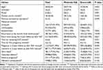
Figure 2 depicts the procedural steps involved in the patients screening for the study, including inclusion and exclusion. A total of 156 individuals diagnosed with IUA were included in the study, wherein 67.31% (105/156) exhibited moderate adhesions, and 32.69% (51/156) presented with severe adhesions as per the AFS grading. All participants concluded a comprehensive 2-year follow-up as of October 2020. The preoperative characteristics of the entire cohort of IUA patients, encompassing parameters such as age, gravidity, parity, disease course (month), and the number of prior intrauterine surgeries, which were heteroscedasticity, are comprehensively depicted in Table 1.
 |
Table 1 Statistical Analysis of Clinical Parameters and Post-Treatment Characteristics of Patients with IUA |
 |
Figure 2 Sample inclusion process. Note: Criteria from American Fertility Society (AFS) was adopted for intrauterine adhesion classification. |
Menstrual Changes
The participants were monitored for enhancements in menstrual patterns 3 months post-surgical intervention. Prior to the procedure, 137 individuals exhibited aberrant menstruation, characterized by diminished menstrual quantity and instances of amenorrhea. We restricted the comparison of menstrual volume to an increase of one-third as “increased.” Subsequent to treatment, 91.97% (126/137) of the participants manifested an increased in menstrual flow. The improvement rates for individuals with moderate and severe IUAs were 92.31% (84/91) and 91.31% (42/46), respectively. However, these differences were not statistically significant (Table 1). Notably, among the cohort of 8 patients experiencing preoperative amenorrhea (comprising 2 cases of moderate IUAs and 6 cases of severe IUAs), menstruation resumed subsequent to the surgical intervention.
Intrauterine Repair and Stent Removal Time
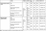
The evaluation of uterine cavity morphology recovery was conducted on two occasions: during the hysteroscopic second exploration and the 2-year follow-up after the stent removal. There was a significant difference at the two as the recurrence rates were 11.54% (18/156) and 32.69% (51/156), respectively. Among those patients, the all 18 cases who recurrent at the hysteroscopic second exploration, received another operations, while during the 2-year follow-up, there were only 23 cases (8 in moderate IUAs and 15 in severe IUAs) received a second or more operations while other 28 women refused for varied reasons, including a sense of hopelessness regarding future childbearing and the absence of additional reproductive requirements post-delivery. Furthermore, patients with severe IUA exhibited a significantly higher recurrence rate compared to those with moderate adhesions at both assessment time points (Table 1). Notably, for most patients with severe IUA, the duration of intrauterine stent placement was markedly longer compared to those with moderate adhesions, as depicted in (Figure 3a). Preoperative severity of IUA emerged as an independent risk factor for uterine re-adhesion within the 2-year period, as determined through both univariate and multivariate logistic regression analyses (Table 2).
 |
Table 2 Univariate and Multivariate Logistic Regression Analysis of Adhesion Recurrence After 2 years of Follow-Up |
Early Pregnancy Outcome
Subsequent to the removal of the stent, the participants were meticulously monitored for pregnancy, and the same was continued for a duration of 24 months. Examination of median statistics revealed that individuals afflicted with moderate to severe IUA typically experienced their first pregnancy subsequent to stent removal within a median period of 5 (3, 12) months from the procedural intervention (Table 1). Notably, a noteworthy 71.79% (112/156) of patients achieved early pregnancy subsequent to the intervention, while 80.36% (90/112) conceived within the initial year. The cumulative pregnancy rate exhibited a prominent increase primarily within the initial 8 months post-stent removal, followed by a plateauing of the rate of increase. A notable difference in pregnancy rates emerged between patients with moderate and severe IUA (P = 0.004), with the former demonstrating a statistically significant superiority over the latter, as depicted in both Table 1 and (Figure 3b).
Obstetric Outcomes
During the observational period, a successful delivery of newborns was achieved by 79.28% (88/111) of the pregnant women, while four of them had previously encountered adverse pregnancy outcomes prior to the successful deliver and only 1 patients had an ongoing pregnancy. Adverse pregnancy meant the pregnancy loss due to various causes including spontaneous abortion, ectopic pregnancy, stillbirth, etc in Figure 4. And there were 27 women (the 4 women mentioned above who were finally achieved successful pregnancy were also included) have once suffered pregnancy loss with 20 cases (24.10%, 20/83) in moderate group and 7 cases (25.00%, 7/28) in severe group which had no significant difference. Among the pregnant patients, 4.50% (5/111) employed assisted reproductive technology (ART) to facilitate reproduction.15 Each pregnant woman underwent delivery only once, all instances involved singleton pregnancies. The incidence of full-term deliveries constituted 70.27% (78/111), whereas cesarean section procedures were notably prevalent at 67.05% (59/88). There was no statistically significant difference in live birth rates between patients exhibiting moderate and severe IUA (P = 0.915). Furthermore, there were no significant differences in the rates of full-term labor (P = 0.201) or spontaneous delivery (P = 0.513) (Table 1). Initial univariate logistic regression analysis was conducted to examine variables influencing the IUA pregnancy rate (Table 3), and subsequently, significant variables were incorporated into a multivariate logistic regression reanalysis. The latter revealed a significant correlation between the severity of preoperative IUA, recurrence of adhesions within 2 years, and pregnancy rates in women with IUA (Table 3). Notably, both the severity and recurrence of IUA emerged as potential risk factors for infertility. The obstetric complications we analysed in the study were placenta implantation, placenta previa, postpartum hemorrhage and hysterectomy. It manifested in 21.59% (19/88) of pregnancies, with no significant difference observed between moderate and severe cases (P = 0.100, Table 1). Placental implantation constituted the most prevalent complication, accounting for 15.91% (14/88), primarily detected during labor and delivery based on patient reports or documented in the medical records of delivery.
 |
Table 3 Pregnancy-Related Univariate and Multivariate Logistic Regression Analysis |
Discussion
The present study reveals a notable improvement of 91.97% in postoperative menstrual flow among patients diagnosed with IUA post the intervention, a result consistent with findings in existing literature.16,17 Despite this commendable outcome, the postoperative pregnancy rate remains suboptimal, suggesting that the improvement in menstrual flow may not comprehensively reflect the therapeutic efficacy of IUA treatment. This departure is noteworthy given that several prior studies limited their assessments to a span of 2 weeks to 3 months post-surgery,18,19 with limited exploration into the incidence of distant recurrence. This study contributes to the existing body of scholarly work by systematically exploring novel modalities to enhance and refine the long-term prognostic landscape for individuals affected with IUA.
Our findings indicate that the recurrence rate of adhesions, evaluated twice in this cohort, is lower compared with previous reports20 (recurrence rate of 30% to 66% with non-stent methods), while the pregnancy rate increased significantly, indicating that this approach has clinical utility. Through a comprehensive analysis of both short- and long-term follow-up data pertaining to IUA, it becomes evident that adhesion recurrence poses a formidable challenge in the course of treatment. Furthermore, the proclivity for recurrence is heightened among patients presenting with severe adhesions.21 When the study was initiated several years ago, we chose the intrauterine stent as the postoperative maintenance treatment for intrauterine adhesion (IUA). Considering the significant bias and uncertainty for using other therapeutic schedule as controls, we finally designed the case control study and self-control study. In spite of these meaningful findings, the retrospective study and the lack of a completely blank control may have weakened the results of our efficacy analysis. In the future, we hope to conduct rigorous case-control studies or large-scale multicenter clinical trials to validate these findings.
In this study, we creatively discover a new modified stent (chitosan anti-adhesion membrane-coated intrauterine device). Prior research findings have suggested that early hysteroscopic examinations can proactively detect nascent membranous adhesions,22 allowing for their timely removal prior to vascularization and robust cicatrization. The timing of the subsequent examination is contingent upon the efficacy and duration of postoperative anti-adhesion therapies. The durability of the stainless-steel ring contributes to sustaining the uterine cavity’s form for an extended period, affording the endometrium ample time for functional repair.13 Additionally, the chitosan anti-adhesion membrane, with a typical degradation period of 2 to 3 months within the cavity, mitigates direct contact between the stainless-steel ring arms and the uterine wall. This preventive measure reduces the risk of the ring arms becoming embedded during the susceptible recurrent adhesion phase.23 Moreover, functioning as a physical barrier, the chitosan anti-adhesion membrane serves to diminish sterile endometrial inflammation induced by conventional stents.24,25 Significantly, this modified stent is readily accessible and straightforward to prepare in a clinical context, although there is an unavoidable disadvantage of the stent that it could not provide support at the bilateral uterine cornua positions which may easily lead to adhesion recurrence at specific sites. Nevertheless, it is imperative to acknowledge that a severely compromised endometrium cannot be solely rectified through the placement of an intrauterine stent. Therefore, a comprehensive treatment approach, which consists of a skillful operation of HA, postoperative placement of intrauterine stents, administration of medications such as estrogen, and the timely hysteroscopic reevaluation, is strongly recommended for patients with IUA.26
In this study, it was discerned that the duration of the intrauterine stent placement procedure was longer in individuals with severe IUA in contrast to those with moderate IUA. This implies that prior attention should be devoted to the restoration of the uterine cavity and endometrium before contemplating the removal of the intrauterine device (IUD). It is advisable that this assessment be conducted concomitantly with the utilization of noninvasive 3-D ultrasound, as 3D ultrasound can three-dimensionally reconstruct the structure of the uterine cavity, visually displaying the presence of adhesions, as well as the location, extent, and severity of intrauterine adhesions.27 What’s more, unlike hysteroscopy, it is simple to operate and non-invasive, making it more suitable for long-term follow-up.28
The collective postoperative pregnancy rates among the participants in this study surpassed those documented in prior studies.29,30 Nevertheless, the attainment of pregnancy proved to be a more difficult task in the severe IUA cohort as opposed to the moderate group. The severity of IUA and the recurrence of adhesions may constitute potential risk factors for secondary infertility. This information serves as a valuable point of reference for clinicians in refining treatment modalities and appraising the fertility outcomes of their patients. For individuals with IUA expressing fertility aspirations, the principal aim of treatment should be the enhancement of live birth rates.31 Nonetheless, studies have uncovered a heightened incidence of adverse pregnancy and obstetric complications among patients with IUA, which were no differences reported between the moderate IUA patients and the severe patients. Thses may result from the limitation of sample size, and the occurrence of abortion and pregnancy complications is associated not only with intrauterine environmental abnormalities but also with other pathogenic mechanisms. Therefore, it is necessary to conduct more extensive clinical and basic research to validate the relationship between intrauterine adhesions and these diseases.
Owing to a history of recurrent miscarriages involving repetitive dilatation and curettage procedures, individuals may undergo multiple intrauterine operations, thereby elevating the likelihood of IUAs.32 Consequently, the presence of IUAs amplifies the susceptibility to recurrent miscarriage, ectopic pregnancy, preterm labor, and placental abnormalities, establishing a cyclic pattern of adverse outcomes. Individuals experiencing pregnancies subsequent to IUAs, which are categorized as high-risk due to obstetric complications, commonly opt for cesarean sections to conclude the pregnancy. Predominant complications encompass placental implantation, placenta previa, and postpartum hemorrhage, some of which may necessitate hysterectomy. Primary causative factors for adverse obstetric and neonatal outcomes are attributed to placental hemodynamic disorders and placental implantation issues. Consequently, it is imperative to enhance both prenatal and intrapartum monitoring for individuals with post-IUA pregnancies. This need arises from the persistence of risks associated with IUAs beyond the gestational stage, as they transition from the infertile stage to the gestational stage.
Conclusions
In this study, we conducted a retrospective analysis of patients presenting with moderate-to-severe IUA to systematically evaluate the pregnancy outcomes following HA surgery combined with a modified intrauterine stent. The intrauterine stent utilized in this study yielded favorable outcomes within the cohort of patients with IUA, especially the startling pregnancy rate data. Besides, the gravity of IUA severity is not only the risk factor for adhesion recurrence but also as a principal determinant of infertility. Nearly half of the patients became pregnant in 3 months after the treatment, suggesting that it is necessary to seize the short time after the stent removal to actively prepare for pregnancy. At the same time, the follow-up obstetric outcomes also remind us to pay attention to the obstetric complications of IUA patients.
The complexities surrounding the post-treatment pregnancy status in patients with IUA introduce a nuanced perspective, necessitating a comprehensive consideration of the incidence of obstetric complications. It is unequivocally apparent that a refinement and supplementation of existing criteria are imperative to judiciously appraise the suitability of the intrauterine environment for gestation in individuals afflicted with IUA. There are also some limitations. This study only has self-control without strict external control group, which limits the wide promotion of the research results. Second, this study retrospectively analyzed the clinical data from 5 years ago. Although the interference scheme of stent therapy is the mainstream idea of IUA treatment and reflects the clinical practicability, there may still be a gap with the effect of new materials such as bioactive materials. In the future, we will hope to make up for this deficiency through multi-center rigorous clinical research and the application of advanced scaffolds.
Data Sharing Statement
All data generated or analysed during this study are included in this article. Further enquiries can be directed to the corresponding author.
Ethics Approval and Consent to Participate
Prior to their participation, all enrolled patients provided informed consent, and ethical approval for the study was obtained from the Ethics Committee of the First Affiliated Hospital of Chongqing Medical University (No.2020-472). This study was conducted in accordance with the declaration of Helsinki.
Acknowledgments
We would like to acknowledge the hard and dedicated work of all the staff that implemented the intervention and evaluation components of the study.
Funding
This study has received grants from Chongqing Health Commission Research funds, No: 2021MSXM153.
Disclosure
The authors declare that they have no competing interests in this work.
References
1. Khan Z, Goldberg JM. Hysteroscopic management of Asherman’s syndrome. J Minim Invasive Gynecol. 2018;25(2):218–228. doi:10.1016/j.jmig.2017.09.020
2. Santamaria X, Isaacson K, Simon C. Asherman’s Syndrome: it may not be all our fault. Hum Reprod. 2018;33(8):1374–1380. doi:10.1093/humrep/dey232
3. Wu F, Lei N, Yang S, et al. Treatment strategies for intrauterine adhesion: focus on the exosomes and hydrogels. Front Bioeng Biotechnol. 2023;11:1264006.
4. Di Spiezio-Sardo A, De Angelis MC, Dimitrios K, et al. restoring fertility of patients with severe asherman’s syndrome in the office setting: a step-by-step recipe for success. J Minim Invasive Gynecol. 2023;30(5):355–356. doi:10.1016/j.jmig.2023.02.002
5. Santamaria X. The American fertility society classifications of adnexal adhesions, distal tubal occlusion, tubal occlusion secondary to tubal ligation, tubal pregnancies, Müllerian anomalies and intrauterine adhesions. Fertil Sterility. 1988;49:944–955.
6. Hooker AB, de Leeuw RA, Emanuel MH, et al. The link between intrauterine adhesions and impaired reproductive performance: a systematic review of the literature. BMC Pregnancy Childbirth. 2022;22(1):837. doi:10.1186/s12884-022-05164-2
7. Rodríguez-Eguren A, Bueno-Fernandez C, Gómez-álvarez M, et al. Evolution of biotechnological advances and regenerative therapies for endometrial disorders: a systematic review. Hum Reprod Update. 2024;30:584–613.
8. Zhao J, Chen Q, Cai D, et al. Dominant factors affecting reproductive outcomes of fertility-desiring young women with intrauterine adhesions. Arch Gynecol Obstet. 2017;295(4):923–927. doi:10.1007/s00404-017-4314-z
9. Mortimer RM, Lanes A, Srouji SS, et al. Treatment of intrauterine adhesions and subsequent pregnancy outcomes in an in vitro fertilization population. Am J Obstet Gynecol. 2024;231(5):536.e1–536.e10. doi:10.1016/j.ajog.2024.05.026
10. Zhao Y, Huang X, Huang R, et al. A retrospective cohort study to examine factors affecting live birth after hysteroscopic treatment of intrauterine adhesions. Fertil Steril. 2024;121(5):873–880. doi:10.1016/j.fertnstert.2024.01.022
11. Luo Y, Sun Y, Huang B, et al. Effects and safety of hyaluronic acid gel on intrauterine adhesion and fertility after intrauterine surgery: a systematic review and meta-analysis with trial sequential analysis of randomized controlled trials. Am J Obstet Gynecol. 2024;231:36–50.
12. wang Y, Song X, Wu S, et al. Comparison of autocross-linked hyaluronic acid gel and intrauterine device for preventing intrauterine adhesions in infertility patients: a randomized clinical trail. Minim Invasiv Ther. 2020;9:1025–1027.
13. Xu P, Xu H, Lu Q, et al. Reproductive outcomes following copper-containing intrauterine device after hysteroscopic lysis for intrauterine adhesions. Exp Ther Med. 2024;27(4):175. doi:10.3892/etm.2024.12463
14. Miguel SP, Moreira AF, Correia IJ. Chitosan based-asymmetric membranes for wound healing: a review. Int J Biol Macromol. 2019;127:460–475.
15. Dan S, Zhao X, Huang H, et al. In vitro fertilization and embryo transfer may improve live birth rate for patients with intrauterine adhesions after hysteroscopic adhesiolysis. Zhong Nan Da Xue Xue Bao Yi Xue Ban. 2022;47:1559–1567.
16. Han L, Shi G, Zheng A, et al. Hysteroscopy for retained products of conception: a single-institution experience. BMC Women's Health. 2023;23(1):25. doi:10.1186/s12905-023-02170-0
17. Wang Y, Yin LL, Sun XF, et al. Retrospective analysis of autologous bone marrow mesenchymal stem cells as adjuvant therapy in recurrent intrauterine adhesions. Arch Gynecol Obstet. 2025;311(3):789–799. doi:10.1007/s00404-025-07952-5
18. Zhang J, Shi C, Sun J, Niu J. Analysis of factors affecting the prognosis of patients with intrauterine adhesions after transcervical resection of adhesions. Fertil Steril. 2024;122(2):365–372. doi:10.1016/j.fertnstert.2024.03.016
19. Dou Y, Yu T, Li Z, et al. Short- and long-term outcomes of postoperative intrauterine application of hyaluronic acid gel: a meta-analysis of randomized controlled trials. J Minim Invasive Gynecol. 2022;29(8):934–942. doi:10.1016/j.jmig.2022.05.006
20. Zhao J, Zhang L, Liang J, et al. The effect of the combined use of an intrauterine device and a Foley balloon in the prevention of adhesion following hysteroscopic adhesiolysis. Women Health. 2023;63(1):1–7. doi:10.1080/03630242.2022.2144984
21. Feng L, Sun Y, Zhang S, et al. A novel intrauterine estrogen-releasing system for preventing the postoperative recurrence of intrauterine adhesion: a multicenter randomized controlled study. BMC Med. 2024;22:395.
22. Dilbaz B. Elevating gynecologic surgery practice report: practice guidelines on intrauterine adhesions developed in collaboration with the European Society of Gynaecological Endoscopy (ESGE). J Minim Invasive Gynecol. 2017;24:695–705.
23. Matsubara S. A novel uterine stent for preventing intrauterine adhesion: not only gynecologic but also obstetric significance. Ann Transl Med. 2020;8(10):614–623. doi:10.21037/atm.2020.03.65
24. Yu Z, Min Y, Ouyang Q, et al. Study on an injectable chitosan-lignin/ poloxamer hydrogel loaded with platelet-rich plasma for intrauterine adhesion treatment. Polymers. 2025;17:474.
25. Zhang H, Shao L, Wang L, et al. Chitosan combined with intrauterine device prevents intrauterine adhesions after hysteroscopic adhesiolysis: a target trial emulation study. J Obstet Gynaecol Res. 2023;49:1571–1578.
26. Guo J, Shi X, Yu F, et al. Adjuvants to prevent reformation of adhesions following adhesiolysis for Asherman syndrome: a systematic review and meta-analysis. Hum Fertil. 2023;26:797–814.
27. Burjoo A, Zhao X, Zou L, et al. The role of preoperative 3D-ultrasound in intraoperative judgement for hysteroscopic adhesiolysis. Ann Transl Med. 2020;8(4):55–61. doi:10.21037/atm.2020.01.06
28. Cao C, Chen Y, Li J, et al. Short-term reproductive outcomes analysis and prediction of the modified uterine stent treatment for mild to moderate intrauterine adhesions: experience at a single institution. BMC Women’s Health. 2024;24.
29. Qing F, Gao BS, Huang H, et al. Obstetrical outcome in the third trimester after hysteroscopic adhesiolysis. Ann Transl Med. 2020;8:51–59.
30. Zhang L, Zhao W, Yang B, Xu Y, Feng L. Clinical results of severe intrauterine adhesion women following a comprehensive management. Chin J Clin Obstet Gynecol. 2017;18:221–223.
31. Zhang D, Du Q, Li C, et al. Functionalized human umbilical cord mesenchymal stem cells and injectable HA/Gel hydrogel synergy in endometrial repair and fertility recovery. ActaBiomater. 2023;167:205–218. doi:10.1016/j.actbio.2023.06.013
32. Chung JPW, Law TSM, Ng K, et al. Intrauterine adhesion in ultrasound- guided manual vacuum aspiration (USG-MVA) versus electric vacuum aspiration (EVA): a randomised controlled trial. BMC Pregnancy Childbirth. 2024;24(1):135. doi:10.1186/s12884-024-06328-y
 © 2025 The Author(s). This work is published and licensed by Dove Medical Press Limited. The
full terms of this license are available at https://www.dovepress.com/terms.php
and incorporate the Creative Commons Attribution
- Non Commercial (unported, 4.0) License.
By accessing the work you hereby accept the Terms. Non-commercial uses of the work are permitted
without any further permission from Dove Medical Press Limited, provided the work is properly
attributed. For permission for commercial use of this work, please see paragraphs 4.2 and 5 of our Terms.


