Back to Journals » Drug Design, Development and Therapy » Volume 19
Rho Kinase Inhibitors in Glaucoma Management: Current Perspectives and Future Directions
Authors Chatzimichail E, Christodoulaki E, Konstas PAG, Tsiropoulos GN , Amaxilati E , Gugleta K, Gatzioufas Z, Panos GD
Received 31 December 2024
Accepted for publication 28 March 2025
Published 2 April 2025 Volume 2025:19 Pages 2519—2531
DOI https://doi.org/10.2147/DDDT.S515166
Checked for plagiarism Yes
Review by Single anonymous peer review
Peer reviewer comments 2
Editor who approved publication: Professor Frank Boeckler
Eleftherios Chatzimichail,1 Eirini Christodoulaki,1 Panagiotis AG Konstas,2 Georgios N Tsiropoulos,3 Efstratia Amaxilati,3 Konstantin Gugleta,1 Zisis Gatzioufas,1,* Georgios D Panos3,4,*
1Department of Ophthalmology, University Hospital of Basel, Basel, 4031, Switzerland; 2Eye Centre of Greece, Pylaia, Thessaloniki, Greece; 3First Department of Ophthalmology, AHEPA University Hospital, School of Medicine, Aristotle University of Thessaloniki, Thessaloniki, Greece; 4Division of Ophthalmology and Visual Sciences, School of Medicine, University of Nottingham, Nottingham, UK
*These authors contributed equally to this work
Correspondence: Georgios D Panos, First Department of Ophthalmology, AHEPA University Hospital, School of Medicine, Aristotle University of Thessaloniki, Kiriakidi 1, Thessaloniki, 54636, Greece, Tel +30 231 330 3110, Email [email protected]
Abstract: Glaucoma is a group of eye conditions characterised by optic nerve damage and visual field loss, representing the leading cause of irreversible blindness worldwide. Glaucoma exerts substantial global impact on visual impairment and blindness. The management of glaucoma has traditionally relied on medications such as prostaglandin analogs, beta-blockers, alpha agonists, and carbonic anhydrase inhibitors, which aim to lower intraocular pressure through various mechanisms. Rho kinase (ROCK) inhibitors have recently emerged as a novel class of antiglaucoma drugs, offering an alternative approach by enhancing aqueous humour outflow through the conventional pathway. Recent clinical studies assessing the efficacy and safety of Ripasudil (K-115) and Netarsudil (AR-13324) have demonstrated promising outcomes in the treatment of various types of glaucoma. Comparative studies have shown that ROCK inhibitors are non-inferior to traditional antiglaucomatous medications, such as beta-blockers and prostaglandins. Additionally, emerging evidence suggests their neuroprotective properties, which may play a role in preserving retinal ganglion cells. Furthermore, positive outcomes have been observed when these agents are used in conjunction with glaucoma filtering surgery, potentially enhancing surgical success rates. Adverse effects, including conjunctival hyperemia, cornea verticillata, conjunctivitis, and blepharitis, have been reported following the use of ROCK inhibitors. However, those side effects appear to be subtle in most cases. This review aims to provide an overview of ROCK inhibitors, focusing on their mechanisms of action, clinical efficacy, safety profiles, and additional benefits for eye health. Furthermore, further potential applications of ROCK inhibitors in glaucoma management are going to be discussed.
Keywords: Rho-associated protein kinase, Rho-kinase, ROCK inhibitors in glaucoma, ripasudil, netarsudil, K-115, AR-13324
Introduction
Glaucoma encompasses a group of conditions defined by the cupping of the optic nerve head and damage to the visual field.1 This eye condition is categorised into primary or secondary types and further into open-angle or closed-angle variants. Glaucoma is the primary cause of irreversible blindness globally.2 It is estimated that 3.5% of individuals aged 40 to 80 worldwide are affected by glaucoma. As of 2010, glaucoma accounted for blindness in one out of every 15 blind individuals and was the cause of visual impairment in one out of every 45 people with vision problems.3
To this day, glaucoma management is primarily based on medications such as prostaglandin analogs, beta-blockers, alpha agonists, and carbonic anhydrase inhibitors, which reduce the production of aqueous humour or increase its outflow. Combination therapies are also common, especially in patients who do not respond adequately to a single medication. However, the use of multiple antiglaucoma medications to lower IOP is often associated with ocular surface impairment, largely due to increased exposure to preservatives, which can also lead to reduced adherence to antiglaucoma therapy.4,5
Rho kinase (ROCK) inhibitors represent a new class of antiglaucoma medications that have been approved by the FDA for the reduction of IOP. ROCK inhibition primarily reduces IOP by enhancing the outflow of aqueous humour through the conventional pathway, which includes the trabecular meshwork and Schlemm’s canal.6
Considering the above, the primary goal of this review is to highlight current and future perspectives on the use of Rho kinase inhibitors in the pharmacological and surgical management of glaucoma. Our review will explore various aspects, including mechanism of action, clinical efficacy, safety profile, additional positive effects for the eye and potential future applications of Rho kinase inhibitors.
Methods
For this narrative review, we conducted a search in PubMed, Embase and Scopus using the keywords “(Rock Kinase Inhibitors) AND (Glaucoma)”. These searches were performed between 20 October 2024, and 20 December 2024. To be included in the review, studies needed to focus on either the properties of ROCK kinase inhibitors or their clinical implementation in glaucomatous conditions. Studies without manuscript in English were excluded.
Rho Kinase Inhibitors, Mechanism of Action in Glaucoma
In humans, both ROCK1 and ROCK2 are present in the eye but have distinct distribution patterns in other tissues. ROCK1 is predominantly found in non-neural tissues such as the heart, lungs, and skeletal muscles, whereas ROCK2 is mainly localised in the brain.7 ROCK1 and ROCK2 have an overall amino acid sequence similarity of 65%, with a 92% similarity within their kinase domain.8
Rho kinase is a serine/threonine kinase that acts as a key downstream effector of Rho guanosine triphosphatase (Rho GTPase).9 The Rho family of GTPases comprises small signaling G proteins, approximately 21 kDa in size, which are guanine nucleotide-binding proteins found in the cytosol. This family is categorised into three main subgroups: Rho, Rac, and Cdc42. The Rho family GTPases are activated by various molecules, including endothelin-1, lysophosphatidic acid, thrombin, angiotensin II, cytokines, transforming growth factor-β, or through integrin interactions with the extracellular matrix In its GTP-bound state, Rho activates downstream effectors such as ROCK, which phosphorylates substrates like Myosin light chain phosphatase, LIM-kinase, CPI-17, and others.10 This cascade regulates actin cytoskeletal dynamics, actin-myosin contraction, cell adhesion, stiffness, morphology, proliferation, apoptosis, and extracellular matrix remodeling. With focus on IOP-lowering effect, ROCK inhibitors impact the cytoskeleton of trabecular meshwork and Schlemm’s canal cells by reducing the density of actin stress fibers.11
The Role of Rho Kinase Inhibitors in the Glaucoma Treatment Algorithm: Clinical Evidence
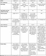
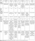
In this section, we provide a comprehensive summary of relevant clinical studies that highlight the efficacy of ROCK inhibitors in the treatment of glaucoma. We specifically examine three key aspects: their effects on IOP reduction, their neuroprotective properties, and their impact on wound healing and their role against postoperative scar formation. Table 1 and Table 2 present an extensive overview of the key findings from these studies. Ripasudil (K-115) and Netarsudil (AR-13324) are the Rho-kinase inhibitor drugs currently available in the market. Netarsudil is primarily used as an adjunct treatment for glaucoma in the United States.
 |
Table 1 Overview of Most Prominent Clinical Studies on Ripasudil (K-115) in Glaucoma |
 |
Table 2 Overview of Most Prominent Clinical Studies on Netarsudil (AR-13324) in Glaucoma |
Ripasudil (K-115)
Tanihara et al in 2013 in a phase 1 clinical trial investigated the effect of K-115 (Ripasudil) in healthy volunteers.12 Ripasudil induced a significant reduction of IOP from baseline across all tested concentrations within 1 to 2 hours, with the highest concentration (0.8%) achieving a reduction of −4.3 mmHg.
The same research group conducted a multicentre, prospective study to elucidate the effects of 0.4% ripasudil on reducing IOP.13 Patients, diagnosed with open-angle glaucoma or ocular hypertension, were administered 0.4% Ripasudil twice daily for a duration of 52 weeks. 388 patients were included in this study. Ripasudil demonstrated significant IOP-lowering effects over 52 weeks across all analyses, including monotherapy, add-on therapy, and both subgroups (baseline IOP ≥21 mmHg and <21 mmHg) of monotherapy. At week 52, the mean reductions in IOP at trough and peak were 2.6 and 3.7 mmHg for monotherapy, and 1.4 and 2.4 mmHg, 2.2 and 3.0 mmHg, and 1.7 and 1.7 mmHg, respectively, for the various add-on therapy groups, demonstrating the IOP-lowering effects of ripasudil.
Moreover, Ripasudil has demonstrated favourable outcomes in achieving long-term IOP reduction. The J-ROCK study was a prospective observational trial, that included 3178 patients to evaluate the effectiveness of Ripasudil in treating glaucoma or ocular hypertension.14 Ripasudil significantly reduced IOP from baseline, with a least-squares mean ± standard error change of −2.6 ± 0.1 mmHg at 24 months (p < 0.001). Significant IOP reductions were observed across four types of glaucoma: primary open-angle glaucoma, normal-tension glaucoma, primary angle-closure glaucoma, secondary glaucoma, and ocular hypertension. Regarding side effects, the most common were blepharitis (8.6%), conjunctival hyperaemia (8.5%), and conjunctivitis (6.3%).
In secondary types of glaucoma, Ripasudil has demonstrated significant efficacy in reducing IOP. ROCK-S was a retrospective multicentre study that included 332 eyes with uveitic, exfoliation, and steroid-induced glaucoma, all of which were treated with 0.04% Ripasudil.6 The average reductions in IOP from baseline were −5.86 ± 9.04 mmHg at 1 month, −6.18 ± 9.03 mmHg at 3 months, and −7.00 ± 8.60 mmHg at 6 months. All reductions in IOP were statistically significant (p < 0.0001), with those observed in uveitic and steroid-induced glaucoma being significantly greater than those in exfoliation glaucoma.
Netarsudil (AR-13324)
A phase 2 clinical study from Araie et al included 215 patients, who after randomisation received either Netarsudil 0.01%, 0.02%, 0.04%, or placebo, and were treated once-daily for overall 4 weeks.16 By week 4, the average decrease in mean diurnal IOP from baseline was 4.10 mmHg (19.8%), 4.80 mmHg (23.5%), 4.81 mmHg (23.8%), and 1.73 mmHg (8.2%) for the respective Netarsudil concentrations, showing statistically significant reductions (p < 0.0001) for all concentrations of Netarsudil compared to placebo. Adverse events were observed in a concentration-dependent pattern, with the highest incidence reported in the Netarsidul 0.04% group (68.6%) and the lowest in the placebo group (9.1%). Conjunctival hyperaemia was the most commonly reported adverse effect.
Furthermore, Netarsudil 0.02% was evaluated in a multicentre phase 4 study in a sample of 261 patients, with open-angle glaucoma or ocular hypertension.15 Netarsudil 0.02% was administered once daily for 12 weeks. Mean IOP decrease in patients who were using Netarsudil as monotherapy was 16.9%. Moreover, IOP decreased 2.5% more in patients treated with Netarsudil compared to those treated with Prostaglandin analogues. It proved to be a well tolerated treatment, since only 11.2% discontinued due to adverse events. Overall, Netarsudil 0.02% solution as monotherapy or as an adjunct to other medications achieved clinically significant sustainable IOP reduction.
In a study conducted by Shiuey et al, 340 eyes from 233 glaucoma patients were analyzed to evaluate the clinical efficacy and safety of Netarsudil 0.02% in a tertiary care hospital setting.17 They found that Netarsudil significantly lowered IOP up to 6 months. In this retrospective study 48% experienced a ≥20% decrease in IOP at the 1-month, maintained through the 6-month visit. It should be noticed that in this study patients with previous laser or filtering surgery were included.
Comparative Studies
Netarsudil is a prodrug that undergoes hydrolysis in the cornea to form its active metabolite. This active compound exhibits a dual mechanism of action, functioning both as a Rho kinase (ROCK) inhibitor and a norepinephrine transporter (NET) inhibitor. Through ROCK inhibition, Netarsudil enhances trabecular meshwork relaxation and increases aqueous outflow. Additionally, NET inhibition contributes to a reduction in episcleral venous pressure through vasodilation and may also decrease aqueous humor production by affecting the ciliary body. This dual mechanism may explain its superior IOP-lowering effect compared to Ripasudil, which acts solely as a ROCK inhibitor.
It is important to acknowledge that many clinical trials evaluating Ripasudil report intraocular pressure (IOP) measurements at peak efficacy, typically around two hours post-instillation. While this approach highlights the maximum effect of the drug, it may overestimate its sustained efficacy throughout the dosing interval. However, long-term studies such as the J-ROCKET and ROCK-S trials have also included IOP measurements taken at trough (pre-dose), providing a more comprehensive assessment of Ripasudil’s effectiveness over time.6,18
Ripasudil (K-115) has demonstrated significant IOP-lowering effects in multiple clinical studies, including phase 1 trials and long-term observational studies. Studies by Tanihara et al and Futakuchi et al showed that Ripasudil effectively reduces IOP both as monotherapy and add-on therapy, with sustained effects over 24 months and notable efficacy in secondary glaucoma types.6,12–14 Common side effects include conjunctival hyperemia, blepharitis, and conjunctivitis. Similarly, Netarsudil (AR-13324) has shown strong IOP-lowering potential in various phase 2–4 studies, with significant reductions observed across different concentrations and treatment regimens, though conjunctival hyperemia remains a common side effect.15–17 Both drugs present promising options for glaucoma management with sustained efficacy and manageable adverse effects.
This part of the review highlights comparative studies, both between ROCK inhibitors and conventional antiglaucoma medications, as well as among different ROCK inhibitors themselves (see also Table 3). The most prominent studies in this field are the ROCKET and the Meteor studies.
 |
Table 3 Overview of the Comparative Studies on ROCK Inhibitors in Glaucoma Treatment |
Netarsudil 0.02% ophthalmic solution was evaluated for its efficacy and safety in treating open-angle glaucoma and ocular hypertension through multiple clinical trials. The ROCKET-1 and ROCKET-2 studies were double-masked, randomized noninferiority trials comparing once-daily (QD) and twice-daily (BID) Netarsudil to timolol 0.5% BID.19 Across 1167 patients, Netarsudil QD significantly reduced IOP and was found to be noninferior to timolol in patients with baseline IOP < 25 mm Hg. The most common adverse event was conjunctival hyperemia, affecting 50%-59% of the patients that received Netarsudil, compared to 8%-11% of timolol users. These results suggest that Netarsudil QD is an effective and well-tolerated treatment option.
ROCKET-2 was a 12-month multicenter trial that further assessed netarsudil’s long-term efficacy and safety in 756 patients.20 The study confirmed sustained IOP reductions, with Netarsudil QD lowering IOP to 17.9–18.8 mm Hg, Netarsudil BID to 17.2–18.0 mm Hg, and timolol to 17.5–17.9 mm Hg. In addition to conjunctival hyperemia (61%-66% for Netarsudil vs 14% for timolol), cornea verticillata (25%-26%) and conjunctival hemorrhage (19%-20%) were observed in Netarsudil users but were mostly mild and did not impact visual function.
ROCKET-4 Study was a randomised phase 3 study that compared Netarsudil 0.02% with timolol 0.5%. 186 patients from each therapeutic group were included.21 Both Netarsudil and timolol demonstrated significant reductions in IOP from baseline (P < 0.001 for both). Overall Netarsudil met the criteria for noninferiority to timolol. Netarsudil has also demonstrated a comparable IOP-lowering effect to brimonidine when used as an adjunctive antiglaucoma treatment.22
Moreover, the findings from the MERCURY-3 study demonstrate that once-daily Netarsudil-latanoprost is non-inferior to bimatoprost/timolol in reducing IOP in patients with open angle glaucoma and ocular hypertension.23 Both treatments exhibited comparable efficacy, with no statistically significant differences in mean diurnal IOP.
With regard to comparative studies between Netarsudil and Ripasudil, J-Rocket study was a randomised phase 3 study, that included 244 Patients, who were randomly assigned in a 1:1 ratio to receive either Netarsudil 0.02% or Ripasudil 0.4% for a duration of 4 weeks.18 At week 4, Netarsudil 0.02% demonstrated a significantly greater IOP-lowering effect compared to Ripasudil 0.4%, achieving a mean difference of −1.74 mmHg. Adverse effects were less common with Netarsudil 0.02% compared to Ripasudil 0.4%, reported at rates of 59.8% and 66.7%, respectively.
In addition, a meta-analysis conducted by Lee et al incorporated four studies to draw conclusions about the IOP-lowering effects of Netarsudil/Latanoprost combination therapy compared to Latanoprost monotherapy.25–29 In patients treated with Netarsudil/Latanoprost combination therapy compared to those on latanoprost monotherapy, the mean difference in IOP reduction was −2.41 mmHg (95% confidence interval [CI], −2.95 to −1.87) after 2 weeks and −1.77 mmHg (95% CI, −2.31 to −1.87) after 4 to 6 weeks of treatment. On the other hand, latanoprost monotherapy demonstrated a greater IOP-lowering effect than Netarsudil monotherapy after 4 to 6 weeks of use, with a mean difference of 0.95 mmHg.
Furthermore, a prospective randomised study evaluated the efficacy of a fixed-dose combination of Ripasudil and Brimonidine compared to Ripasudil or Brimonidine administered individually.24 A total of 18 subjects (six per group) were included. The combination of Ripasudil and brimonidine significantly reduced IOP from baseline at 1 hour on days 1 and 8 (12.7 vs 9.1 and 9.0 mmHg) and achieved greater IOP reductions than Ripasudil or brimonidine at multiple time points. Mild conjunctival hyperaemia was the most common adverse reaction, peaking 15 minutes post-instillation with combination of Ripasudil and brimonidine or Ripasudil. Overall, the combination of Ripasudil and brimonidine significantly lowered IOP compared to either agent used individually.
Comparative studies have shown that ROCK inhibitors, such as Netarsudil and Ripasudil, are effective in lowering intraocular pressure (IOP) and can be comparable to conventional glaucoma treatments like timolol and brimonidine. Some studies indicate that Netarsudil may provide superior IOP reduction compared to Ripasudil, with fewer adverse effects. Combination therapies, such as ROCK inhibitors with prostaglandin analogs or alpha agonists, have demonstrated enhanced IOP-lowering efficacy compared to monotherapy.
Rho Kinase Inhibitors as Neuroprotective Agents
Although the intraocular pressure-lowering effects of ROCK inhibitors have been thoroughly documented over the past decade, increasing emphasis has recently been placed on their neuroprotective potential. Research over the last several years suggests that ROCK inhibitors may offer additional benefits beyond IOP reduction, such as enhancing optic nerve blood flow, reducing retinal ganglion cell stress, and promoting cellular survival pathways—factors that collectively support neuroprotection in glaucoma patients.30,31
A series of studies have investigated the impact of ROCK inhibition on various neurodegenerative diseases. Reboussin et al investigated the neuroprotective potential of three ROCK inhibitors—Y-27632, Y-33075, and H-1152—using an ex-vivo retinal explant model.32 The ROCK inhibitor Y-33075 significantly improved RGC survival, reduced microglial activation, and downregulated pro-inflammatory and glial marker expression compared to other ROCK inhibitors. RNA-seq analysis revealed that Y-33075 suppressed the expression of M1 microglial markers (Tnfα, Il-1β, Nos2) and glial markers (Gfap, Itgam, Cd68). Additionally, it reduced processes such as apoptosis, ferroptosis, inflammasome activation, complement system activation, TLR signaling pathways, and the expression of genes like P2rx7 and Gpr84. In contrast, Y-27632 and H-1152 exerted no neuroprotective role in retinal ganglion cells. These findings suggest Y-33075’s potential as a promising neuroprotective and anti-inflammatory therapy for glaucoma.
Ripasudil has been shown to increase ocular blood flow in rabbits. In these studies ocular blood flow was measured through Laser speckle flowgraphy. In a study from Ohta et al. Ripasudil caused a concentration-dependent relaxation in isolated rabbit ciliary arteries that had been precontracted using a high-potassium solution.33 Similar results delivered another study with Ripasudil administered in the form of eye drops in rabbits.34
Application of Rho Kinase Inhibitors in Glaucoma Filtration Surgery
Failure of filtration surgery in glaucoma due to scarring with subsequent rise in intraocular pressure still constitutes a challenge in glaucoma surgery. Scar formation is the consequence of tissue remodeling with Tenon fibroblasts exerting the major effect35 Proliferation of subconjunctival fibroblasts begins as early as the third postoperative day.36 Fibroblasts promote the crosslinking of type I collagen and elastin, resulting in the formation of collagen supercoils and the development of dense scar tissue.37
Regarding the role of Rho kinase inhibitors in scar formation, in vitro studies have shown that the incubation of human tenon fibroblast with AMA0526 suppressed the differentiation of fibroblasts into myofibroblasts induced by TGF-β1.38 Y-27632, another rock inhibitor has shown also positive effect against postoperative scarring. Histological examination of rabbit tissues revealed that the topical application of Y-27632 markedly decreased subconjunctival collagen deposition.39
Clinical studies have further illuminated the efficacy of ROCK inhibitors in managing the postoperative course following filtration surgery. Muhlisah et al carried out a multicentre randomised clinical trial to assess the impact of Ripasudil on patients with open-angle glaucoma who underwent either trabeculectomy alone or trabeculectomy combined with cataract surgery, followed by a 3-month course of postoperative Ripasudil treatment. Participants were randomly assigned to either a group receiving the ROCK inhibitor Ripasudil or a group not receiving Ripasudil (non-Ripasudil group). The Ripasudil group demonstrated a significant reduction in the number of intraocular pressure- lowering medications required after trabeculectomy compared to the control group at both 24 months (p=0.010) and 36 months (p=0.016). However, the 3-year cumulative probability of surgical success did not differ significantly between the two groups.40 In addition, Mizuno et al investigated the effect of Ripasudil in needling procedures.41 A total of 27 eyes from 27 glaucoma patients who underwent a needling procedure with mitomycin C were included in the study. These patients were divided into two groups: those treated with Ripasudil (Ripasudil group), and those who did not receive Ripasudil (control group). Results showed that at 12 months post-needling, mean IOP decreased from 16.9 ± 4.5 to 12.6 ± 1.1 mmHg in the control group and from 16.0 ± 5.3 to 12.2 ± 1.2 mmHg in the Ripasudil group (p=0.77). Success rates at 12 months were 60.00% for the control group and 56.25% for the Ripasudil group (p=0.98). Overall, treatment with Ripasudil, following the needling procedure with mitomycin C did not yield superior outcomes compared to the needling procedure with mitomycin C alone at 12 months post-procedure.
Safety Profile
As with any new medication entering the market, the safety profile of ROCK inhibitors is of paramount importance. Clinicians aim to provide patients with well-tested treatments that have fully clarified safety profiles. The safety profile of ROCK inhibitors has been extensively documented in the scientific literature. Therefore, we present only a brief summary of their adverse effects in this review (see also Table 4).
 |
Table 4 Rock Inhibitors and Reported Adverse Effects |
In general, ROCK inhibitors have been linked to adverse events such as conjunctival hyperaemia, conjunctival hemorrhage, cornea verticillata, conjunctivitis, allergic conjunctivitis and blepharitis.42 Conjunctival hyperemia appears to reach its peak within 10 to 15 minutes after instillation and gradually resolves within 120 minutes.43,44 No major adverse effects after administration of ROCK Inhibitors has been documented. Ripasudil has been also associated with Honeycomb epithelial oedema and blepharitis more often in comparison to Netarsudil.45,46 Cornea verticillata has been almost exclusively linked with Netarsudil.47 A case of crystalline keratopathy secondary to Netarsudil administration has also been published.48 Further reported adverse effects after use of Netarsudil include punctal stenosis, transient myopic shift and bullous epithelial edema.42,49
Future Perspectives
ROCK inhibitors show great potential in glaucoma treatment. However, their topical application is often associated with conjunctival hyperaemia and limited intraocular bioavailability like mentioned above. To address these challenges, Mietzner et al proposed the use of poly (lactide-co-glycolide) (PLGA) microspheres loaded with fasudil (also known as HA-1077) in the form of depot formulation for intravitreal injection. Fasudil release from the microspheres was sustained for as long as 45 days (Figure 1).50 The microspheres varied in size from 3 to 67 µm. The release of fasudil led to a reduction in actin stress fibers within trabecular meshwork cells, Schlemm’s canal cells, and fibroblasts. However, recent scientific literature reveals a lack of significant progress in this area in recent years.
 |
Figure 1 Fasudil-loaded microspheres injected into the vitreous body. Notes: Reprinted from Mietzner R, Kade C, Froemel F et al. Fasudil Loaded PLGA Microspheres as Potential Intravitreal Depot Formulation for Glaucoma Therapy. Pharmaceutics. 2020;12(8):706. Creative Commons.50 |
Over the past decade, research on ROCK inhibitors has demonstrated promising results in the treatment of corneal diseases, particularly in conditions such as Fuchs Endothelial Dystrophy and procedures like Descemet Stripping Only.42 ROCK inhibitors appear to play a protective role in preserving endothelial cells, a benefit that extends also to the field of cataract surgery. Specifically, the perioperative and postoperative administration of ROCK inhibitors have shown efficacy in preventing endothelial cell loss following cataract surgery.51,52 Given these perspectives, we anticipate that ROCK inhibitors will experience broader application in managing a range of corneal diseases in the near future.
To date, the only available combination of ROCK inhibitors with conventional antiglaucoma eye drops is the combination of Netarsudil with latanoprost, marketed as ROCKLATAN (Netarsudil and latanoprost ophthalmic solution) 0.02%/0.005%. However, there is ongoing anticipation for further combinations of ROCK inhibitors with other glaucoma medications. Such combinations could offer a more comprehensive treatment strategy, enhancing IOP control while potentially reducing the need for surgical interventions.
Conclusion
ROCK inhibitors have emerged as promising therapeutic agents in glaucoma management, offering unique mechanisms of action that extend beyond IOP reduction. By enhancing trabecular meshwork outflow and exerting cytoprotective effects on ocular tissues, ROCK inhibitors offer a novel and complementary approach to traditional glaucoma treatments. Clinical studies have demonstrated their efficacy, showing significant reductions in IOP, noninferiority to conventional antiglaucoma therapies, and a manageable safety profile. Furthermore, the promising results of ROCK inhibitors in postoperative treatment following glaucoma filtration surgery along with their neuroprotective role highlight a crucial area of clinical research. Further studies are needed to determine whether ROCK inhibitors can be firmly established as part of the post-surgical regimen.
Looking towards the future, ROCK inhibitors hold great promise not only for enhancing glaucoma treatment but also for broader applications in ocular diseases, including corneal disorders and cataract surgery. The corneal endothelial cell-promoting and corneal clarity-enhancing properties of Ripasudil and Netarsudil suggest that these substances hold significant potential for a broad spectrum of applications in corneal surgery. Their ability to support endothelial cell function and maintain corneal transparency may pave the way for innovative therapeutic approaches, ultimately improving surgical outcomes and expanding treatment options in corneal surgery.
Future research should focus on refining formulations, reducing adverse effects, and exploring combination therapies to maximise therapeutic benefits. Moreover, advancements in drug delivery systems and long-term safety studies will be pivotal in expanding the clinical utility of ROCK inhibitors. As our understanding of ROCK pathways deepens, these inhibitors are poised to play a significant role in shaping the future of glaucoma management and beyond.
Disclosure
The authors declare no conflicts of interest related to this work.
References
1. Jonas JB, Aung T, Bourne RR, Bron AM, Ritch R, Panda-Jonas S. Glaucoma. Lancet. 2017;390(10108):2183–2193. doi:10.1016/S0140-6736(17)31469-1
2. Kang JM, Tanna AP. Glaucoma. Med Clin North Am. 2021;105(3):493–510. doi:10.1016/j.mcna.2021.01.004
3. Bourne RRA, Taylor HR, Flaxman SR, et al. Number of people blind or visually impaired by glaucoma worldwide and in world regions 1990 – 2010: a meta-analysis. Leung YF, ed. PLoS One. 2016;11(10):e0162229. doi:10.1371/journal.pone.0162229
4. Andole S, Senthil S. Ocular surface disease and anti-glaucoma medications: various features, diagnosis, and management guidelines. Semin Ophthalmol. 2023;38(2):158–166. doi:10.1080/08820538.2022.2094714
5. Zhang X, Vadoothker S, Munir WM, Saeedi O. Ocular surface disease and glaucoma medications: a clinical approach. Eye Contact Lens Sci Clin Pract. 2019;45(1):11–18. doi:10.1097/ICL.0000000000000544
6. Futakuchi A, Morimoto T, Ikeda Y, et al. Intraocular pressure-lowering effects of ripasudil in uveitic glaucoma, exfoliation glaucoma, and steroid-induced glaucoma patients: ROCK-S, a multicentre historical cohort study. Sci Rep. 2020;10(1):10308. doi:10.1038/s41598-020-66928-4
7. Wang J, Wang H, Dang Y. Rho-kinase inhibitors as emerging targets for glaucoma therapy. Ophthalmol Ther. 2023;12(6):2943–2957. doi:10.1007/s40123-023-00820-y
8. Liao JK, Seto M, Noma K. Rho Kinase (ROCK) Inhibitors. J Cardiovasc Pharmacol. 2007;50(1):17–24. doi:10.1097/FJC.0b013e318070d1bd
9. American Academy of Ophthalmology. 2023-2024 Basic and Clinical Science Coursetm, Section 2. American Academy of Ophthalmology; 2023.
10. Wu J, Wei J, Chen H, Dang Y, Lei F. Rho Kinase (ROCK) Inhibitors for the Treatment of Glaucoma. Curr Drug Targets. 2024;25(2):94–107. doi:10.2174/0113894501286195231220094646
11. Tanna AP, Johnson M. Rho kinase inhibitors as a novel treatment for glaucoma and ocular hypertension. Ophthalmology. 2018;125(11):1741–1756. doi:10.1016/j.ophtha.2018.04.040
12. Tanihara H. Phase 1 clinical trials of a selective rho kinase inhibitor, K-115. JAMA Ophthalmol. 2013;131(10):1288. doi:10.1001/jamaophthalmol.2013.323
13. Tanihara H, Inoue T, Yamamoto T, et al. One‐year clinical evaluation of 0.4% ripasudil (K‐115) in patients with open‐angle glaucoma and ocular hypertension. Acta Ophthalmol (Copenh). 2016;94(1). doi:10.1111/aos.12829
14. Tanihara H, Kakuda T, Sano T, Kanno T, Kurihara Y. Long-term intraocular pressure-lowering effects and adverse events of ripasudil in patients with glaucoma or ocular hypertension over 24 months. Adv Ther. 2022;39(4):1659–1677. doi:10.1007/s12325-021-02023-y
15. Zaman F, Gieser SC, Schwartz GF, Swan C, Williams JM. A multicenter, open-label study of netarsudil for the reduction of elevated intraocular pressure in patients with open-angle glaucoma or ocular hypertension in a real-world setting. Curr Med Res Opin. 2021;37(6):1011–1020. doi:10.1080/03007995.2021.1901222
16. Araie M, Sugiyama K, Aso K, et al. Phase 2 randomized clinical study of netarsudil ophthalmic solution in Japanese patients with primary open-angle glaucoma or ocular hypertension. Adv Ther. 2021;38(4):1757–1775. doi:10.1007/s12325-021-01634-9
17. Shiuey EJ, Mehran NA, Ustaoglu M, et al. The effectiveness and safety profile of netarsudil 0.02% in glaucoma treatment: real-world 6-month outcomes. Graefes Arch Clin Exp Ophthalmol. 2022;260(3):967–974. doi:10.1007/s00417-021-05410-x
18. Araie M, Sugiyama K, Aso K, et al. Phase 3 clinical trial comparing the safety and efficacy of netarsudil to ripasudil in patients with primary open-angle glaucoma or ocular hypertension: japan rho kinase elevated intraocular pressure treatment trial (J-ROCKET). Adv Ther. 2023;40(10):4639–4656. doi:10.1007/s12325-023-02550-w
19. Serle JB, Katz LJ, McLaurin E, et al. Two phase 3 clinical trials comparing the safety and efficacy of netarsudil to timolol in patients with elevated intraocular pressure: rho kinase elevated IOP treatment trial 1 and 2 (ROCKET-1 and ROCKET-2). Am J Ophthalmol. 2018;186:116–127. doi:10.1016/j.ajo.2017.11.019
20. Kahook MY, Serle JB, Mah FS, et al. Long-term safety and ocular hypotensive efficacy evaluation of netarsudil ophthalmic solution: Rho kinase elevated IOP treatment trial (ROCKET-2). Am J Ophthalmol. 2019;200:130–137. doi:10.1016/j.ajo.2019.01.003
21. Khouri AS, Serle JB, Bacharach J, et al. Once-daily netarsudil versus twice-daily timolol in patients with elevated intraocular pressure: the randomized phase 3 ROCKET-4 Study. Am J Ophthalmol. 2019;204:97–104. doi:10.1016/j.ajo.2019.03.002
22. Pham AT, Bradley C, Casey C, Jampel HD, Ramulu PY, Yohannan J. Effectiveness of netarsudil versus brimonidine in eyes already being treated with glaucoma medications at a single academic tertiary care practice: a comparative study. Curr Ther Res. 2023;98:100689. doi:10.1016/j.curtheres.2022.100689
23. Stalmans I, Lim KS, Oddone F, et al. MERCURY-3: a randomized comparison of netarsudil/latanoprost and bimatoprost/timolol in open-angle glaucoma and ocular hypertension. Graefes Arch Clin Exp Ophthalmol. 2024;262(1):179–190. doi:10.1007/s00417-023-06192-0
24. Tanihara H, Yamamoto T, Aihara M, et al. Crossover randomized study of pharmacologic effects of ripasudil–brimonidine fixed-dose combination versus ripasudil or brimonidine. Adv Ther. 2023;40(8):3559–3573. doi:10.1007/s12325-023-02534-w
25. Lee JW, Ahn HS, Chang J, et al. Comparison of netarsudil/latanoprost therapy with latanoprost monotherapy for lowering intraocular pressure: a systematic review and meta-analysis. Korean J Ophthalmol. 2022;36(5):423–434. doi:10.3341/kjo.2022.0061
26. Walters TR, Ahmed IIK, Lewis RA, et al. once-daily netarsudil/latanoprost fixed-dose combination for elevated intraocular pressure in the randomized phase 3 MERCURY-2 study. Ophthalmol Glaucoma. 2019;2(5):280–289. doi:10.1016/j.ogla.2019.03.007
27. Bacharach J, Dubiner HB, Levy B, Kopczynski CC, Novack GD. Double-masked, randomized, dose–response study of AR-13324 versus latanoprost in patients with elevated intraocular pressure. Ophthalmology. 2015;122(2):302–307. doi:10.1016/j.ophtha.2014.08.022
28. Asrani S, Robin AL, Serle JB, et al. Netarsudil/latanoprost fixed-dose combination for elevated intraocular pressure: three-month data from a randomized phase 3 trial. Am J Ophthalmol. 2019;207:248–257. doi:10.1016/j.ajo.2019.06.016
29. Lewis RA, Levy B, Ramirez N, et al. Fixed-dose combination of AR-13324 and latanoprost: a double-masked, 28-day, randomised, controlled study in patients with open-angle glaucoma or ocular hypertension. Br J Ophthalmol. 2016;100(3):339–344. doi:10.1136/bjophthalmol-2015-306778
30. Koch JC, Tatenhorst L, Roser AE, Saal KA, Tönges L, Lingor P. ROCK inhibition in models of neurodegeneration and its potential for clinical translation. Pharmacol Ther. 2018;189:1–21. doi:10.1016/j.pharmthera.2018.03.008
31. Moskal N, Riccio V, Bashkurov M, et al. ROCK inhibitors upregulate the neuroprotective Parkin-mediated mitophagy pathway. Nat Commun. 2020;11(1):88. doi:10.1038/s41467-019-13781-3
32. Reboussin É, Bastelica P, Benmessabih I, et al. Evaluation of Rho kinase inhibitor effects on neuroprotection and neuroinflammation in an ex-vivo retinal explant model. Acta Neuropathol Commun. 2024;12(1):150. doi:10.1186/s40478-024-01859-z
33. Ohta Y, Takaseki S, Yoshitomi T. Effects of ripasudil hydrochloride hydrate (K-115), a Rho-kinase inhibitor, on ocular blood flow and ciliary artery smooth muscle contraction in rabbits. Jpn J Ophthalmol. 2017;61(5):423–432. doi:10.1007/s10384-017-0524-y
34. Wada Y, Higashide T, Nagata A, Sugiyama K. Effects of ripasudil, a rho kinase inhibitor, on blood flow in the optic nerve head of normal rats. Graefes Arch Clin Exp Ophthalmol. 2019;257(2):303–311. doi:10.1007/s00417-018-4191-6
35. Skuta GL, Parrish RK. Wound healing in glaucoma filtering surgery. Surv Ophthalmol. 1987;32(3):149–170. doi:10.1016/0039-6257(87)90091-9
36. Wong J, Wang N, Miller JW, Schuman JS. Modulation of human fibroblast activity by selected angiogenesis inhibitors. Exp Eye Res. 1994;58(4):439–451. doi:10.1006/exer.1994.1037
37. Van Bergen T, Van De Velde S, Vandewalle E, Moons L, Stalmans I. Improving patient outcomes following glaucoma surgery: state of the art and future perspectives. Clin Ophthalmol. 2014;8:857. doi:10.2147/OPTH.S48745
38. Van De Velde S, Van Bergen T, Vandewalle E, et al. Rho kinase inhibitor AMA0526 improves surgical outcome in a rabbit model of glaucoma filtration surgery. Progress in Brain Research. Vol 220. Elsevier; 2015:283–297. 10.1016/bs.pbr.2015.04.014
39. Honjo M, Tanihara H, Kameda T, Kawaji T, Yoshimura N, Araie M. Potential role of Rho-associated protein kinase inhibitor Y-27632 in glaucoma filtration surgery. Invest Opthalmol Vis Sci. 2007;48(12):5549. doi:10.1167/iovs.07-0878
40. Muhlisah A, Hirooka K, Nurtania A, et al. Effect of ripasudil after trabeculectomy with mitomycin C: a multicentre, randomised, prospective clinical study. BMJ Open Ophthalmol. 2024;9(1):e001449. doi:10.1136/bmjophth-2023-001449
41. Mizuno Y, Komatsu K, Tokumo K, et al. Safety and efficacy of the rho-kinase inhibitor (ripasudil) in bleb needling after trabeculectomy: a prospective multicenter study. J Clin Med. 2023;13(1):75. doi:10.3390/jcm13010075
42. Futterknecht S, Chatzimichail E, Gugleta K, Panos G, Gatzioufas Z. The role of rho kinase inhibitors in corneal diseases. Drug Des Devel Ther. 2024;18:97–108. doi:10.2147/DDDT.S435522
43. Sakamoto E, Ishida W, Sumi T, et al. Evaluation of offset of conjunctival hyperemia induced by a Rho-kinase inhibitor; 0.4% Ripasudil ophthalmic solution clinical trial. Sci Rep. 2019;9(1):3755. doi:10.1038/s41598-019-40255-9
44. Terao E, Nakakura S, Fujisawa Y, et al. Time course of conjunctival hyperemia induced by a rho-kinase inhibitor anti-glaucoma eye drop: ripasudil 0.4%. Curr Eye Res. 2017;42(5):738–742. doi:10.1080/02713683.2016.1250276
45. Jain N, Singh A, Mishra DK, Murthy SI. Honeycomb epithelial oedema due to ripasudil: clinical, optical coherence tomography and histopathological correlation. BMJ Case Rep. 2022;15(7):e251074. doi:10.1136/bcr-2022-251074
46. Tran JA, Jurkunas UV, Yin J, et al. Netarsudil-associated reticular corneal epithelial edema. Am J Ophthalmol Case Rep. 2022;25:101287. doi:10.1016/j.ajoc.2022.101287
47. Rivera SS, Radunzel N, Boese EA. Symptomatic netarsudil-induced verticillata. JAMA Ophthalmol. 2023;141(11):e232949. doi:10.1001/jamaophthalmol.2023.2949
48. Cummings OW, Meléndez-Montañez JM, Naraine L, Yavuz Saricay L, El Helwe H, Solá-Del Valle D. Crystalline keratopathy following long-term netarsudil therapy. Am J Ophthalmol Case Rep. 2024;35:102069. doi:10.1016/j.ajoc.2024.102069
49. Lin JB, Harris JM, Baldwin G, Goss D, Margeta MA. Ocular effects of Rho kinase (ROCK) inhibition: a systematic review. Eye. 2024;38(18):3418–3428. doi:10.1038/s41433-024-03342-4
50. Mietzner R, Kade C, Froemel F, et al. Fasudil loaded PLGA microspheres as potential intravitreal depot formulation for glaucoma therapy. Pharmaceutics. 2020;12(8):706. doi:10.3390/pharmaceutics12080706
51. Antonini M, Coassin M, Gaudenzi D, Di Zazzo A. Rho-associated kinase inhibitor eye drops in challenging cataract surgery. Am J Ophthalmol Case Rep. 2022;25:101245. doi:10.1016/j.ajoc.2021.101245
52. Alkharashi M, Abusayf MM, Otaif W, Alkharashi A. The protective effect of rho-associated kinase inhibitor eye drops (ripasudil) on corneal endothelial cells after cataract surgery: a prospective comparative study. Ophthalmol Ther. 2024;13(6):1773–1781. doi:10.1007/s40123-024-00950-x
 © 2025 The Author(s). This work is published and licensed by Dove Medical Press Limited. The
full terms of this license are available at https://www.dovepress.com/terms.php
and incorporate the Creative Commons Attribution
- Non Commercial (unported, 4.0) License.
By accessing the work you hereby accept the Terms. Non-commercial uses of the work are permitted
without any further permission from Dove Medical Press Limited, provided the work is properly
attributed. For permission for commercial use of this work, please see paragraphs 4.2 and 5 of our Terms.
Recommended articles
Late-Onset Ocular Hypotensive Effect of Ripasudil on Primary Open-Angle Glaucoma
Sano K, Terauchi R, Fukai K, Ogawa S, Noro T, Tatemichi M, Nakano T
Clinical Ophthalmology 2024, 18:3905-3912
Published Date: 24 December 2024

