")
Back to Journals » International Journal of Nanomedicine » Volume 20
Smart Thrombosis Care: The Rise of Closed-Loop Diagnosis-to-Treatment Nano Systems
Authors Wu J, Zhang Y, Chen W, Hao T, Ran C, Zhou Y, Shen Y, You W, Wang T
Received 27 March 2025
Accepted for publication 10 June 2025
Published 19 June 2025 Volume 2025:20 Pages 7851—7868
DOI https://doi.org/10.2147/IJN.S530884
Checked for plagiarism Yes
Review by Single anonymous peer review
Peer reviewer comments 2
Editor who approved publication: Prof. Dr. RDK Misra
Jiong Wu,1,* Yuanyuan Zhang,2,* Wu Chen,1,* Tianjiao Hao,1 Chuanjiang Ran,1 Yuanyuan Zhou,1 Yan Shen,1 Wei You,3 Tao Wang4
1Department of Pharmaceutics, School of Pharmacy, China Pharmaceutical University, Nanjing, Jiangsu Province, 210009, People’s Republic of China; 2Department of Pharmacy, Jiangsu Cancer Hospital, Jiangsu Institute of Cancer Research, Nanjing Medical University Affiliated Cancer Hospital, Nanjing, People’s Republic of China; 3Department of Cardiology, Nanjing First Hospital, Nanjing Medical University, Nanjing, People’s Republic of China; 4Department of Clinical Laboratory, Second People’s Hospital of Taixing City, Taixing, Jiangsu Province, 225400, People’s Republic of China
*These authors contributed equally to this work
Correspondence: Wei You, Email [email protected] Tao Wang, Email [email protected]
Abstract: Thrombosis continues to be a leading cause of morbidity and mortality worldwide, presenting complex pathophysiological challenges that complicate effective diagnosis and treatment. A holistic approach to thrombosis management, incorporating integrated diagnostic and therapeutic systems, is essential for improving patient outcomes. This review explores the emerging concept of closed-loop diagnosis-to-treatment nanosystems in thrombosis care, with a focus on integrating advanced technologies. Specifically, we examine the targeting of critical components involved in thrombosis, including platelets, coagulation factors, endothelial cells, the fibrinolytic system, and the immune system. Techniques such as platelet aggregation assays, coagulation function tests, biomarker detection, and nanotechnology-based therapies are discussed. Moreover, the application of these integrated systems is reviewed in both the acute and chronic phases of thrombosis, covering conditions such as acute coronary syndrome, acute pulmonary embolism, chronic deep vein thrombosis, and post-surgical thrombosis prevention. Finally, the review highlights potential future developments in integrated thrombosis care, with an emphasis on personalized treatment strategies and the role of emerging technologies in enhancing clinical outcomes. These insights underscore the transformative potential of closed-loop nano-systems in achieving more precise, timely, and effective thrombosis management.
Keywords: thrombosis, nanotechnology, integrated management
Graphical Abstract:
Introduction
Thrombosis-related diseases, including deep vein thrombosis (DVT), pulmonary embolism (PE), and coronary artery thrombosis, impose a substantial global health burden. These conditions contribute to high morbidity and mortality rates, with venous thromboembolism (VTE) alone affecting millions of individuals annually.1 In the United States and Europe, VTE accounts for an estimated 300,000 to 600,000 deaths per year, often due to complications such as PE. The economic burden is equally significant, as healthcare systems allocate substantial resources to hospitalization, long-term anticoagulation therapy, and the management of recurrent thrombotic events.2 Development of diagnosis and treatment of thrombosis is shown in Figure 1. Despite advancements in thrombosis management, major challenges persist in early diagnosis, risk stratification, and individualized treatment, underscoring the urgent need for more effective and integrated approaches.3
 |
Figure 1 Development of diagnosis and treatment of thrombosis. |
One of the primary challenges in thrombosis management is the difficulty of early diagnosis. The process of thrombus formation is illustrated in Figure 2. Thrombotic events often develop asymptomatically or present with nonspecific clinical manifestations, leading to frequent misdiagnoses or delayed interventions.4 Current diagnostic methods, such as D-dimer testing and imaging modalities like computed tomography (CT) and ultrasound, have inherent limitations. While D-dimer testing exhibits high sensitivity, its low specificity frequently results in false-positive findings, necessitating additional confirmatory imaging. However, imaging techniques require specialized equipment and trained personnel, limiting their availability and timely application, particularly in resource-constrained settings.5 The lack of real-time monitoring further complicates disease management, as thrombosis progresses dynamically and necessitates continuous assessment rather than single-point diagnostics.
 |
Figure 2 The process of thrombosis. |
Treatment strategies for thrombosis are also hindered by uncertainties in therapeutic decision-making.6 Anticoagulants, antiplatelet agents, and thrombolytic therapies serve as the cornerstone of pharmacological interventions, yet their clinical application must be highly individualized. Patient-specific factors, including genetic predispositions, comorbidities, and drug metabolism variability, significantly influence treatment efficacy and safety. Improper dosing or inadequate therapy can lead to severe complications, such as hemorrhagic events or recurrent thrombosis.7 The lack of precise, real-time guidance often results in suboptimal treatment outcomes, highlighting the necessity for personalized, adaptive therapeutic strategies that integrate diagnostic and therapeutic processes into a seamless, closed-loop system.
A closed-loop thrombosis management system is essential to overcoming the limitations of conventional approaches.8 Traditional thrombosis care relies on static assessments and empirical treatment decisions, which fail to capture the dynamic nature of thrombus formation and resolution. Furthermore, the fragmented nature of current diagnostic and therapeutic workflows leads to delays in clinical decision-making and potential treatment inefficacies.9 A smart thrombosis care system, integrating real-time monitoring, data-driven decision-making, and automated therapeutic adjustments, has the potential to revolutionize disease management. Leveraging multimodal biosensing technologies, such a system can continuously monitor key thrombosis-related parameters, including platelet activation, coagulation cascade dynamics, endothelial function, fibrinolysis, and immune responses.10 The incorporation of artificial intelligence (AI) and big data analytics enables precise risk stratification and individualized therapeutic modulation, optimizing treatment while minimizing adverse effects.11 Additionally, smart drug delivery systems can facilitate targeted, controlled-release therapies, ensuring that anticoagulation and thrombolysis are finely tuned to the patient’s physiological state.
The realization of a closed-loop thrombosis care system necessitates advancements across multiple technological domains. Multimodal biosensors play a crucial role in capturing real-time biological signals associated with thrombosis, allowing for continuous assessment of hemostatic balance.12 Nanotechnology and smart drug delivery platforms hold the potential to enable precision therapeutics by delivering anticoagulants, antiplatelet agents, or thrombolytics in response to physiological cues. AI-driven decision support systems, leveraging predictive analytics and machine learning algorithms, facilitate dynamic treatment adjustments based on evolving patient data.13 The integration of these components into a cohesive framework paves the way for next-generation thrombosis management, bridging the gap between diagnosis and treatment through automation and intelligence.
This review will examine the technological landscape of integrated thrombosis management, beginning with an in-depth discussion of key targets in thrombosis care, including platelets, coagulation factors, endothelial function, the fibrinolytic system, and immune responses. Subsequently, we will explore the application of these integrated technologies in both acute and chronic thrombosis management, emphasizing their potential to improve clinical outcomes. Finally, we will discuss future perspectives, addressing challenges and opportunities in the development and implementation of smart thrombosis care systems. Through this comprehensive analysis, we aim to elucidate the transformative potential of closed-loop diagnosis-to-treatment systems in modern thrombosis management.
Nanotechnology Enables Precision Targeting for Thrombosis
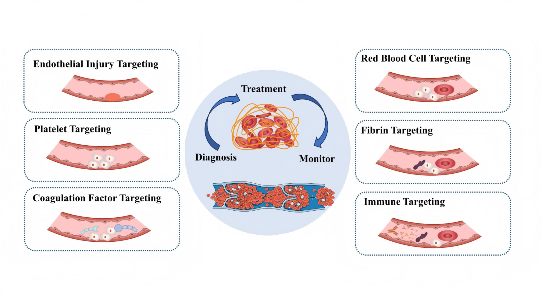
Given the clinical urgency and challenges of thrombosis, integrated nano-based management platforms are essential for improving patient outcomes. Conventional diagnostics and therapies often operate separately, causing delays and reducing efficacy. In contrast, nano-enabled theranostic systems integrate real-time thrombus detection with targeted therapy, enhancing precision and efficiency.14 Nanoparticles improve thrombus imaging through MRI, PAI, and fluorescence, while nanocarriers enable controlled, site-specific drug delivery, minimizing systemic side effects. This section explores recent advances in nano-based thrombosis management, focusing on six key targeting strategies: endothelial injury, platelets, coagulation factors, red blood cell–thrombus interactions, fibrin, and inflammation (Table 1). These nanoscale strategies enable more accurate diagnosis, personalized treatment, and real-time thrombus monitoring, advancing patient-centered care.
 |
Table 1 The Six Targets of the Integrated Thrombosis Diagnosis and Treatment Technology |
Endothelial Injury Targeting
Endothelial injury plays a pivotal role in thrombus formation by initiating platelet adhesion, leukocyte recruitment, and activation of the coagulation cascade.21 Dysfunctional endothelial cells upregulate adhesion molecules such as vascular cell adhesion molecule-1 (VCAM-1) and intercellular adhesion molecule-1 (ICAM-1), facilitating interactions with circulating immune cells and platelets and thereby accelerating thrombus development. Furthermore, endothelial dysfunction reduces the bioavailability of nitric oxide (NO) and prostacyclin, exacerbating vascular inflammation and fostering a pro-thrombotic state.22–24 Given its central role in thrombogenesis, targeting endothelial dysfunction represents a promising strategy for integrated thrombus diagnosis and therapy.25–28
Current theranostic approaches for endothelial injury encompass both imaging and therapeutic interventions.29 Molecular imaging techniques utilize fluorescent probes targeting endothelial adhesion molecules, such as VCAM-1 and ICAM-1, enabling noninvasive visualization of endothelial activation and early thrombus formation.30 Concurrently, NO-releasing nanocarriers have emerged as multifunctional platforms capable of restoring endothelial function while serving as contrast agents for imaging.31 NO possesses vasodilatory, anti-inflammatory, and antithrombotic properties, making it an attractive therapeutic agent for vascular protection.32 By incorporating NO donors into nanocarriers, these systems achieve controlled NO release at sites of endothelial dysfunction, mitigating oxidative stress, inhibiting platelet aggregation, and enhancing imaging contrast.33 Shi et al developed NO-loaded lipid microbubbles (NO-MBs) combined with ultrasound-targeted microbubble destruction (UTMD) for precise thrombolysis.34 This strategy enables targeted NO release under real-time ultrasound imaging, enhancing thrombus dissolution while minimizing systemic effects, as shown in Figure 3A. Notably, cavitation stimulated endothelial cells to produce endogenous NO, further promoting thrombolysis and vascular repair. NO-MBs also alleviated oxidative stress and inflammation, demonstrating excellent biosafety. These findings highlight the endothelial-targeted potential of NO-MBs for arterial thrombosis treatment.
 |
Figure 3 Schematic Representations of Advanced Nano-Platforms for Thrombosis Diagnosis and Therapy. (A) Diagram of the structure, synthesis, and mechanism of action of NO-MBs. Reprinted from Europ J Pharm Biopharm. Volume 205. Shi B, Yang Q, Liang Z, et al. Accelerating thrombolysis of arterial thrombus with NO-MBs UTMD therapy. 114566. Copyright 2024, with permission from Elsevier. Creative Commons.34 (B) Schematic illustration of PNP-PA design and its characterization. The red arrow indicates that the platelet membrane has successfully transferred to the desired coating direction. Reprinted from Xu J, Zhang Y, Xu J, et al. Engineered nanoplatelets for targeted delivery of plasminogen activators to reverse thrombus in multiple mouse thrombosis models. Adv Mate. 2020;32(4):1905145. © 2019 WILEY-VCH Verlag GmbH & Co. KGaA, Weinheim.35 (C) Role of TOSP-3 in the diagnosis and treatment of thrombosis. Reprinted from Inter J Biolog Macromolecules. Volume: 306. George AM, Chakraborty K, Paulose SK, Jalal S, Pai AA, Dhara S. Anticoagulant potential of sulfated galactofucan from Turbinaria ornata: targeting coagulation pathways and thrombin signaling in human umbilical vein endothelial cells. 141499. Copyright 2025, with permission from Elsevier.36 (D)Therapeutic Mechanisms of USIO/UK@EM for Thrombosis Removal. Reprinted from Zhu L, Lian W, Yu N, et al. Erythrocyte-membrane-camouflaged magnetic nanocapsules with photothermal/magnetothermal effects for thrombolysis. Adv Healthc Mater. 2024;13(20):e2400127. © 2024 Wiley-VCH GmbH.37 (E)Schematic illustration of an ultrasound-responsive theranostic platform. Reprinted from Lin L, Ba Z, Tian H, et al. Ultrasound-responsive theranostic platform for the timely monitoring and efficient thrombolysis in thrombi of tPA resistance. Nat Commun. 2024;15(1):6610. Creative Commons.38 Note: *p<0.05 |
Despite these advancements, several challenges remain in optimizing endothelial-targeted theranostic platforms.39 A key limitation is the need for enhanced targeting specificity to minimize off-target effects and improve diagnostic precision. Functionalizing nanocarriers with endothelial-specific ligands, such as peptides or antibodies against VCAM-1 or ICAM-1, could enhance selective accumulation at sites of endothelial injury.40 Additionally, extending systemic circulation time through polyethylene glycol (PEG) modification or biomimetic coatings could improve in vivo stability and prolong therapeutic effects. Another promising strategy is the development of stimuli-responsive nanocarriers that release NO in response to pathological cues, such as oxidative stress or shear stress alterations in thrombogenic regions. Refining these approaches could lead to more precise, efficient, and clinically translatable endothelial-targeted theranostic platforms for thrombus management.
Platelet Targeting
Platelets play a central role in primary thrombus formation by adhering to the injured vascular endothelium, undergoing activation, and facilitating the deposition of coagulation factors.41 Upon activation, platelets release procoagulant molecules and express surface receptors that promote aggregation, leading to the formation of a platelet-rich thrombus core.42–45 This process forms a scaffold for stabilizing the fibrin network, highlighting platelets as a key target for thrombus theranostics.46 Strategies aimed at detecting and modulating platelet activity hold significant potential for enabling early diagnosis and intervention, thereby reducing the risk of thrombotic complications.
Current platelet-targeted theranostic technologies integrate molecular imaging with therapeutic functionalities to achieve precise thrombus detection and treatment.47 Platelet membrane-coated biomimetic nanoparticles represent an advanced strategy for targeting thrombi, as they retain the natural adhesion properties of platelets, allowing for selective accumulation at thrombotic sites.48 These biomimetic nanocarriers can be engineered to carry contrast agents for imaging and thrombolytic agents for therapy, enabling dual functionality.49 Xu et al developed platelet membrane-coated polymer nanoparticles for targeted delivery of recombinant tissue plasminogen activator (rt-PA) to the thrombus site for thrombolytic therapy.35 In vitro targeting and thrombolytic efficacy of PNP-PA are shown in Figure 3B. Additionally, fluorescence-based platelet aggregation probes have been developed to detect activated platelets in real-time. These probes selectively bind to surface markers expressed on activated platelets, providing a noninvasive approach to visualize thrombus formation and assess platelet function in vivo.
Despite these advancements, challenges persist in enhancing the resolution and specificity of platelet-targeted imaging and therapy.50 A key limitation is the need for improved imaging sensitivity to distinguish thrombotic lesions from surrounding vascular structures with high precision.51,52 Future strategies may focus on the development of multimodal imaging probes that integrate fluorescence, magnetic resonance, and photoacoustic imaging to improve diagnostic accuracy. Additionally, optimizing the stability and biocompatibility of platelet-mimicking nanocarriers could extend their circulation time and enhance therapeutic efficacy.53 Addressing these limitations could facilitate the development of next-generation platelet-targeted theranostic platforms, offering more effective and personalized approaches for thrombus diagnosis and treatment.
Coagulation Factor Targeting
The coagulation cascade is a tightly regulated process in which coagulation factors, particularly thrombin and factor Xa (FXa), play a pivotal role in amplifying clot formation.54 Thrombin, a key serine protease, catalyzes the conversion of fibrinogen to fibrin and further activates platelets, thereby enhancing thrombus stability.55–58 FXa, positioned at the convergence of the intrinsic and extrinsic coagulation pathways, acts as a central mediator of thrombin generation, making it a key target for anticoagulant strategies.59 Given the cascading nature of coagulation factor activation, precise modulation is critical to prevent excessive thrombosis while minimizing the risk of bleeding.60 George et al identified TOSP-3, a sulfated polysaccharide from Turbinaria ornata, as a potent anticoagulant that targets coagulation factors.36 TOSP-3 significantly prolonged clotting times and suppressed factor Xa expression by 89% in endothelial cells, demonstrating its regulatory effects on both intrinsic and extrinsic pathways, as shown in Figure 3C. It effectively inhibited thrombin-catalyzed fibrin polymerization and platelet aggregation while reducing thrombin production by 33%. Through electrostatic interactions, TOSP-3 modulated the coagulation cascade more efficiently than heparin, highlighting its potential as a natural anticoagulant for thrombotic disorder management.61
Current theranostic approaches targeting coagulation factors integrate imaging with anticoagulant therapy, enabling real-time thrombus monitoring and controlled drug release.62,63 Thrombin-responsive nanocarriers have been developed to achieve adaptive anticoagulation, wherein thrombin-mediated cleavage triggers the localized release of anticoagulant agents in a self-regulated manner.64 Similarly, FXa inhibitor-loaded nanoparticles have been designed to prolong drug circulation, enhance therapeutic efficacy, and reduce systemic bleeding risks.65 These nanosystems employ targeted delivery strategies to localize anticoagulant effects at thrombotic sites, thereby improving safety and efficacy compared to conventional anticoagulant therapies.
Despite these advancements, challenges remain in optimizing coagulation factor-targeted theranostics.66 A primary concern is the risk of excessive anticoagulation, which may lead to hemorrhagic complications. Future research should focus on developing highly selective and dynamically responsive drug delivery systems that adapt to pathological coagulation factor levels while preserving hemostatic balance.67 Additionally, improving the biocompatibility of anticoagulant nanocarriers and hydrogels is crucial to minimizing immune responses and enhancing long-term safety.68 Strategies such as surface modification with biomimetic coatings or the incorporation of responsive polymeric architectures could further refine these platforms. Addressing these limitations will be critical for advancing next-generation coagulation factor-targeted theranostic technologies, ultimately enabling safer and more effective thrombus diagnosis and treatment.
Red Blood Cell–Thrombus Interaction Targeting
Red blood cells (RBCs) play a critical role in thrombus stability by influencing clot architecture and modulating fibrinolysis.6 Within the thrombus microenvironment, RBCs become enmeshed in the fibrin network, increasing thrombus density and mechanical resistance to degradation. Additionally, RBC-derived microparticles contribute to procoagulant activity by exposing phosphatidylserine, which serves as a catalytic surface for coagulation factor assembly.69–72 These interactions not only enhance thrombus stability but also affect the efficacy of thrombolytic therapies, making RBC-thrombus interactions a promising target for theranostic strategies to enhance thrombus detection and facilitate efficient clot dissolution.
Emerging theranostic technologies targeting RBC-thrombus interactions integrate both imaging and therapeutic functions, enabling real-time thrombus visualization and controlled thrombolysis.73 RBC membrane-coated nanoparticles have been developed to exploit the prolonged circulation time of native RBCs, thereby enhancing thrombus imaging and drug delivery.74 These biomimetic carriers improve thrombus-targeting efficiency while reducing immune clearance, ultimately enhancing diagnostic accuracy and therapeutic efficacy.75 Furthermore, hemoglobin-based nanocomposites have been engineered for photoacoustic imaging and thrombolysis, leveraging hemoglobin’s strong optical absorption properties to enhance imaging contrast while simultaneously mediating oxygen delivery to accelerate fibrinolysis.76 Zhu et al developed erythrocyte membrane (EM)-camouflaged nanocapsules (USIO/UK@EM) for targeted thrombolysis, integrating ultra-small iron oxide (USIO) and urokinase (UK).37 Therapeutic Mechanisms of USIO/UK@EM for Thrombosis Removal are shown in Figure 3D. The EM coating enhanced circulation time (t₁/2 = 3.28h), enabling prolonged thrombus targeting. Leveraging photothermal and magnetothermal effects, USIO/UK@EM rapidly increased local temperature under laser or magnetic stimulation, significantly improving thrombolytic efficiency (82.4% vs ~15% for UK alone). In vivo studies demonstrated effective dissolution of venous and arterial thrombi in mice and rabbits. This biomimetic platform presents a promising erythrocyte-targeted nanomedicine approach for thrombolytic therapy.
Despite these advancements, several challenges remain in optimizing RBC-targeted theranostics for thrombus management.77 Enhancing the specificity of these platforms for large thrombi and deeply embedded clots remains a critical focus, as current targeting strategies may be limited by the heterogeneous composition of thrombi.78,79 Additionally, optimizing photothermal thrombolysis efficiency requires improved light penetration and controlled energy deposition to prevent collateral tissue damage.80 Future research should prioritize the development of multifunctional nanocomposites that combine RBC-mimetic properties with enhanced photothermal conversion efficiency or stimulus-responsive drug release mechanisms.81 Addressing these challenges will be essential for advancing RBC-targeted theranostic technologies and improving precision thrombus imaging and therapy.
Fibrin Targeting
Fibrin plays a central role in the structural integrity of mature thrombi, serving as the primary scaffold that stabilizes clot formation and resists degradation.82 As the final product of the coagulation cascade, fibrin polymerizes into a dense network that entraps platelets and red blood cells, reinforcing thrombus stability and hindering fibrinolysis.83–86 Given its critical role in thrombus persistence, fibrin represents a key therapeutic target for thrombolysis, particularly in pathological conditions where excessive clot formation leads to vascular occlusion and ischemic complications.87 Strategies that selectively target fibrin enable precise thrombus imaging and efficient clot dissolution while minimizing off-target effects.
Current theranostic technologies designed for fibrin targeting integrate advanced imaging modalities with fibrinolytic therapy, enabling real-time thrombus visualization and controlled degradation.88 Fibrin-specific fluorescent probes have been developed for MRI and CT imaging, providing high-contrast thrombus detection through selective fibrin binding.89 These imaging agents enhance diagnostic accuracy and allow clinicians to monitor thrombus progression and therapeutic response. Additionally, fibrin-targeted nanoscale thrombolytics have been engineered to improve clot dissolution efficiency. These nanocarriers encapsulate fibrinolytic agents such as tissue plasminogen activator (tPA) and are designed to release their therapeutic payload in response to external stimuli, such as ultrasound. By incorporating targeted delivery mechanisms, these nanosystems enhance thrombolytic efficacy while reducing systemic bleeding risks associated with conventional fibrinolytic therapy. Lin et al developed a noninvasive theranostic platform integrating sonodynamic and mechanical thrombolysis for treating tPA-resistant thrombi under ultrasonic imaging guidance.38 Analysis of patient-derived thrombi revealed that fibrin scaffolds, neutrophil extracellular traps (NETs), and ε-(γ-glutamyl) lysine isopeptide bonds form a structural network conferring resistance to tPA, as shown in Figure 3E. Targeting this fibrin-based framework, the proposed strategy achieved over 90% recanalization in a rat model, with sustained vascular reconstruction and no thrombosis recurrence. Successful application in pigs and thrombosis-prone tissue-engineered vascular grafts underscores its translational potential for fibrin-targeted thrombolysis in clinical settings.
Despite these advancements, several challenges remain in optimizing fibrin-targeted theranostic strategies.90 Enhancing the thrombolytic efficiency of these systems requires improvements in fibrin affinity and drug release kinetics to ensure rapid and complete clot dissolution.91 Additionally, minimizing off-target fibrinolysis is essential to reduce hemorrhagic risk. Future research should focus on developing multifunctional nanocarriers that integrate fibrin targeting with stimuli-responsive drug release while exploring alternative thrombus imaging techniques with higher resolution and real-time monitoring capabilities.92,93 Addressing these challenges will be crucial for advancing fibrin-targeted theranostic platforms and improving personalized thrombolytic therapy.
Inflammation and Immune Targeting
Inflammation and immune responses play a critical role in thrombus stabilization and post-thrombotic complications, with neutrophil extracellular traps (NETs) serving as key contributors to thrombus persistence.94–97 NETs, composed of DNA, histones, and granular proteins, form a fibrous network that reinforces thrombus structure, promotes platelet aggregation, and exacerbates vascular occlusion.98,99 Moreover, NETs impede fibrinolysis and contribute to endothelial dysfunction, thereby hindering vessel recanalization and increasing the risk of thrombosis recurrence.100,101 Given their pivotal role in thrombus pathophysiology, targeting NETs and modulating the inflammatory microenvironment represent promising strategies for integrated thrombus diagnosis and therapy.102
Current theranostic approaches focus on NET-targeted nano-delivery systems that integrate anti-inflammatory and thrombolytic functions.103,104 These nanoparticles are designed to selectively bind NET components, facilitating precise drug delivery while simultaneously serving as imaging agents for thrombus detection.105 By incorporating thrombolytic agents such as DNase I or anti-NET antibodies, these nanosystems promote NET degradation, enhance clot resolution, and attenuate the inflammatory response.106 Additionally, ultrasound-triggered anti-inflammatory microbubbles have been developed to provide both imaging and therapeutic effects.1 These microbubbles, loaded with anti-inflammatory agents, can be selectively activated at the thrombus site through ultrasound stimulation, enabling controlled drug release and real-time monitoring of thrombus-associated inflammation.
Despite these advancements, challenges remain in optimizing the efficacy and specificity of inflammation-targeted thrombolytic strategies.107 Improving the precision of thrombotic microenvironment modulation is essential for minimizing off-target effects and improving therapeutic outcomes.108 Additionally, the long-term safety and biocompatibility of NET-targeted nanoparticles and microbubble formulations require further investigation.109 Future research should focus on refining delivery systems to respond dynamically to inflammatory signals and integrating multi-modal imaging techniques for comprehensive thrombus characterization.110 By addressing these challenges, inflammation-targeted theranostic strategies hold significant potential for advancing thrombus management and improving clinical outcomes in thrombin inflammatory disorders.
Nano-Based Thrombosis Therapy
Thrombosis is a complex and dynamic pathological process that progresses through multiple stages, including initiation, propagation, and resolution. Thrombosis can cause many diseases (Table 2). The advent of integrated nano-based delivery systems has introduced a closed-loop approach that seamlessly links diagnosis and treatment, thereby enhancing therapeutic efficacy while minimizing systemic side effects.111,112 These advanced systems utilize multifunctional nanoplatforms to improve the precision and adaptability of thrombosis management.
 |
Table 2 Diseases Caused by Thrombus |
A fundamental component of these systems is intelligent diagnosis, which integrates advanced imaging modalities, biosensors, and real-time monitoring techniques. These technologies facilitate early detection, precise localization, and continuous assessment of thrombus evolution, enabling timely therapeutic intervention.113 Concurrently, targeted therapy is achieved through nanocarriers specifically designed to interact with thrombosis-associated components, such as fibrin networks, activated platelets, coagulation factors, and inflamed vascular endothelium.114,115 By enhancing drug accumulation at the thrombotic site, these systems improve treatment efficacy while minimizing off-target effects. To enable real-time and synergistic modulation of the thrombotic microenvironment, advanced nano-systems are increasingly being engineered to both sense pathological cues and initiate targeted therapeutic responses. A notable example involves polydopamine-based nanomotors designed for the treatment of inferior vena cava thrombosis.116 These nanomotors feature a mesoporous polydopamine core that serves as a photothermal-responsive matrix. Upon near-infrared (NIR) irradiation, the system generates localized heat to enhance thrombus dissolution while simultaneously propelling the nanomotors to penetrate deeper into vascular tissue. In parallel, the nanomotors are loaded with the fibrinolytic agent urokinase and functionalized with RGD peptides for thrombus-specific recognition. Moreover, the RGD motif interacts with elevated levels of reactive oxygen species (ROS) in the thrombotic niche to catalyze the generation of nitric oxide (NO)—a multifunctional molecule that not only augments nanomotor propulsion but also promotes endothelial regeneration. Through this multifunctional design, the nanosystem achieves spatiotemporally controlled drug release, targeted navigation, and concurrent regulation of inflammatory and pro-thrombotic signals, exemplifying a closed-loop theranostic strategy with strong translational promise. There are many integrated nano-therapy strategies for thrombosis management (Table 3).
 |
Table 3 Comprehensive Nano-Therapy Strategies for Thrombosis Management |
In addition to precise targeting, adaptive treatment strategies further optimize thrombosis management by enabling controlled drug release in response to pathological cues. Stimuli-responsive nano-systems can react to microenvironmental factors such as pH variations, reactive oxygen species, enzymatic activity, or shear stress, ensuring on-demand therapeutic action aligned with disease progression.117 Through the integration of these capabilities, nano-based platforms present a promising strategy for both acute and chronic thrombosis management, which will be explored in the following sections.
Application in Acute Phase
Acute thrombosis is marked by rapid platelet aggregation, activation of coagulation factors, and fibrin network formation, leading to vascular occlusion and potentially life-threatening complications.118–121 The primary therapeutic objectives during this phase include prompt thrombolysis, platelet inhibition, and anticoagulation while minimizing the risk of hemorrhagic events. Nano-based integrated thrombosis management systems provide innovative strategies to achieve these goals by enhancing drug targeting, real-time monitoring, and controlled therapeutic release.122
Targeted thrombolytic delivery systems have been developed to improve the precision and efficacy of clot dissolution.123–126 These nanosystems are specifically engineered to interact with key thrombotic components, including fibrin, coagulation factors (eg, FXa, FIIa), and platelet membrane receptors (eg, GP IIb/IIIa). By promoting site-specific drug accumulation, these approaches minimize systemic exposure and reduce adverse effects.127 For instance, stimuli-responsive nanoparticles sensitive to pH changes or shear stress can selectively release thrombolytic agents, such as tPA, at the thrombotic site, enhancing clot degradation while mitigating systemic bleeding risks.128,129 A representative example is a platelet-mimetic nano platform functionalized with Annexin V and loaded with tPA, which selectively binds to phosphatidylserine-exposing activated platelets at the thrombus site, as shown in Figure 4A.130 This biomimetic system significantly improved thrombolytic efficacy and neurological outcomes in a mouse model of acute ischemic stroke, while reducing the risk of intracerebral hemorrhage.
 |
Figure 4 (A)Schematic of APLT-PA preparation and thrombus-targeted therapy for acute stroke. APLT-PA was prepared via freeze–thaw and thin-film hydration, with platelet membranes and Annexin V enabling Ca²+-responsive targeting and thrombolysis in ischemic stroke mice. Reprinted from Quan X, Han Y, Lu P, et al. Annexin V-Modified Platelet-Biomimetic Nanomedicine for Targeted Therapy of Acute Ischemic Stroke. Adv Healthcare Mate. 2022;11(16):2200416. © 2022 Wiley-VCH GmbH.130 (B). Schematic of supramolecular nanomedicine for vascular injury therapy. Constructed via α-CD–PEG-peptide interactions, NP@PBA&NO inhibits platelet activation and smooth muscle cell responses, reducing thrombosis and intimal hyperplasia. Reprinted from Zhou K, Huang C, Li J, et al. Multifunctional NO supramolecular nanomedicine for thrombus risk reduction and intimal hyperplasia inhibition. J Mater Chem B. 2025;13(5):1811–1822. Copyright Royal Society of Chemistry.25 |
Beyond drug delivery, intelligent imaging and theranostic platforms play a critical role in acute thrombosis management by enabling real-time thrombus detection and treatment monitoring.131–134 Nanoprobes incorporating fluorescence, magnetic resonance imaging (MRI), or photoacoustic imaging (PAI) modalities facilitate high-resolution visualization of thrombus formation, supporting timely clinical decision-making.135–138 A notable example includes platelet membrane-coated nanoparticles loaded with superparamagnetic iron oxide (SPIO), which enhance MRI/PAI contrast while providing simultaneous diagnostic and therapeutic capabilities.139,140
Additionally, mechanical stimulation and external stimulus-assisted therapies have emerged as promising strategies to enhance thrombolysis.141–144 Nanocarriers integrated with mechanosensitive or externally activatable components, such as ultrasound-responsive nanobubbles, magnetic nanoparticles, or electrically charged drug carriers, facilitate clot dissolution upon activation.145 For example, ultrasound-triggered nanobubbles can not only release thrombolytic agents but also exert mechanical disruption on the thrombus, improving clot degradation efficiency.146
By integrating targeted drug delivery, intelligent diagnostics, and external stimulus-responsive strategies, nano-based thrombosis management systems represent a promising approach for improving thrombolytic efficacy while mitigating complications associated with conventional anticoagulant therapies.147,148 These advancements lay the foundation for the next generation of personalized and adaptive interventions in acute thrombotic events.
Application in Chronic Phase
Chronic thrombosis is characterized by persistent inflammation, endothelial dysfunction, fibrosis, and vascular remodeling, all of which contribute to long-term complications and an increased risk of thrombus recurrence.149–152 The primary therapeutic goals in this phase include preventing thrombosis, restoring endothelial function, and minimizing chronic vascular damage.153 Nano-based therapeutic strategies offer innovative solutions by integrating anti-inflammatory, endothelial-regenerative, and long-acting anticoagulant approaches.
A key aspect of chronic thrombosis management involves nano-enabled anti-inflammatory and immunomodulatory therapies. Since inflammation-driven vascular injury plays a crucial role in thrombus recurrence, targeted drug delivery is essential for sustained vascular protection. Nanocarriers loaded with anti-inflammatory agents, such as interleukin-10 (IL-10) or transforming growth factor-beta (TGF-β) modulators, have been developed to mitigate vascular inflammation and promote endothelial homeostasis.154–157 For instance, leukocyte membrane-coated nanoparticles (eg, neutrophil-mimicking nanoparticles) can home to inflamed sites, delivering small-molecule anti-inflammatory drugs or siRNA to suppress atherosclerosis-associated thrombotic risk.158,159 An illustrative example involves a supramolecular nanomedicine co-delivering nitric oxide and antioxidants via a cyclodextrin-based system, enabling targeted accumulation at vascular injury sites, as shown in Figure 4B.25 By synergistically reducing oxidative stress and enhancing NO-mediated endothelial repair, this platform effectively mitigated thrombotic risk and intimal hyperplasia, offering a versatile approach for chronic vascular inflammation control.
Beyond inflammation control, targeted endothelial repair strategies are critical for mitigating chronic thrombosis risk.160–162 Nanotherapeutics designed to improve endothelial cell (EC) proliferation and function facilitate vascular healing and reduce long-term complications. Nanocarriers encapsulating vascular endothelial growth factor (VEGF), NO donors, or microRNAs have been shown to promote endothelial regeneration and restore vascular integrity. By improving EC function, these nanosystems stabilize the vascular environment, thereby reducing the likelihood of recurrent thrombotic events.163,164
Long-acting anticoagulant and antiplatelet nano delivery systems provide another promising approach for chronic thrombosis management.165–167 Conventional anticoagulant therapies often require frequent dosing, increasing the risks of systemic bleeding. In contrast, nanoformulations enable controlled, site-specific drug release triggered by pathological stimuli such as pH shifts, enzymatic activity, or reactive oxygen species (ROS). For example, self-regulating antiplatelet nanoparticles can release therapeutic agents such as aspirin or clopidogrel in response to high-shear stress conditions, ensuring localized and sustained antithrombotic effects while minimizing systemic adverse reactions.168,169
By integrating anti-inflammatory modulation, endothelial repair, and stimulus-responsive anticoagulant delivery, nano-based thrombosis management systems provide a multifaceted approach to reducing the long-term burden of thrombotic diseases.170 These advancements hold significant potential for preventing recurrent thrombotic events and improving vascular health in patients with chronic thrombosis.
Conclusion
Integrated nano-enabled systems for thrombosis management represent a significant advancement in the diagnosis and treatment of thrombotic disorders. By integrating intelligent diagnostics, targeted therapeutics, and real-time feedback mechanisms, these platforms improve treatment precision while minimizing systemic toxicity. Key innovations in endothelial injury targeting, platelet modulation, coagulation factor regulation, red blood cell–thrombus interactions, fibrinolysis, and inflammation control have demonstrated substantial potential for managing both acute and chronic thrombosis.
Despite these advances, several challenges impede clinical translation. Major barriers include concerns about biocompatibility, potential immunogenicity, long-term toxicity, and suboptimal thrombus-targeting specificity. Moreover, the scalability and reproducibility of nanocarrier fabrication, along with stringent regulatory requirements, pose significant obstacles to widespread clinical adoption.
Future Perspectives
To enable clinical translation, future research must overcome current limitations through technological innovation and interdisciplinary collaboration. One key challenge in translating Smart Thrombosis Care platforms into clinical practice lies in integrating these technologies into conventional workflows, which still rely heavily on anticoagulants and standard diagnostic tools.171 Demonstrating superior efficacy, safety, and cost-effectiveness over conventional strategies is essential for regulatory and clinical acceptance.
Personalized treatment strategies enabled by artificial intelligence (AI) and machine learning hold considerable promise.172,173 These technologies can analyze patient-specific thrombotic profiles—including clot location, fibrin composition, and systemic biomarkers—to guide individualized drug selection, dosing, and release schedule.174 Such algorithm-assisted optimization could substantially improve therapeutic outcomes while minimizing adverse effects.
Biomimetic nanotechnology, particularly the development of cell membrane-coated nanoparticles, offers further potential by enhancing immune evasion and targeting specificity.175 Meanwhile, stimuli-responsive and programmable drug delivery systems capable of adjusting therapeutic release in response to environmental signals (eg, pH, ROS, thrombin) may enable precise, on-demand treatment in dynamic thrombotic microenvironments.
In addition, multimodal theranostic platforms integrating real-time imaging modalities such as PET or MRI with targeted nano therapy could enhance diagnostic accuracy and allow for spatiotemporally controlled drug delivery. RNA-based strategies—such as siRNA- and mRNA-loaded nanoparticles—also present new opportunities for endothelial protection and thrombus resolution at the genetic level.
Ultimately, the advancement of these technologies will require close collaboration among materials scientists, biomedical engineers, computational biologists, clinicians, and regulatory bodies.176 Establishing standardized protocols for nanoparticle characterization, safety assessment, and clinical validation will be critical to ensuring reproducibility and accelerating clinical translation. Through such coordinated efforts, nano-based thrombosis management systems have the potential to reshape the future of personalized medicine for thrombotic disorders.
Disclosure
The authors report no conflicts of interest in this work.
References
1. Wang C, Liang F, Wang L. et al. Bilayer vascular grafts separately composited with nitric oxide-releasing keratin conjugates and hydrogen sulfide-releasing heparin conjugates. Int J Biol Macromol. 2025;307:141887. doi:10.1016/j.ijbiomac.2025.141887
2. Wen J, Huang Q, Chen X, Zhang K, Peng L. Impact of aortic branch retention strategies on thrombus growth prediction in type B aortic dissection: a hemodynamic study. Comput Methods Programs Biomed. 2025;263:108679. doi:10.1016/j.cmpb.2025.108679
3. Maekawa K, Nakamura E, Saito Y, et al. Inflammatory stimuli and hypoxia on atherosclerotic plaque thrombogenicity: linking macrophage tissue factor and glycolysis. PLoS One. 2025;20(3):e0316474. doi:10.1371/journal.pone.0316474
4. Van Steenberghe M, Perret F, Myers PO, Khatchatourov G. Subclinical leaflet thrombosis in Ozaki procedure. Interdiscip Cardiovasc Thorac Surg. 2025;40(3):ivaf051. doi:10.1093/icvts/ivaf051
5. Zhang Y, Wang Z, Zhou P, Zhang H. From reticulated platelets to immature platelet fraction: structure, function, and clinical applications. Platelets. 2025;36(1):2467383. doi:10.1080/09537104.2025.2467383
6. Traets MJM, Bos JF, van der Veen S, et al. Pyruvate Kinase Function Correlates With Red Blood Cell Properties and Clinical Manifestations in Sickle Cell Disease. Am J Hematol. 2025;100(5):785–796. doi:10.1002/ajh.27644
7. Rodriguez Alvarez AA, Patel SS, Cieri IF, et al. Single versus dual antiplatelet therapy impact on coagulation/thrombosis post PAD revascularization. Sci Prog. 2025;108(1):368504251324332. doi:10.1177/00368504251324332
8. Hu S, Gu Y, Zhao T, et al. Steroids combined with anticoagulant in acute/subacute severe cerebral venous thrombosis. Chin Med J. 2025. doi:10.1097/CM9.0000000000003502
9. Zhu S, Zou R, Lu Z, et al. Application of virtual deployment and hemodynamic simulation in treatment of a basal arterial dissecting aneurysm using flow diverter: a case report and literature review. Heliyon. 2025;11(4):e42545. doi:10.1016/j.heliyon.2025.e42545
10. Wang D, Yang M, Li S, Tang C, Ai J, Liu D. Efficacy and safety of low-molecular-weight-heparin plus citrate in nephrotic syndrome during continuous kidney replacement therapy: retrospective study. PeerJ. 2025;13:e18919. doi:10.7717/peerj.18919
11. Pal R, Le J, Rudas A, et al. A review of machine learning methods for non-invasive blood pressure estimation. J Clin Monit Comput. 2025;39(1):95–106. doi:10.1007/s10877-024-01221-7
12. Kern AY, Kreinin Y, Charle L, Epshrein M, Korin N, Mangin PH. A macrofluidic model to investigate the intrinsic thrombogenicity of clinically used stents and develop less thrombogenic stents. Heliyon. 2024;10(5):e26550. doi:10.1016/j.heliyon.2024.e26550
13. Cha MJ, An D-G, Kang M, et al. Correct closure of the left atrial appendage reduces stagnant blood flow and the risk of thrombus formation: a proof-of-concept experimental study using 4d flow magnetic resonance imaging. Korean J Radiol. 2023;24(7):647–659. doi:10.3348/kjr.2023.0173
14. Qureshi A, Melidoro P, Balmus M, et al. MRI-based modelling of left atrial flow and coagulation to predict risk of thrombogenesis in atrial fibrillation. Med Image Anal. 2025;101:103475. doi:10.1016/j.media.2025.103475
15. Komi DEA, Khomtchouk K, Santa Maria PL. A review of the contribution of mast cells in wound healing: involved molecular and cellular mechanisms. Clin Rev Allergy Immunol. 2020;58(3):298–312. doi:10.1007/s12016-019-08729-w
16. Hu G, Niu F, Liao K, et al. HIV-1 tat-induced astrocytic extracellular vesicle mir-7 impairs synaptic architecture. J Neuroimmune Pharmacol. 2020;15(3):538–553. doi:10.1007/s11481-019-09869-8
17. Andrews JPM, Portal C, Walton T, et al. Non-invasive in vivo imaging of acute thrombosis: development of a novel factor XIIIa radiotracer. Eur Heart J Cardiovasc Imaging. 2020;21(6):673–682. doi:10.1093/ehjci/jez207
18. Nielly H, Mathian A, Cazenave M, et al. Safety and effectiveness of transjugular renal biopsy for systemic lupus erythematosus and antiphospholipid antibody syndrome patients taking antithrombotics. Nephrol Dial Transplant. 2020;35(10):1721–1729. doi:10.1093/ndt/gfz085
19. Gligorijević N, Vasović T, Lević S, Miljević Č, Nedić O, Nikolić M. Atypical antipsychotic clozapine binds fibrinogen and affects fibrin formation. Int J Biol Macromol. 2020;154:142–149. doi:10.1016/j.ijbiomac.2020.03.119
20. Aksentijevich M, Lateef SS, Anzenberg P, Dey AK, Mehta NN. Chronic inflammation, cardiometabolic diseases and effects of treatment: psoriasis as a human model. Trends Cardiovasc Med. 2020;30(8):472–478. doi:10.1016/j.tcm.2019.11.001
21. Du J-H, Zhao -L-L, Yang B, Dai K-S. Influencing factors of FeCl3 induced mouse carotid artery thrombosis model. Zhongguo Shi Yan Xue Ye Xue Za Zhi. 2025;33(1):193–197. doi:10.19746/j.cnki.issn.1009-2137.2025.01.028
22. Gondil VS, Ashcraft M, Ghalei S, et al. Anti-infective bacteriophage immobilized nitric oxide-releasing surface for prevention of thrombosis and device-associated infections. ACS Appl Bio Mater. 2025;8(2):1362–1376. doi:10.1021/acsabm.4c01638
23. Kurhaluk N, Tkaczenko H. L-arginine and nitric oxide in vascular regulation-experimental findings in the context of blood donation. Nutrients. 2025;17(4):665. doi:10.3390/nu17040665
24. Li S, Yang S, Sun X, Ma T, Zheng Y, Liu X. Nitric oxide distribution correlates with intraluminal thrombus in abdominal aortic aneurysm: a computational study. Bioengineering. 2025;12(2):191. doi:10.3390/bioengineering12020191
25. Zhou K, Huang C, Li J, et al. Multifunctional NO supramolecular nanomedicine for thrombus risk reduction and intimal hyperplasia inhibition. J Mater Chem B. 2025;13(5):1811–1822. doi:10.1039/d4tb02271h
26. Khalaf ML, Soliman AM, Fahmy SR, Mohamed AS. Anti-thrombotic mechanisms of echinochrome a on arterial thrombosis in rats: in-silico, in-vitro and in-vivo studies. Cardiovasc Hematol Agents Med Chem. 2024. doi:10.2174/0118715257332064241104114546
27. Souissi A, Dergaa I, Hajri SE, Chamari K, Saad HB. A new perspective on cardiovascular function and dysfunction during endurance exercise: identifying the primary cause of cardiovascular risk. Biol Sport. 2024;41(4):131–144. doi:10.5114/biolsport.2024.134757
28. Kokoris S, Polyviou A, Evangelidis P, et al. Thrombosis in paroxysmal nocturnal hemoglobinuria (PNH): from pathogenesis to treatment. Int J Mol Sci. 2024;25(22):12104. doi:10.3390/ijms252212104
29. Yang H, Ma X, Li X. The diagnosis of DIC: a current overview. Front Med. 2025;12:1502628. doi:10.3389/fmed.2025.1502628
30. Yang K-J, Huang -J-J, Xuan C-X. Association of stent thrombectomy and conventional treatment with neuroprotection, complications, anxiety, and depression in acute ischemic stroke patients. World J Psychiatry. 2025;15(1):101182. doi:10.5498/wjp.v15.i1.101182
31. Yu X-Y, Jia X-Y, Wang T-Y, et al. Inhibition of IP3 (inositol 1,4,5-trisphosphate) receptors retards SARS-CoV-2-induced endothelial von Willebrand factor secretion and thrombosis. Thromb Haemost. 2025. doi:10.1055/a-2471-8767
32. Blauenfeldt RA, Waller J, Drasbek KR, et al. Effect of remote ischemic conditioning on the form and function of red blood cells in patients with acute ischemic stroke. Stroke. 2025;56(3):603–612. doi:10.1161/STROKEAHA.124.048976
33. Bernardi N, Neep BF, Garibaldi S, et al. The lncRNA DSCR9 is modulated in pulmonary arterial hypertension endothelial cell models and is associated with alterations in the nitric oxide pathway. Vascul Pharmacol. 2025;158:107464. doi:10.1016/j.vph.2025.107464
34. Shi B, Yang Q, Liang Z, et al. Accelerating thrombolysis of arterial thrombus with NO-MBs UTMD therapy. Europ J Pharm Biopharm. 2024;205:114566. doi:10.1016/j.ejpb.2024.114566
35. Xu J, Zhang Y, Xu J, et al. Engineered nanoplatelets for targeted delivery of plasminogen activators to reverse thrombus in multiple mouse thrombosis models. Adv Mate. 2020;32(4):1905145. doi:10.1002/adma.201905145
36. George AM, Chakraborty K, Paulose SK, Jalal S, Pai AA, Dhara S. Anticoagulant potential of sulfated galactofucan from Turbinaria ornata: targeting coagulation pathways and thrombin signaling in human umbilical vein endothelial cells. Inter J Biolog Macromolecules. 2025;306:141499. doi:10.1016/j.ijbiomac.2025.141499
37. Zhu L, Lian W, Yu N, et al. Erythrocyte-membrane-camouflaged magnetic nanocapsules with photothermal/magnetothermal effects for thrombolysis. Adv Healthc Mater. 2024;13(20):e2400127. doi:10.1002/adhm.202400127
38. Lin L, Ba Z, Tian H, et al. Ultrasound-responsive theranostic platform for the timely monitoring and efficient thrombolysis in thrombi of tPA resistance. Nat Commun. 2024;15(1):6610. doi:10.1038/s41467-024-50741-y
39. Cooke JP, Connor JH, Jain A. Acute and chronic cardiovascular manifestations of COVID-19: role for endotheliopathy. Methodist DeBakey Cardiovasc J. 2021;17(5):53–62. doi:10.14797/mdcvj.1044
40. Barbon E, Kawecki C, Marmier S, et al. Development of a dual hybrid AAV vector for endothelial-targeted expression of von Willebrand factor. Gene Ther. 2023;30(3–4):245–254. doi:10.1038/s41434-020-00218-6
41. Alarabi AB, Khasawneh FT, Alshbool FZ. Managing thrombus formation with EL2-5HTVac: a selective vaccination-based approach targeting the platelet serotonin 5-HT2AR. J Pharmacol Exp Ther. 2025;392(4):103399. doi:10.1016/j.jpet.2025.103399
42. Delaney A, Kritsotakis EI, Horner K, et al. High prevalence of platelet function disorders in women referred for surgical management of refractory heavy menstrual bleeding. Haemophilia. 2025. doi:10.1111/hae.70016
43. Liu W, Li G, Shi J, et al. NR4A1 acts as a novel regulator of platelet activation and thrombus formation. Circ Res. 2025. doi:10.1161/CIRCRESAHA.124.325645
44. Zhang Y-M, Luo Q, Lu M, et al. Pharmacological effects and mechanism of Ilexsaponin A1 in modulating platelet function. J Ethnopharmacol. 2025;344:119564. doi:10.1016/j.jep.2025.119564
45. Wang H, Tan Y, Liu Q, Yang S, Cui L. Ubiquitin-proteasome system: a potential participant and therapeutic target in antiphospholipid syndrome. Front Immunol. 2025;16:1523799. doi:10.3389/fimmu.2025.1523799
46. Sachs UJ, Reich M, Qiu D, Bayat B, Cooper N, Bein G. Platelet autoantibodies have an impact on the platelet count in patients. J Thromb Haemost. 2025;S1538-7836(25):00114–X. doi:10.1016/j.jtha.2025.02.016
47. Gao C, Dai Y, Spezza PA, et al. Megakaryocytes transfer mitochondria to bone marrow mesenchymal stromal cells to lower platelet activation. J Clin Invest. 2025;135(8):e189801. doi:10.1172/JCI189801
48. Suarez Ferreira SP, Rodriguez Alvarez AA, Cieri IF, et al. Racial variability in platelet response among patients with peripheral artery disease. J Surg Res. 2025;307:107–115. doi:10.1016/j.jss.2025.01.022
49. Setarehaseman A, Mohammadi A, Maitta RW. Thrombocytopenia in Sepsis. Life. 2025;15(2):274. doi:10.3390/life15020274
50. Zhang Y, Ye J, Sun S, et al. Role of platelets and NETs in arterial thrombosis. Naunyn Schmiedebergs Arch Pharmacol. 2025. doi:10.1007/s00210-025-03921-6
51. Yang L, Liu Y, Tao C, et al. Bionic nanovesicles sequentially treat flaps with different durations of ischemia by thrombolysis and prevention of ischemia-reperfusion injury. Mater Today Bio. 2025;31:101529. doi:10.1016/j.mtbio.2025.101529
52. Shuai Y, Qian Y, Zheng M, et al. Injectable platelet-mimicking silk protein-peptide conjugate microspheres for hemostasis modulation and targeted treatment of internal bleeding. J Nanobiotechnology. 2025;23(1):128. doi:10.1186/s12951-025-03180-w
53. Keovilay JA, Howard KC, Taylor KA, et al. Development of zafirlukast analogues for improved antithrombotic activity through thiol isomerase inhibition. Arterioscler Thromb Vasc Biol. 2025;45(4). doi:10.1161/ATVBAHA.124.321579
54. Ieko M, Ohmura K, Naito S, Yoshida M, Kumano O. Development of new anticoagulants targeting coagulation factor XI and prospects for clinical use. J Cardiol. 2025;S0914-5087(25):00061. doi:10.1016/j.jjcc.2025.02.013
55. Tong J, Zhao Y, Jin Y, Hao Z, Li S, Sun M. A mini review on the regulation of coagulation homeostasis through interfering with vitamin K-dependent coagulation/anticoagulation factors. Biochem Biophys Res Commun. 2025;753:151494. doi:10.1016/j.bbrc.2025.151494
56. Zhou Y, Wang D, Wu J, et al. Discovery of the low-hemorrhagic antithrombotic effect of montelukast by targeting FXIa in mice. Arterioscler Thromb Vasc Biol. 2025;45(4). doi:10.1161/ATVBAHA.124.322145
57. Wang W, Zhang Y, Fang Y, et al. Targeting the contact-kinin system: a cyclopeptide with anti-thromboinflammatory properties against stroke. Eur J Pharmacol. 2025;998:177497. doi:10.1016/j.ejphar.2025.177497
58. Ferdinande K, Raevens S, Decaestecker J, et al. Unravelling the coagulation paradox in liver cirrhosis: challenges and insights. Acta Clin Belg. 2025:1–11. doi:10.1080/17843286.2025.2469906.
59. Strijbis V, Cheung KL, Veizaj D, et al. Modifications of the prothrombin active site S4 subpocket confer resistance to dabigatran. Thromb Haemost. 2025. doi:10.1055/a-2537-6037
60. Wang JJ, Warkentin TE, Schönborn L, et al. VITT-like monoclonal gammopathy of thrombotic significance. N Engl J Med. 2025;392(10):995–1005. doi:10.1056/NEJMoa2415930
61. Zhao Y, Hu J, Sun X, et al. Loss of m6A demethylase ALKBH5 promotes post-ischemic angiogenesis via post-transcriptional stabilization of WNT5A. Clin Transl Med. 2021;11(5):e402. doi:10.1002/ctm2.402
62. Arachchillage DJ, Mobayen G, Laffan M, Randi AM, Ahnström J, Pericleous C. A cell-based model to study mechanisms of endothelial-dependent thrombin generation in response to inflammation and its modulation by hydroxychloroquine. Res Pract Thromb Haemost. 2025;9(1):102665. doi:10.1016/j.rpth.2024.102665
63. Habibi A, Ruf W, Schurgers L. Protease-activated receptors in vascular smooth muscle cells: a bridge between thrombo-inflammation and vascular remodelling. Cell Commun Signal. 2025;23(1):57. doi:10.1186/s12964-025-02066-6
64. Zhu L, Dong H, Li L, Liu X. The mechanisms of sepsis induced coagulation dysfunction and its treatment. J Inflamm Res. 2025;18:1479–1495. doi:10.2147/JIR.S504184
65. Fredenburgh JC, Weitz JI. Exosite crosstalk in thrombin. J Thromb Haemost. 2025;S1538-7836(25):00009. doi:10.1016/j.jtha.2025.01.003
66. Liu K, Cheng C, Yan J, et al. Polydatin mitigates thrombosis by inhibiting PHD2-induced proline hydroxylation on collagen, reducing platelet adhesion. Phytomedicine. 2025;138:156392. doi:10.1016/j.phymed.2025.156392
67. Barnes GD. New targets for antithrombotic medications: seeking to decouple thrombosis from hemostasis. J Thromb Haemost. 2024;S1538-7836(24):00723. doi:10.1016/j.jtha.2024.12.003
68. Ouyang Y, Yue Y, Wu N, Wang J, Geng L, Zhang Q. Identification and anticoagulant mechanisms of novel factor XIa inhibitory peptides by virtual screening of a in silico generated deep-sea peptide database. Food Res Int. 2024;197(Pt 2):5. doi:10.1016/j.foodres.2024.115308
69. Singh AK, Warbal P, Basterrechea KF, Bader K, Shekhar H. Enhancing passive cavitation imaging using pth root compression delay, sum, and integrate beamforming: in vitro and in vivo studies. IEEE Trans Biomed Eng. 2025;1–12. doi:10.1109/TBME.2025.3540101
70. Weisel JW, Litvinov RI. Exploring the thrombus niche: lessons learned and potential therapeutic opportunities. Blood. 2025;2024025319. doi:10.1182/blood.2024025319
71. Wu R, Kabir MS, Truskey GA, Randles A. Investigating the impact of sickle cell disease on red blood cell transport in complex capillary networks. Annu Int Conf IEEE Eng Med Biol Soc. 2024;2024:1–4. doi:10.1109/EMBC53108.2024.10781578
72. Luo J-Y, Zhou C, Shi S-X, et al. Use of tranexamic acid in hepatectomy under controlled low central venous pressure: a randomized controlled study. BMC Anesthesiol. 2025;25(1):94. doi:10.1186/s12871-025-02935-0
73. Nawal CL, Singh A, Saini HL, Rankawat G. Impact of blood glucose level on hematological indices in patients with type 2 diabetes mellitus. J Assoc Physicians India. 2025;73(2):16–20. doi:10.59556/japi.73.0851
74. Gąsecka A, Błażejowska E, Konieczka A, et al. Branched endovascular aortic aneurysm repair decreases platelet reactivity and platelet-rich thrombus formation - a prospective, cohort study. Platelets. 2025;36(1):2458622. doi:10.1080/09537104.2025.2458622
75. Zhan H, Cheng L, Chen H, et al. Evaluation of inflammatory-thrombosis panel as a diagnostic tool for vascular Behçet’s disease. Clin Rheumatol. 2025;44(3):1279–1291. doi:10.1007/s10067-025-07301-6
76. Zhang J, Wang C, He C, Yang Y. Lower red blood cell count is a risk factor for higher D-dimer level in patients with spinal cord injury: a five year retrospective cross-sectional study. J Spinal Cord Med. 2025;1–11. doi:10.1080/10790268.2025.2452685
77. Dinçer BT, Urgancı N, Bayrak AH, Durmaz Ö, Özden İ. The role of partial splenic artery embolization in the management of refractory esophageal variceal bleeding due to portal vein thrombosis. BMC Pediatr. 2025;25(1):49. doi:10.1186/s12887-025-05414-0
78. Zhang C, Wang C, Cha R, et al. Rapid preparation of collagen/red blood cell membrane tubes for stenosis-free vascular regeneration. ACS Nano. 2025;19(3):3293–3311. doi:10.1021/acsnano.4c11919
79. Price AD, Becker ER, Chae RC, et al. Factors affecting the direct red cell effect on thrombosis: hematocrit dilution and injury patterns. J Trauma Acute Care Surg. 2025;98(2):197–203. doi:10.1097/TA.0000000000004513
80. Gwozdzinski L, Pieniazek A, Gwozdzinski K. The roles of oxidative stress and red blood cells in the pathology of the varicose vein. Int J Mol Sci. 2024;25(24):13400. doi:10.3390/ijms252413400
81. Fregona V, Luraghi G, Fereidoonnezhad B, et al. Impact of thrombus composition on virtual thrombectomy procedures using human clot analogues mechanical data. J Mech Behav Biomed Mater. 2025;163:106886. doi:10.1016/j.jmbbm.2025.106886
82. Ji J, Xu X, Zhang L, et al. Dedicator of cytokinesis 2 regulates cytoskeletal actin dynamics and is essential for platelet biogenesis and functions. Cardiovasc Res. 2025:cvaf009. doi:10.1093/cvr/cvaf009.
83. Nakadate K, Saitoh H, Sakaguchi M, et al. Advances in understanding lipopolysaccharide-mediated hepatitis: mechanisms and pathological features. Curr Issues Mol Biol. 2025;47(2):79. doi:10.3390/cimb47020079
84. Papakonstantinou PE, Rivera-Caravaca JM, Chiarito M, et al. Atrial fibrillation versus atrial myopathy in thrombogenesis: two sides of the same coin? Trends Cardiovasc Med. 2025;S1050-1738(25):00007. doi:10.1016/j.tcm.2025.01.002
85. Ervando H, Ridwan LS, Dilogo IH. Factors related to deep vein thrombosis as a complication of post-total Hip arthroplasty patients: a systematic review. Eur J Orthop Surg Traumatol. 2025;35(1):82. doi:10.1007/s00590-025-04209-4
86. Lazarus B, Lok CE, Moist L, Polkinghorne KR. Strategies to prevent hemodialysis catheter dysfunction. J Am Soc Nephrol. 2025;36(5):952–966. doi:10.1681/ASN.0000000666
87. Tang K-T, Chen Y-S, Chen -T-T, et al. Inhibiting tyrosine kinase 2 ameliorates antiphospholipid syndrome nephropathy. Mediators Inflamm. 2024;2024(1):5568822. doi:10.1155/mi/5568822
88. de La Taille T, Sarfati P, Aid R, et al. Microemulsion-inspired polysaccharide nanoparticles for an advanced targeted thrombolytic treatment. ACS Nano. 2025;19(2):2944–2960. doi:10.1021/acsnano.4c17049
89. Fini E, Argento FR, Borghi S, et al. Fibrinogen structural changes and their potential role in endometriosis-related thrombosis. Antioxidants. 2024;13(12):1456. doi:10.3390/antiox13121456
90. Li Q, Ye J, Li Z, et al. The role of neutrophils in tPA thrombolysis after stroke: a malicious troublemaker. Front Immunol. 2024;15:1477669. doi:10.3389/fimmu.2024.1477669
91. Risman RA, Sen M, Tutwiler V, Hudson NE. Deconstructing fibrin(ogen) structure. J Thromb Haemost. 2025;23(2):368–380. doi:10.1016/j.jtha.2024.10.024
92. Ząbczyk M, Natorska J, Undas A. Novel factors affecting fibrin clot formation and their clinical implications. Pol Arch Intern Med. 2024;134(12):16884. doi:10.20452/pamw.16884
93. Nencini F, Bettiol A, Argento FR, et al. Post-translational modifications of fibrinogen: implications for clotting, fibrin structure and degradation. Mol Biomed. 2024;5(1):45. doi:10.1186/s43556-024-00214-x
94. Zhong S, Yi J, Chen S, et al. Combining immune checkpoint inhibitors and molecular-targeted agents with hepatic arterial infusion chemotherapy for hepatocellular carcinoma with inferior vena cava and/or right atrium tumor thrombus. Hepatol Int. 2025;19(3):560–575. doi:10.1007/s12072-025-10777-8
95. Ayyoub S, Dhillon NK, Tura-Ceide O. Genetics of long COVID: exploring the molecular drivers of persistent pulmonary vascular disease symptoms. Infect Dis Rep. 2025;17(1):15. doi:10.3390/idr17010015
96. Ni H, Ge Y, Zhuge Y, et al. LncRNA MIR181A1HG deficiency attenuates vascular inflammation and atherosclerosis. Circ Res. 2025;136(8):862–883. doi:10.1161/CIRCRESAHA.124.325196
97. Khene Z-E, Bhanvadia R, Tachibana I, et al. Surgical outcomes of radical nephrectomy and inferior vena cava thrombectomy following preoperative systemic immunotherapy: a propensity score analysis. Clin Genitourin Cancer. 2025;23(2):102307. doi:10.1016/j.clgc.2025.102307
98. Serrano-Gonzalo I, Menéndez-Jandula B, Franco-García E, et al. Neutrophil extracellular traps and macrophage activation contibute to thrombosis and post-covid syndrome in SARS-CoV-2 infection. Front Immunol. 2025;16:1507167. doi:10.3389/fimmu.2025.1507167
99. Moussa MD, Abou-Arab O, Staessens S, et al. Comparison of the effects of phosphorylcholin versus heparin-based surface coating on clinical and histological outcomes during veno-arterial ECMO support: a propensity score weighted analysis. J Thromb Haemost. 2025;S1538-7836(25):00118. doi:10.1016/j.jtha.2025.02.020
100. Kim J-Y, Han H-J. Case report: in vivo detection of neutrophil extracellular traps in a dog with thrombosis induced by bacterial vasculitis. Front Vet Sci. 2025;12:1470605. doi:10.3389/fvets.2025.1470605
101. Mi J, Guo J, Kang K, Wang S, Huang M. Advances in targeting neutrophil extracellular traps as a promising approach for breast cancer treatment. Comb Chem High Throughput Screen. 2025;28. doi:10.2174/0113862073376243250130060239.
102. Peng L, Wu Z, Sun W, Wang C. Clinical characteristics, treatment, and outcomes of nivolumab induced immune thrombocytopenia. Invest New Drugs. 2024;42(5):575–580. doi:10.1007/s10637-024-01472-w
103. Zhang W, Xie Y, Chen F, Xie B, Yin Z. Development and validation of a neutrophil extracellular traps-related gene signature for lower-grade gliomas. Comput Biol Med. 2025;188:109844. doi:10.1016/j.compbiomed.2025.109844
104. Von meijenfeldt FA, Lisman T, Pacheco A, Zen Y, Bernal W. Histologic evidence of neutrophil extracellular traps and fibrin(ogen) deposition in liver biopsies from patients with inflammatory liver disease. Res Pract Thromb Haemost. 2025;9(1):102666. doi:10.1016/j.rpth.2024.102666
105. Semerano A, Dell’Acqua B, Genchi A, et al. Cerebral thrombus analysis as a useful diagnostic tool for infective endocarditis in ischemic stroke patients. Eur Stroke J. 2025:23969873251320449. doi:10.1177/23969873251320449.
106. Perez-Toledo M, Beristain-Covarrubias N, Pillaye J, et al. Discrete and conserved inflammatory signatures drive thrombosis in different organs after Salmonella infection. Nat Commun. 2025;16(1):2356. doi:10.1038/s41467-025-57466-6
107. Jha M, McCarthy IR, Gelfand EV. Lipoprotein(a) - from biomarker to therapy: a review for the clinician. Am J Cardiol. 2025;S0002-9149(25):00120. doi:10.1016/j.amjcard.2025.02.034
108. Heldal TF, Åsberg A, Ueland T, et al. Systemic inflammation is an important risk factor and predictor of graft loss and mortality one year after kidney transplantation. Front Immunol. 2025;16:1529812. doi:10.3389/fimmu.2025.1529812
109. Liao J, Duan Y, Liu Y, et al. Simvastatin alleviates glymphatic system damage via the VEGF-C/VEGFR3/PI3K-Akt pathway after experimental intracerebral hemorrhage. Brain Research Bulletin. 2024;216:111045. doi:10.1016/j.brainresbull.2024.111045
110. Rehill AM, McCluskey S, Ledwith AE, et al. Trained immunity causes myeloid cell hypercoagulability. Sci Adv. 2025;11(10):eads0105. doi:10.1126/sciadv.ads0105
111. Feng Y, Mo S, Li X, et al. Development and validation of a score for clinical deterioration in patients with cerebral venous thrombosis. Neurosurg Rev. 2025;48(1):56. doi:10.1007/s10143-025-03224-7
112. Murariu-Gligor EE, Mureșan S, Cotoi OS. From cell interactions to bedside practice: complete blood count-derived biomarkers with diagnostic and prognostic potential in venous thromboembolism. J Clin Med. 2025;14(1):205. doi:10.3390/jcm14010205
113. B PL, V S. Innovative modified-net architecture: enhanced segmentation of deep vein thrombosis. Sci Rep. 2024;14(1):30835. doi:10.1038/s41598-024-81703-5
114. Yu NH, Shin D, Ryu IH, Yoo TK, Koh K. Retinal vein occlusion risk prediction without fundus examination using a no-code machine learning tool for tabular data: a nationwide cross-sectional study from South Korea. BMC Med Inform Decis Mak. 2025;25(1):118. doi:10.1186/s12911-025-02950-8
115. Scott M, Ghazanfar M, Windsor J, Ramsay G, Bekheit M. Splanchnic vein in acute pancreatitis study group the management of splanchnic vein thrombosis in acute pancreatitis: a global DELPHI consensus study. HPB. 2025;27(3):343–351. doi:10.1016/j.hpb.2024.12.002
116. Fang D, Li T, Wu Z, et al. Dual drive mode polydopamine nanomotors for continuous treatment of an inferior vena cava thrombus. J Mater Chem B. 2021;9(41):8659–8666. doi:10.1039/d1tb01202a
117. Speranza G, Mischkewitz S, Al-Noor F, Kainz B. Value of clinical review for AI-guided deep vein thrombosis diagnosis with ultrasound imaging by non-expert operators. NPJ Digit Med. 2025;8(1):135. doi:10.1038/s41746-025-01518-0
118. Ahmed A, Nayak S, Hoteit M, et al. Acute coronary syndrome in patients with autosomal dominant polycystic kidney disease: a systematic review and meta-analysis. Intern Med J. 2025;55(3):493–502. doi:10.1111/imj.16659
119. Shang Q, Zhou J, Ye J, Chen M. Adverse events reporting of edaravone: a real-world analysis from FAERS database. Sci Rep. 2025;15(1):8148. doi:10.1038/s41598-025-92605-5
120. Wang H, Lv B, Li W, et al. Anatomic distribution and analysis of influencing factors on deep vein thrombosis in patients with spinal fractures caused by high-energy injuries. Eur J Trauma Emerg Surg. 2025;51(1):128. doi:10.1007/s00068-025-02801-1
121. Zhang T, Li Z, Mei Q, et al. Cardiovascular outcomes in long COVID-19: a systematic review and meta-analysis. Front Cardiovasc Med. 2025;12:1450470. doi:10.3389/fcvm.2025.1450470
122. Fang W, Sun W, Fang W, Zhao S, Wang C. Clinical features, treatment, and outcomes of patients with carfilzomib induced thrombotic microangiopathy. Inter Immunopharmacol. 2024;134:112178. doi:10.1016/j.intimp.2024.112178
123. Compagnucci P, Dello Russo A, Mohanty S, et al. Catheter ablation of atrial fibrillation in patients with psoriasis: a multicenter study. J Am Heart Assoc. 2025:e038882. doi:10.1161/JAHA.124.038882.
124. Liu Q, Zhang X, Lv L, et al. Chidamide in combination with DCAG with or without venetoclax for relapsed/refractory acute myeloid leukemia. Cancer Med. 2025;14(5):e70734. doi:10.1002/cam4.70734
125. van Kan C, van Es J, Idrissi HE, et al. Clinical and radiological characteristics of CTEPH patients without a history of acute venous thromboembolism. J Thromb Haemost. 2025;S1538-7836(25):00132. doi:10.1016/j.jtha.2025.02.033
126. Yasmin F, Zaidi SF, Moeed A, et al. Clinical outcomes of immediate versus staged revascularization of nonculprit arteries in patients with acute coronary syndrome: a systematic review and meta-analysis. Clin Cardiol. 2025;48(3):e70105. doi:10.1002/clc.70105
127. Zhang C, Zhang S, Yin Y, et al. Clot removAl with or without decompRessive craniectomy under ICP monitoring for supratentorial intracerebral hemorrhage (CARICH): a randomized controlled trial. Inter J Surg. 2024;110(8):4804. doi:10.1097/JS9.0000000000001466
128. Nilsen DWT, Aarsetoey R, Poenitz V, et al. Sex-related differences in the prognostic utility of inflammatory and thrombotic cardiovascular risk markers in patients with chest pain of suspected coronary origin. Int J Cardiol Heart Vasc. 2025;56:101600. doi:10.1016/j.ijcha.2025.101600
129. Daggumati L, Gu C-S, Kolluri R, Kavali P, Vedantham S. The effect of thrombolysis of deep vein thrombosis on late symptoms of post-pulmonary embolism syndrome. J Vasc Interv Radiol. 2025;S1051-0443(25):00216. doi:10.1016/j.jvir.2025.02.024
130. Quan X, Han Y, Lu P, et al. Annexin V-Modified Platelet-Biomimetic Nanomedicine for Targeted Therapy of Acute Ischemic Stroke. Adv Healthcare Mate. 2022;11(16):2200416. doi:10.1002/adhm.202200416
131. Kasiviswanathan G, Sivashanmugam S, Bakthavatchalam R, Gaur A, Sugunakar Reddy K, Varatharajan S. Clinical profile of cerebral sinus venous thrombosis: a prospective observational study in South India. Kurume Med J. 2025. doi:10.2739/kurumemedj.MS7112012
132. Allam NAT, Abdelsalam ME, Elsharkawy HI, et al. Comprehensive epidemiological evaluation of ruminant brucellosis and associated risk factors in some Egyptian Governorates. Vet World. 2024;17(12):2780–2796. doi:10.14202/vetworld.2024.2780-2796
133. Zhang J, Tong H, Jiang L, Hu J. Dietary patterns associated with heat retention in blood vessel syndrome (HRBVS) in coronary heart disease: a cross-sectional study. Int J Gen Med. 2025;18:1283–1294. doi:10.2147/IJGM.S510507
134. Couturaud F, Schmidt J, Sanchez O, et al. Extended treatment of venous thromboembolism with reduced-dose versus full-dose direct oral anticoagulants in patients at high risk of recurrence: a non-inferiority, multicentre, randomised, open-label, blinded endpoint trial. Lancet. 2025;405(10480):725–735. doi:10.1016/S0140-6736(24)02842-3
135. Sławek-Szmyt S, Stępniewski J, Kurzyna M, et al. Multicentre, real-world data of next-generation computer-assisted vacuum aspiration thrombectomy in acute pulmonary embolism. Respir Res. 2025;26(1):87. doi:10.1186/s12931-025-03162-4
136. Ranade M, Foster MT, Brady PS, et al. Novel mechanical aspiration thrombectomy in patients with acute pulmonary embolism: results from the prospective APEX-AV trial. J Soc Cardiovasc Angiogr Interv. 2025;4(1):102463. doi:10.1016/j.jscai.2024.102463
137. Kerber B, Hüllner M, Maurer A, et al. Photon-counting detector ct iodine maps versus SPECT/CT: advancing lung perfusion imaging in chronic thromboembolic pulmonary hypertension. Invest Radiol. 2025. doi:10.1097/RLI.0000000000001163
138. Gitto M, Sartori S, Vogel B, et al. Potent P2Y12 inhibitors versus clopidogrel in cancer patients undergoing percutaneous coronary intervention. Can J Cardiol. 2025;S0828-282X(25):00179. doi:10.1016/j.cjca.2025.02.035
139. Chauhan W, Sj S, Ferdowsi S, Sohel A, Zennadi R. RBC Rpl13a snoRNAs guides 2’-O-methylation on peroxidasin mRNA promoting venous thrombosis in aging. J Thromb Haemost. 2025;S1538-7836(25):00135. doi:10.1016/j.jtha.2025.02.036
140. Harmouch KM, Hamza M, Kumar N, et al. Revascularization strategies for multivessel disease in acute coronary syndrome: network meta-analysis. J Soc Cardiovasc Angiogr Interv. 2025;4(1):102449. doi:10.1016/j.jscai.2024.102449
141. Byrne RA, Valgimigli M, Bhatt DL, et al. Great debate: default duration of dual antiplatelet treatment after percutaneous coronary intervention in acute coronary syndrome should be 12 months. Eur Heart J. 2025:ehaf070. doi:10.1093/eurheartj/ehaf070.
142. Zhao F, Zhang L, Chen X, Wang C. In reply to the letter to the editor regarding “building and verifying a prediction model for deep vein thrombosis among spinal cord injury patients undergoing inpatient rehabilitation. World Neurosurg. 2025;123859. doi:10.1016/j.wneu.2025.123859
143. Davenport MH, Christopher S, Deering RE, et al. International Delphi study of clinical and exercise professionals’ opinion of physical activity prescreening and contraindications for participating in postpartum physical activity. Br J Sports Med. 2025:bjsports–2024–109104. doi:10.1136/bjsports-2024-109104.
144. Baltsen CD, Ellegaard MS, Nørholt C, et al. Mechanical ventilation in acute pulmonary embolism: a randomised, experimental, crossover study. Eur Heart J Acute Cardiovasc Care. 2025:zuaf036. doi:10.1093/ehjacc/zuaf036.
145. Ai W, Li F, Yang Q, et al. Urine output as a novel predictor for in-hospital mortality in acute pulmonary embolism patients: training with the MIMIC database and validation with independent cohort. Cardiovasc Ther. 2025;2025(1):7907049. doi:10.1155/cdr/7907049
146. Vergallo R, Park S-J, Stone GW, et al. Vulnerable or high-risk plaque: a JACC: cardiovascular imaging position statement. JACC Cardiovasc Imaging. 2025;S1936-878X(25):00028. doi:10.1016/j.jcmg.2024.12.004
147. Lin S, Zhu N, Zhang S. The role of controlling nutritional status score in predicting postthrombotic syndrome in patients with lower extremity deep venous thrombosis. Thromb J. 2025;23(1):19. doi:10.1186/s12959-025-00701-3
148. Luo L, Zheng D, Da L, Cheng J, Cao Y, Wang N. Thrombin generation indices and Wells score predict pulmonary embolism in patients with acute exacerbation of chronic obstructive pulmonary disease. Clinics. 2025;80:100582. doi:10.1016/j.clinsp.2025.100582
149. Martin GP, Pate A, Bladon S, Sperrin M, Riley RD. A decision-analytical perspective on incorporating multiple outcomes in the production of clinical prediction models: defining a taxonomy of risk estimands. BMC Med. 2025;23(1):142. doi:10.1186/s12916-025-03978-3
150. Lombardi M, Glass J, Wasan S, Marston W. Acute iliofemoral deep venous thrombosis in adolescents and young adults is associated with hypercoagulable states and a significant incidence of post-thrombotic syndrome. J Vasc Surg Venous Lymphat Disord. 2025;13(4):102211. doi:10.1016/j.jvsv.2025.102211
151. Flumignan RL, Nakano LC, Flumignan CD, Baptista-Silva JC. Angioplasty or stenting for deep venous thrombosis. Cochrane Database Syst Rev. 2025;2(2):CD011468. doi:10.1002/14651858.CD011468.pub2
152. Puchsaka P, Rattanavipanon W, Phetroong S, et al. Anticoagulation intensity and outcomes among southeast-Asians with moderate-to-severe mitral valve stenosis. Br J Clin Pharmacol. 2025. doi:10.1002/bcp.70034
153. Zhang C, Ge H, Zhang S, et al. Hematoma evacuation via image-guided para-corticospinal tract approach in patients with spontaneous intracerebral hemorrhage. Neurol Ther. 2021;10(2):1001–1013. doi:10.1007/s40120-021-00279-8
154. Miao Q, Zheng Z, Piao M, Cao H, Wang B, Liu W. Case report: upper gastrointestinal bleeding associated with pancreatic segmental portal hypertension: six case reports and literature review. Front Med. 2025;12:1522413. doi:10.3389/fmed.2025.1522413
155. Acosta-Medina AA, Sridharan M, Go RS, et al. Clinical outcomes and treatment strategies of adult transplant-associated thrombotic microangiopathy: external validation of harmonizing definitions and high-risk criteria. Am J Hematol. 2025;100(5):830–839. doi:10.1002/ajh.27651
156. Leimi L, Vettenranta K, Meri S. Complement activation and vascular complications after pediatric allogeneic hematopoietic stem cell transplantation. Sci Rep. 2025;15(1):7073. doi:10.1038/s41598-025-91455-5
157. Zhou M, Xu Y, Zhang L, Yang Y, Zheng J. Comprehensive bioinformatics analysis of hub genes in ischemic heart failure and atrial fibrillation. Front Cardiovasc Med. 2025;12:1499065. doi:10.3389/fcvm.2025.1499065
158. Thapar A, Lawton R, Bradbury A, et al. Compression hosiery to avoid post-thrombotic syndrome: CHAPS randomized clinical trial. Br J Surg. 2025;112(2):znaf018. doi:10.1093/bjs/znaf018
159. Kamihara T, Kaneko S, Omura T, et al. Cross-sectional study demonstrating specificity of iron kinetics in atrial fibrillation. Circ Rep. 2025;7(3):160–167. doi:10.1253/circrep.CR-24-0123
160. Ohmori T, Toda K, Taoka T, Ishihara T, Ito Y. Effectiveness of aspirin in preventing deep vein thrombosis following proximal femoral fracture surgery in Japan. Sci Rep. 2025;15(1):4325. doi:10.1038/s41598-025-88493-4
161. Baig T, Wickramasinghe S. Idiopathic concurrent pulmonary embolism and pulmonary vein thrombosis: a rare entity. Cureus. 2025;17(2):e78584. doi:10.7759/cureus.78584
162. Goret M, Pluchon K, Le Mao R, et al. Impact of non-invasive ventilation before and after cardiac surgery for preventing cardiac and pulmonary complications: a clinical randomized trial. Chest. 2025;S0012-3692(25):00167. doi:10.1016/j.chest.2025.02.010
163. El Ghoneimy AM, Mahmoud Kotb TA, Rashad I, Elgalaly D, AlFarsi K, Khalil MA. Intraoperative tranexamic acid infusion reduces perioperative blood loss in pediatric limb-salvage surgeries: a double-blinded randomized placebo-controlled trial. J Bone Joint Surg Am. 2025;107(5):437–443. doi:10.2106/JBJS.24.00261
164. Ma C, Xiao X, Chen Q, et al. Intraprocedural activated clotting time and heparin dosage in pulsed field ablation of paroxysmal atrial fibrillation. Front Cardiovasc Med. 2025;12:1501716. doi:10.3389/fcvm.2025.1501716
165. Java A, Sparks MA, Kavanagh D. Post-transplant thrombotic microangiopathy. J Am Soc Nephrol. 2025;36(5):940–951. doi:10.1681/ASN.0000000645
166. Henry AC, Smits FJ, Daamen LA, et al. Root-cause analysis of mortality after pancreatic resection in a nationwide cohort. HPB. 2024;S1365-182X(24):02429. doi:10.1016/j.hpb.2024.11.014
167. Yang Y, Li X-J, Yuan H-Y, Xiong -J-J, Li P-F, Wang Z. Severe pregnancy-associated atypical hemolytic uremia syndrome in the context of the COVID-19 pandemic: a novel survival case report. BMC Pregnancy Childbirth. 2025;25(1):93. doi:10.1186/s12884-025-07212-z
168. Thaher O, Wendt E, Hukauf M, Croner RS, Stroh C. Thromboembolic prophylaxis in bariatric and metabolic surgery: state-of-the-art according to the results of a nationwide registry study. Updates Surg. 2025. doi:10.1007/s13304-025-02073-9
169. Giertz M, Aarnivala H, Michelsen SW, et al. Venous thromboembolism in children with Hodgkin lymphoma - A population-based study in Sweden, Finland, and Denmark. Thromb Res. 2025;248:109287. doi:10.1016/j.thromres.2025.109287
170. Karakoc A, Ozguler Y, Ozdede A, et al. Venous ulcers in Behçet syndrome. Semin Arthritis Rheum. 2025;71:152643. doi:10.1016/j.semarthrit.2025.152643
171. Sethi R, Vishwakarma P, Pradhan A. Evidence for aldosterone antagonism in heart failure. Card Fail Rev. 2024;10:e15. doi:10.15420/cfr.2024.10
172. Jiang H, Liao Y, Zhu M, et al. Innovative atherosclerosis models: advancing pathophysiology and translational research. Research. 2025;8:0617. doi:10.34133/research.0617
173. Chen G-B, Wu F, Tang R-M, Chen L-J. Limitations and enhancement opportunities for variceal rebleeding prediction model in patients with cirrhosis. World J Gastroenterol. 2025;31(8):102841. doi:10.3748/wjg.v31.i8.102841
174. Chaudhary R, Nourelahi M, Thoma FW, et al. Machine learning predicts bleeding risk in atrial fibrillation patients on direct oral anticoagulant. Am J Cardiol. 2025;S0002-9149(25):00115. doi:10.1016/j.amjcard.2025.02.030
175. Wu F, Wang S, Xu X, et al. Pharmacovigilance analysis of polatuzumab plus bendamustine and rituximab treatment protocol: identifying comprehensive safety signals using FDA database. Front Pharmacol. 2025;16:1459067. doi:10.3389/fphar.2025.1459067
176. Lanting VR, van Bergen En Henegouwen K, Bosch FTM, et al. Treatment and outcomes after on-treatment recurrent venous thromboembolism in patients with cancer: a post-hoc analysis of the Hokusai VTE cancer study. J Thromb Haemost. 2025;S1538-7836(25):00120. doi:10.1016/j.jtha.2025.02.022
 © 2025 The Author(s). This work is published and licensed by Dove Medical Press Limited. The
full terms of this license are available at https://www.dovepress.com/terms.php
and incorporate the Creative Commons Attribution
- Non Commercial (unported, 4.0) License.
By accessing the work you hereby accept the Terms. Non-commercial uses of the work are permitted
without any further permission from Dove Medical Press Limited, provided the work is properly
attributed. For permission for commercial use of this work, please see paragraphs 4.2 and 5 of our Terms.
© 2025 The Author(s). This work is published and licensed by Dove Medical Press Limited. The
full terms of this license are available at https://www.dovepress.com/terms.php
and incorporate the Creative Commons Attribution
- Non Commercial (unported, 4.0) License.
By accessing the work you hereby accept the Terms. Non-commercial uses of the work are permitted
without any further permission from Dove Medical Press Limited, provided the work is properly
attributed. For permission for commercial use of this work, please see paragraphs 4.2 and 5 of our Terms.