Back to Journals » Infection and Drug Resistance » Volume 18
Successful Treatment of Fournier’s Gangrene in Child with Relapsed Acute Lymphoblastic Leukemia: Case Report and Review of the Literature
Authors Kołodziejczyk J, Czarny J, Królak S, Rutkowska S, Moryciński S, Mańkowski P, Bartkowska-Śniatkowska A, Wachowiak J, Derwich K , Zając-Spychała O
Received 7 October 2024
Accepted for publication 22 January 2025
Published 1 April 2025 Volume 2025:18 Pages 1667—1673
DOI https://doi.org/10.2147/IDR.S490240
Checked for plagiarism Yes
Review by Single anonymous peer review
Peer reviewer comments 2
Editor who approved publication: Dr Sandip Patil
Julia Kołodziejczyk,1,* Jakub Czarny,1,* Stanisław Królak,1 Sandra Rutkowska,2 Sebastian Moryciński,3 Przemysław Mańkowski,3 Alicja Bartkowska-Śniatkowska,4 Jacek Wachowiak,2 Katarzyna Derwich,2 Olga Zając-Spychała2
1Student Scientific Society, Poznan University of Medical Sciences, Poznan, Poland; 2Department of Pediatric Oncology, Hematology and Transplantology, Carol Jonscher’s Clinical Hospital, Poznan University of Medical Sciences, Poznan, Poland; 3Department of Pediatric Surgery, Traumatology and Urology, Carol Jonscher’s Clinical Hospital, Poznan University of Medical Sciences, Poznan, Poland; 4Department of Pediatric Anesthesiology and Intensive Therapy, Carol Jonscher’s Clinical Hospital, Poznan University of Medical Sciences, Poznan, Poland
*These authors contributed equally to this work
Correspondence: Julia Kołodziejczyk, Student Scientific Society, Poznan University of Medical Sciences, Szpitalna Str. 27/33, 60-572, Poznan, Poland, Email [email protected]
Abstract: Neutropenia associated with onco-hematological treatment may contribute to high-mortality infections, especially caused by multidrug-resistant pathogens. In a 4-year-old girl treated due to early isolated central nervous system (CNS) relapse of B-cell acute lymphoblastic leukemia, skin lesions with traits of Fournier’s gangrene caused by Klebsiella pneumoniae carbapenemase (KPC)-producing Pseudomonas aeruginosa (PsA). The patient was treated with broad-spectrum antibiotic therapy and the wound was debrided and treated with VAC (vacuum-assisted closure) successfully. Despite further intensive anticancer treatment complicated by reactivation of PsA infection, there was no other episode of invasive infection anymore and, nowadays, the patient has been in complete remission for 13 months. The aim of this report is to mention that Fournier’s gangrene is a rapid and potentially fatal infectious complication of chemotherapy in onco-hematological pediatric patients, especially if caused by MDR (multidrug-resistant) pathogens. Successful treatment of this necrotizing fasciitis saves a patient’s life and allows to continue an effective anticancer therapy.
Keywords: Fournier’s gangrene, Pseudomonas aeruginosa, multidrug resistance, therapy, child, acute lymphoblastic leukemia
Introduction
Acute lymphoblastic leukemia (ALL) is the most common type of the pediatric neoplasm characterized by clonal proliferation of B-cell or T-cell progenitors.1 Neutropenia associated with anticancer treatment may contribute to high-mortality infections, especially those caused by multidrug-resistant (MDR) pathogens.2 Bacterial infections indicate the highest mortality rate in onco-hematological patients (68%), followed by fungal (20%) and viral (12%) infections.3,4 Pseudomonas aeruginosa (PsA) is a Gram-negative pathogen in the ESKAPE group of highly virulent and antibiotic-resistant pathogens. As their MDR rate increases, they can evade commonly used antibiotics and become the major cause of life-threatening infections in immunocompromised patients.5,6
Fournier’s gangrene (FG) is a rare, rapid, progressively necrotizing, and potentially fatal perineum inflammation developed as an infectious complication of chemotherapy or neoplasm itself in cancer patients.7 It is classified as type 1 necrotizing fasciitis of polymicrobial etiology.8 The underlying cause is a mixed oxygen-anaerobic bacterial flora with different bacterial species.9 Mortality rates related to the Fournier’s gangrene vary from 4% to 88%, usually range from 20% to 40%.10 Despite its rarity, the disease is well-known from an unfavorable prognosis, but its course depends mostly on the timing. It is known that the latency of the first dose of antibiotics is a main risk factor of serious complications in neutropenic fever treated in an ICU. Treatment delay is usually accompanied by a high lethality (reaching 90%), not only because of the septic shock but also associated complications.11
Here, we present a case of Fournier’s gangrene disease in a child with relapsed ALL during intensive chemotherapy.
Case Report
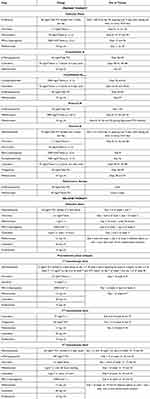
Twenty-seven months after the primary diagnosis of common preB-ALL, the 4-year-old girl was diagnosed with an isolated CNS (central nervous system) relapse of leukemia and started therapy according to the IntReALL SR 2010 protocol (Table 1). Two days after the 4th course of intensive chemotherapy, the CRP (C-reactive protein) level suddenly elevated and other inflammation parameters were increasing, including white blood cells rising from neutropenic to the level of 3.76 × 10⁹/L. The empirical antibacterial treatment of piperacillin-tazobactam was administered and within the next few days inflammation parameters decreased, just to increase again a week later. Then, subsequent antibiotic therapy, including meropenem and linezolid was administered. One day later fever appeared (39°C), and the patient’s condition started deteriorating rapidly. She experienced increasing respiratory failure with such symptoms as tachypnoea shallow breathing with a great respiratory effort and oxygen saturation below 90%, circulatory failure (tachycardia, decreased left ventricular contractility and positive fluid balance despite administered diuretics). Moreover, the liver was enlarged and laboratory findings indicated abnormalities in the coagulation panel. Capillary blood gas analysis conjointly with lactate blood level depicted lactic acidosis. Immediately, metronidazole and micafungin were added. Nevertheless, fever persisted and skin lesions with traits of Fournier’s gangrene appeared in the left groin. It was swollen and painful. The bacteriological blood cultures and wound swab confirmed Klebsiella pneumoniae carbapenemase (KPC)-producing PsA, which the girl had not hosted earlier. Despite the antibacterial treatment, the patient developed multiple-organ dysfunction syndrome (MODS) and was transferred to the pediatric intensive care unit, where additionally to intensive antimicrobial treatment, mechanical ventilation and hemodiafiltration were administered. After the final microbiological result, due to resistance of PsA to meropenem, antibacterial therapy was replaced with ceftazidime-avibactam. The targeted antibiotic therapy lasted for 33 days. Due to concurrent probable invasive pulmonary aspergillosis, empirical antifungal treatment of micafungin was replaced by preemptive therapy with voriconazole. The wound was surgically debrided and treated with vacuum-assisted closure (VAC) (Figure 1). After clinical and hematological recovery, the girl continued anticancer treatment held due to the infection for 4 weeks. Unfortunately, in another episode of neutropenia, the subsequent wound on the girl’s neck appeared after wearing a necklace. Microbiological wound swab confirmed KPC-producing PsA, but this time resistant to both meropenem and also ceftazidime-avibactam. Luckily, PsA strain was sensitive to ceftolozane-tazobactam, which was administered subsequently, and the generalized infection did not occur. Due to the fact of constant immunosuppression and further therapy in front of the patient, anti-PsA vaccine was applied successfully. Neither PsA colonization nor infection has been observed since then. After CAR-T cell-based consolidation of ALL relapse treatment, the patient has been in complete remission for 13 months.
 |
Table 1 Chemotherapy Regimen with Drug Names and Doses |
Discussion
Hematologic patients belong to a high-risk group for bacterial infections, to which they are particularly predisposed due to complex immune deficiencies resulting from their underlying disease and treatment.12,13 The following factors qualified our patient to the highest group of infectious complications, including Fournier’s gangrene, ie relapse of ALL, neutropenia, recently applied steroid and chemotherapy.12–14
Fournier’s gangrene belongs to necrotizing fasciitis associated with a high mortality rate, involving both skin and subcutaneous tissue on the external genitalia and in the perianal region.7 Moreover, the frequency is higher in adults, including 10–40 times higher in males.11 However, despite its usual infrequency in children, this has been also reported as one of the potential consequences of treatment in pediatric onco-hematological population.15 Immunosuppression, including this phenomenon arising from hematological malignancies, such as treated relapsed leukemia in our patient, is a recognized risk factor for necrotizing fasciitis.16,17 However, the diagnosis and therapy of Fournier’s gangrene still remain challenging and troublesome.
The etiological factor of a Fournier’s gangrene is a mixed oxygen-anaerobic bacterial flora,9 mostly the physiological flora of the urogenital or anorectal region, such as enteric rods (Escherichia coli, Klebsiella spp., Proteus spp)., Gram-positive cocci (staphylococci, streptococci, enterococci) and obligate anaerobic bacteria (Clostridium spp., Bacteroides spp., Fusobacterium),8 and in rare cases fungi, including Candida spp. or molds.18,19 However, the cultures of patients with concurrent Fournier’s gangrene and hematological malignancies usually contain Gram-negative bacteria, such as PsA and Escherichia coli. Moreover, in patients with onco-hematological diseases monomicrobial PsA infection is more common than in the general population of Fournier’s gangrene patients.7
The causative microflora is often polymicrobial (aerobic and anaerobic); hence, drugs of choice for Fournier’s gangrene treatment are III–IV generation cephalosporins with antibiotics of the group of nitroimidazole, fluoroquinolones, aminoglycosides. Carbapenems should be introduced within the complex antibacterial therapy, providing the refractory forms.11 In our patient due to rapidly deteriorating general condition, the de-escalation regimen of antibiotic therapy was chosen in the management of Fournier’s gangrene.12,14 Unfortunately, the presence of KPC-PsA, the MDR strain predisposed by multiple previous antibiotic treatments, prolonged hospitalization, immunosuppression, underlying disease and damage to the mucosal barrier, led to the development of septic shock.12,20,21 An appropriate combination of antimicrobial drugs, especially in combination with ceftazidime-avibactam recommended for MDR PsA infection, followed by V generation cephalosporin + beta-lactamase inhibitor, ie ceftolozane-tazobactam, consolidated with subsequent administration of an anti-PsA vaccine enabled therapeutic success and continuation of anticancer treatment without recurrence of invasive PsA infection.21–23
A significantly relevant issue to consider in the treatment of Fournier’s gangrene is an emergency surgical intervention concomitant with antibacterial therapy. It is known that the most effective way to complement systemic treatment of Fournier’s gangrene is using negative pressure wound therapy with VAC.24 It accelerates healing of difficult wounds by accelerating granulation of the wound bed. It is also an effective method of wound debridement, cutting off the fasciitis process.25,26 This may provide significant treatment benefits, leading to improved clinical outcomes and patient quality of life, as well as shorter hospital stays, less analgesic use, faster wound closure, reduced markers of sepsis, fewer operations and postoperative dressing changes.27–30 Interestingly, lower percentages of surgical treatments in immunocompromised patients have been reported by some authors compared with non-immunocompromised patients. Albasanz-Puig A. et al observed that only 62.5% of hematological patients underwent surgical treatment of necrotizing fasciitis, compared with 100% of non-hematological patients.16 The hypothesised reason could be the fear of increased intraoperative bleeding associated with severe pancytopenia, eventually leading to high intraoperative mortality.16 Fortunately, results from the literature review depict that most hematological patients affected by Fournier’s gangrene undergo surgical debridement with long-term benefits.7
Conclusions
The presented case is an example of a patient with Fournier’s gangrene, which developed due to the burden of intensive oncological treatment of a childhood ALL relapse, especially after chemotherapy followed by the neutropenic phase. Results from the presented case and review of the literature have relevant clinical implications. High index of suspicion followed by early diagnosis, target antimicrobial therapy, and early surgery are the key factors of successful Fournier’s gangrene management. Fournier’s gangrene is well-known for its aggressiveness, especially in onco-hematological patients, but surprisingly a high recovery rate in pediatric patients has been reported. Therefore, Gram-negative bacteria, mainly PsA, must be included in the spectrum of antibiotics administered in these group of patients.
Acknowledgments
Oral and written consent was obtained for publication of this case report and photos from the patient’s parents.
Disclosure
The authors report no conflicts of interest in this work.
References
1. Tran TH, Hunger SP. The genomic landscape of pediatric acute lymphoblastic leukemia and precision medicine opportunities. Semin Cancer Biol. 2022;84:144–152. doi:10.1016/j.semcancer.2020.10.013
2. Dale DC. How I diagnose and treat neutropenia. Curr Opin Hematol. 2016;23(1):1–4. doi:10.1097/MOH.0000000000000208
3. Villeneuve S, Aftandilian C. Neutropenia and infection prophylaxis in childhood cancer. Curr Oncol Rep. 2022;24(6):671–686. doi:10.1007/s11912-022-01192-5
4. Zawitkowska J, Drabko K, Lejman M, et al. Incidence of bacterial and fungal infections in Polish pediatric patients with acute lymphoblastic leukemia during the pandemic. Sci Rep. 2023;13(1):22619. doi:10.1038/s41598-023-50093-5
5. Sharma G, Rao S, Bansal A, Dang S, Gupta S, Gabrani R. Pseudomonas aeruginosa biofilm: potential therapeutic targets. Biologicals. 2014;42(1):1–7. doi:10.1016/j.biologicals.2013.11.001
6. Kim HS, Park BK, Kim SK, et al. Clinical characteristics and outcomes of Pseudomonas aeruginosa bacteremia in febrile neutropenic children and adolescents with the impact of antibiotic resistance: a retrospective study. BMC Infect Dis. 2017;17(1):500. doi:10.1186/s12879-017-2597-0
7. Creta M, Sica A, Napolitano L, et al. Fournier’s gangrene in patients with oncohematological diseases: a systematic review of published cases. Healthcare. 2021;9(9):1123. doi:10.3390/healthcare9091123
8. Wróblewska M, Kuzaka B, Borkowski T, Kuzaka P, Kawecki D, Radziszewski P. Fournier’s gangrene - current concepts. Pol J Microbiol. 2014;63(3):267–273.
9. Pilarska B, Nartowicz M, Jaworska-Czerwińska A, Zukow W. Fournier gangrene - diagnostic and treatment. J Educ Health Sport. 2018;8(20):446–457.
10. Creta M, Longo N, Arcaniolo D, et al. Hyperbaric oxygen therapy reduces mortality in patients with Fournier’s Gangrene. Results from a multi-institutional observational study. Minerva Urol Nephrol. 2020;72(2):223–228.
11. Tarasconi A, Perrone G, Davies J, et al. Anorectal emergencies: WSES-AAST guidelines. World J Emerg Surg. 2021;16(1):48. doi:10.1186/s13017-021-00384-x
12. Te Poele EM, Tissing WJE, Kamps WA, de Bont ESJM. Risk assessment in fever and neutropenia in children with cancer: what did we learn? Crit Rev Oncol Hematol. 2009;72(1):45–55. doi:10.1016/j.critrevonc.2008.12.009
13. Lynn -J-J, Chen KF, Weng Y-M, Chiu T-F. Risk factors associated with complications in patients with chemotherapy-induced febrile neutropenia in emergency department. Hematol Oncol. 2013;31(4):189–196. doi:10.1002/hon.2040
14. Zając-Spychała O, Gryniewicz-Kwiatkowska O, Salamonowicz M, et al. Standards of diagnostic and therapeutic management of bacterial infections in children: recommendations of polish society of pediatric oncology and hematology. Przeglad Pediatryczny. 2018;47(2):62–75.
15. Ekingen G, Isken T, Agir H, Oncel S, Günlemez A. Günlemez A Fournier’s gangrene in childhood: a report of 3 infant patients. J Pediatr Surg. 2008;43(12):e39–42. doi:10.1016/j.jpedsurg.2008.09.014
16. Albasanz-Puig A, Rodríguez-Pardo D, Pigrau C, et al. Necrotizing fasciitis in haematological patients: a different scenario. Ann Hematol. 2020;99(8):1741–1747. doi:10.1007/s00277-020-04061-y
17. Patrizi A, Bandini G, Cavazzini G, Sommariva F, Veronesi S. Acute gangrene of the scrotum and penis in a patient with acute promyelocytic leukemia. A case of acute necrotizing gangrene. Dermatologica. 1983;167(3):148–151. doi:10.1159/000249770
18. Kumar S, Pushkarna A, Sharma V, Ganesamoni R, Nada R. Fournier’s gangrene with testicular infarction caused by mucormycosis. Indian J Pathol Microbiol. 2011;54(4):847–848. doi:10.4103/0377-4929.91520
19. Rutchik S, Sanders M. Fungal Fournier gangrene. Infect Urol. 2003;16:54–56.
20. Matics TJ, Sanchez-Pinto LN. Adaptation and validation of a pediatric sequential organ failure assessment score and evaluation of the sepsis-3 definitions in critically III children. JAMA Pediatrics. 2017;171(10):e172352. doi:10.1001/jamapediatrics.2017.2352
21. Aguilera-Alonso D, Escosa-García L, Saavedra-Lozano J, Cercenado E, Baquero-Artigao F. Carbapenem-resistant gram-negative bacterial infections in children antimicrobial agents and chemotherapy. Antimicrob Agents Chemother. 2020;64(3):e02183–02190. doi:10.1128/AAC.02183-19
22. Shirley M. Ceftazidime-avibactam: a review in the treatment of serious gram-negative bacterial infections. Drugs. 2011;78(6):675–692. doi:10.1007/s40265-018-0902-x
23. Matesanz M, Mensa J. Ceftazidime-avibactam. Rev Esp Quimioter. 2021;34(Suppl1):38–40. doi:10.37201/req/s01.11.2021
24. Schneidewind L, Kiss B, Stangl FP, et al. Practice patterns in Fournier’s gangrene in Europe and implications for a prospective registry study. Antibiotics. 2023;12(2):197. doi:10.3390/antibiotics12020197
25. Cuccia G, Mucciardi G, Morgia G, et al. Vacuum-assisted closure for the treatment of Fournier’s gangrene. Urol Int. 2009;82(4):426–431. doi:10.1159/000218532
26. Ozturk E, Ozguc H, Yilmazlar T. The use of vacuum assisted closure therapy in the management of Fournier’s gangrene. Am J Surg. 2009;197(5):660–665. doi:10.1016/j.amjsurg.2008.04.018
27. He R, Li X, Xie K, Wen B, Qi X. Characteristics of Fournier gangrene and evaluation of the effects of negative-pressure wound therapy. Front Surg. 2023;6(9):1075968. doi:10.3389/fsurg.2022.1075968
28. Chen JH, Li YB, Li DG, et al. Vacuum sealing drainage to treat Fournier’s gangrene. BMC Surg. 2023;23(1):211. doi:10.1186/s12893-023-02109-0
29. Iacovelli V, Cipriani C, Sandri M, et al. The role of vacuum-assisted closure (VAC) therapy in the management of FOURNIER’S gangrene: a retrospective multi-institutional cohort study. World J Urol. 2021;39(1):121–128. doi:10.1007/s00345-020-03170-7
30. Tanwar S, Paruthy SB, Singh A, Pandurangappa V, Kumar D, Pal S. Evaluation of negative pressure wound therapy in the management of Fournier’s gangrene. Cureus. 2023;15(11):e48300. doi:10.7759/cureus.48300
 © 2025 The Author(s). This work is published and licensed by Dove Medical Press Limited. The
full terms of this license are available at https://www.dovepress.com/terms.php
and incorporate the Creative Commons Attribution
- Non Commercial (unported, 4.0) License.
By accessing the work you hereby accept the Terms. Non-commercial uses of the work are permitted
without any further permission from Dove Medical Press Limited, provided the work is properly
attributed. For permission for commercial use of this work, please see paragraphs 4.2 and 5 of our Terms.


