")
Back to Journals » International Journal of Nanomedicine » Volume 20
Therapeutic Potential of AgNP-Infused Patches in Periodontal Disease: An Observational Study in Albino Rats
Authors Barik B, Satapathy BS, Acharya B, Pattnaik G , Sillanpää M, Al-Farraj SA, Alkahtane AA
Received 14 January 2025
Accepted for publication 8 April 2025
Published 26 April 2025 Volume 2025:20 Pages 5337—5352
DOI https://doi.org/10.2147/IJN.S515396
Checked for plagiarism Yes
Review by Single anonymous peer review
Peer reviewer comments 2
Editor who approved publication: Dr Kamakhya Misra
Binapani Barik,1 Bhabani Sankar Satapathy,2 Biswajeet Acharya,1 Gurudutta Pattnaik,1 Mika Sillanpää,3,4 Saleh A Al-Farraj,5 Abdullah A Alkahtane5
1School of Pharmacy and Life Sciences, Centurion University of Technology and Management, Odisha, India; 2GITAM School of Pharmacy, GITAM Deemed to be University, Hyderabad Campus, Telangana, 502329, India; 3Saveetha School of Engineering, Saveetha Institute of Medical and Technical Sciences, Saveetha University, Chennai, Tamil Nadu, 602105, India; 4Centre of Research Impact and Outcome, Chitkara University Institute of Engineering and Technology, Chitkara University, Rajpura, Punjab, 140401, India; 5Department of Zoology, College of Science, King Saud University, Riyadh, 11451, Saudi Arabia
Correspondence: Gurudutta Pattnaik, School of Pharmacy and Life Sciences, Centurion University of Technology and Management, Odisha, 752050, India, Tel +9348110393, Email [email protected] Mika Sillanpää, Saveetha School of Engineering, Saveetha Institute of Medical and Technical Sciences, Saveetha University, Chennai, Tamil Nadu, 602105, India, Email [email protected]
Introduction: Silver nanoparticles (AgNPs) have emerged as a promising therapeutic modality in periodontal disease management due to their potent antimicrobial activity and ability to promote tissue regeneration. This study aimed to evaluate the efficacy of AgNP-infused patches in ligature-induced periodontitis rat model.
Methods: AgNPs were synthesized using a green synthesis method with neem (Azadirachta indica) leaf extract followed by incorporation into polyvinyl alcohol (PVA)-based patches. In vivo efficacy of experimental AgNP patch was tested in albino rats vs marketed formulation and control groups, following ARRIVE guidelines.
Results: Characterization by Atomic Force Microscopy revealed spherical AgNPs with an average size of 85 nm, while Field Emission Scanning Electron Microscopy confirmed uniform morphology and size distribution. Production of microporous AgNP patches with uniform nanoparticle dispersion was also validated by Scanning electron microscopy and Energy Dispersive X-ray Analysis. Observational and histopathological analysis revealed significant improvements in gingival and periodontal tissue restoration, inflammation reduction in the AgNP-treated groups compared to untreated and standard-treated groups.
Discussion: Notably, the high-dose AgNP patch (1000 mg/kg) treated group exhibited near-complete tissue restoration. These findings highlight the potential of AgNP patch as a localized treatment alternative for periodontal disease.
Keywords: silver nanoparticles, AgNPs, green synthesis, patch, periodontal disease, tissue regeneration, antimicrobial activity
Graphical Abstract:

Introduction
Periodontal disease, commonly referred to as gum disease, is a chronic inflammatory condition that affects the supporting structures of teeth, including the gums, bones, and connective tissues. Initially manifesting as gingivitis (gum inflammation), it can progress to periodontitis if untreated, leading to bone and tissue damage and, potentially, tooth loss in adults.1 Globally, periodontal disease is highly prevalent, affecting 45–50% of adults, with 10–15% experiencing severe forms. The disease’s prevalence rises with age, particularly among individuals over 35.2 Risk factors, including smoking, diabetes, genetic predispositions, and inadequate oral hygiene, increase susceptibility.3 Additionally, individuals in low-income groups often experience higher prevalence due to limited dental care access.
Standard treatments for periodontitis combine professional interventions with patient-led oral care, including scaling and root planning (deep cleaning), antibiotic therapy, and, in advanced cases, surgical or laser procedures to remove infected tissue and stimulate healing.4 Maintenance therapy with regular cleanings is also critical. However, treatment faces challenges, such as patient adherence to oral care, the recurrence of disease, antibiotic resistance, and the high costs of periodontal care, which limit access, especially for underserved populations.5
Silver nanoparticles (AgNPs) are promising antimicrobial agents due to their broad-spectrum efficacy against bacteria, including antibiotic-resistant strains, and their ability to disrupt biofilms; structured bacterial communities that are resistant to standard treatments.6 AgNPs act through multiple mechanisms, such as membrane disruption, reactive oxygen species (ROS) generation, and interference with DNA and protein synthesis, making bacterial resistance less likely.7 They effectively penetrate biofilms, which is advantageous for treating chronic infections. The extracellular matrix (ECM), synthesized by cells, constitutes a component of biomolecules inside a particular tissue. Further, owing to their facile synthesis methods and stability, they can be integrated into medical device coatings or wound dressings. These properties make AgNPs particularly valuable in managing multidrug-resistant infections and biofilm-associated conditions.8
The well-hydrated nanocomposite known as ECM comprises a number of proteins (including signaling proteins, adhesion proteins, and proteoglycans) and fibers (such as collagen fibers, reticular fibers, and elastic fibers), the majority of which enhance its stiffness. Moreover, a selectively replicated natural ECM scaffold has shown the ability to enhance cytoskeletal organization, cellular signaling, motility, proliferation, adhesion, and differentiation.9 The presence of nanoscale components in the ECM thus renders the cell structure of nanoscale scaffolds more analogous to the natural ECM compared to larger-scale scaffolds. Nanomaterials may be engineered that resembles certain extracellular matrices to enhance cell migration, adhesion, and proliferation in injured tissues.10 Tissue engineering scaffolds that accurately replicate the porosity and surface area of natural extracellular matrix at the nanoscale provide an environment that enhances cellular responses, such as adhesion, proliferation, and differentiation.11
The purpose of this study is to evaluate the in vivo effectiveness of AgNP-infused patches in periodontal infections. Specifically, it aims to assess the antimicrobial efficacy of AgNPs in ligature-induced periodontitis model in terms of their impact on inflammation and tissue healing. By applying AgNP-infused patches directly to affected periodontal pockets, the study seeks to determine their therapeutic potential for managing periodontal disease compared to conventional treatments. Though antimicrobial effects AgNPs have already been reported. But the present work is not just a mere repetition of the published reports. A green synthesis method was followed for production of experimental AgNPs using Azadirachta indica (commonly known as Neem) leaf extract. In view of the established antimicrobial effects of the neem leaves and their long history in periodontal applications, their use as green precursor in AgNPs would synergize the antimicrobial effect of the formulation. Further, fabrication of AgNP infused patch system for periodontal infections are scarcely reported. In vivo efficacy analysis of experimental AgNP patch in ligature-induced periodontitis rat model further sum up novelty to the study.
Materials and Methods
Materials Required
Fresh Neem (Azadirachta indica) leaves were collected from medicinal plant garden of Centurion University of Technology & Management, Odisha, India. Silver nitrate (AgNO3), Polyvinyl alcohol (PVA), Polyethylene glycol (PEG) was procured HiMedia, Hyderabad, India. Ligatures or silk sutures were procured from local medical shop for the induction of periodontal infection. Sodium carboxymethyl cellulose (Na-CMC), Guar gum was procured from Bharat Biochem, Gujarat, India. Ketamine used to anaesthetize the rats was procured from Nephron Pharmaceutical Corporation, Mumbai, India. All other chemicals/solvents used for the work were of analytical grade.
Plant Material Identification
Identification of the plant was done and authenticated by Dr. Santanu Bhattacharya, Botanist, Centurion University of Technology & Management, Odisha, India, having Voucher specimen number as CUTM/BOT/2023/01612 (Herbarium sheet has been provided as Figure S1).
Animals
The study employed three-month-old Wistar albino rats, weighing between 200 g and 300 g, obtained from the National Institute of Science Education and Research (NISER), Bhubaneswar, Odisha. All the animal experiments were conducted following approval of the protocol from the Institutional Animal Ethics Committee, Centurion University of Technology and Management, Odisha, India. Both male and female rats were included in the study. Before the experiment, the animals were acclimatized to the laboratory environment for 2 weeks under controlled conditions, including a temperature of 24 ± 2°C, humidity of 45–55%, and a 12-hour light/dark cycle.13 The rats have unrestricted access to standard laboratory chow and water during this period.14 All animal experiments were conducted strictly in alliance with ARRIVE guidelines.
Methods
Synthesis of AgNPs
Silver nanoparticles (AgNPs) were synthesized by a green synthesis method utilizing neem leaves as both a reducing and capping agent as per our previously published report.15 The preparation of an aqueous extract from Azadirachta indica leaves included the collection of the leaves followed by their thorough drying in the shade. 5 g of Azadirachta indica coarse powder were boiled with 50 mL of distilled water for 30 minutes in a beaker to prepare the extract. Upon cooling to room temperature, the liquid extract was filtered and stored for future use. A solution of silver nitrate was prepared by dissolving 10 mg of silver nitrate in 50 mL of double-distilled water. The solution was heated to a temperature range of 50°C to 60°C, and Azadirachta indica extract was gradually added dropwise until a colour change occurred. The appearance of a pale-yellow colour in the solution indicated the formation of Ag nanoparticles due to its surface plasmon resonance property, which was further validated by the visible absorbance peak at 438 nm. The synthesis method of experimental AgNPs has been summarized in Figure 1.
 |
Figure 1 Green synthesis of experimental silver nanoparticles (AgNPs) using Azadirachta indicaas precursor. |
FESEM
The morphology and shape of the silver nanoparticles were analysed using field emission electron microscopy SUPRA55 (CARL ZEISS, Germany). EDAX analysis of silver nanoparticles was conducted utilizing a SUPRA55 (CARL ZEISS, Germany) equipped with a FESEM and an EDAX attachment.16
AFM
The surface morphology of AgNPs was analysed using AFM (Agilent Technologies AFM- 5500). AFM, or scanning probe microscopy, uses a sharp probe that engages with the surface of the sample to obtain three-dimensional data at the nanoscale and can measure the height of the nanoparticle.17
Preparation of AgNPs Loaded Patch
Subsequently, the obtained AgNPs were incorporated into a PVA-based patch using the solvent casting technique to ensure efficient delivery of the nanoparticles. Briefly, PVA (0.5 g) was dissolved in 10 mL of double distilled water and heating to 80–90°C while stirring until a clear solution was formed. 0.01 g Guar gum (thickeners) and 0.05 g Na-CMC (stabilizers, film-former) were added to the prepared PVA solution. To this, synthesized AgNPs (0.5mL) was added while stirring continuously. Glycerol (0.05mL) and polyethylene glycol (0.5mL) were included as plasticizers to the dispersion by vigorous agitation. The prepared mixture was poured into Petri dishes to form patches of the uniform thickness and shape followed by drying in a hot air oven at 40–60°C for 5 h. Dried patches were carefully removed and cut into uniform dimensions.18
SEM
The study used SEM micrographs to examine the morphology, surface texture, and distribution of AgNPs within a patch.19 Elemental composition analysis was performed using an Energy-Dispersive X-ray (EDAX) Spectroscopy attachment to confirm the presence of silver (Ag) in the patch. The images assessed surface morphology, dispersion, and uniformity of AgNPs within the polymer matrix. EDAX spectra confirmed Ag peaks and distribution. Multiple regions of the patch were examined to ensure reproducibility and compared with control samples to highlight structural and compositional differences induced by AgNP incorporation.20
Antimicrobial Study
The materials used in the microbiological experiments included Mueller Hinton agar (Thermo Scientific, Leicestershire, UK), 94 mm-diameter Petri dishes, and 96% ethyl alcohol (AB Stumbras, Kaunas, Lithuania). The bacterial strains utilized were a referential strain of S. aureus and P. gingivalis.21
Microbiological Methods
The current microbiological experiments aimed to evaluate the antimicrobial activity of AgNP-infused patch: Ag thin-film-coated samples and silver-ion-saturated water against the referential S. aureus and P. gingivalis. The spread plate method was employed to distribute bacteria on the agar surface using a swab or a microbiological loop bent at a 90° angle. This was conducted following serial dilutions of the bacterial suspension. Experimental data were analyzed using Excel software, with further data fitting conducted using exponential and logarithmic functions in Origin Lab software. To ensure statistical reliability, the experiments were repeated four times.22
In-vivo Study
Experimental Design
The study involved a total of 30 Wistar albino rats. The rats were randomly assigned into five groups (n = 6 per group): Group I (Control) received no treatment, Group II (Periodontitis) underwent periodontitis induction without treatment, Group III received 1% chlorhexidine gel, Group IV was treated with 500 mg/kg silver nanoparticle (AgNP) patches, and Group V received 1000 mg/kg AgNP patches. Periodontitis was induced in Groups II–V using a sterile 4–0 silk ligature around the mandibular first molar for 14 days. Treatments were then administered daily for 21 days. Chlorhexidine gel was topically applied in Group III, while AgNP patches were applied in Groups IV and V.23 Treatments were administered daily for a specific duration following ligature placement to evaluate therapeutic outcomes.
Throughout the study, behavioural alterations were systematically monitored to assess the effects of the treatments. Parameters such as respiration were observed for irregularities like shallow or rapid breathing, and piloerection, indicative of stress or discomfort, was recorded. Gait was monitored for abnormalities such as limping or unsteady walking. Catalepsy was assessed by evaluating the animals’ ability to maintain abnormal postures, and the righting reflex was tested by observing their ability to return to an upright position when placed on their backs. The pinna reflex was evaluated for responsiveness to light touch or sound stimuli near the ear. Levels of arousal and alertness, along with signs of sedation such as reduced activity or lethargy, were documented. Spontaneous activity, including general movements and exploratory behaviour, was carefully observed for changes in natural activity levels. Additional observations included redness or irritation, localized swelling, particularly in the oral or gingival regions, and abnormal secretions such as excessive salivation, lacrimation, or nasal discharge. These assessments were conducted daily (From 12h to 14 days post-treatment) to comprehensively understand the treatments’ effects on the animals’ overall health and well-being.
Histopathological Examination
The study was conducted on the selected Wister rats in different treatment groups as discussed above. Following the experimental treatment period, rats were euthanized with an overdose of ketamine/xylazine. Ethical guidelines were strictly followed to minimize animal suffering, ensuring proper postoperative care and pain management.24 Gingival and periodontal tissues were collected and were fixed in 10% neutral-buffered formalin for 48 h, dehydrated, and embedded in paraffin. Serial 5-μm sections of tissue were stained with Hematoxylin and Eosin (H&E) for histological observations. Details of the tissue processing have been illustrated in Figure 2. Two blinded pathologists examined sections under a light microscope (Olympus BX53). Histopathological scoring assessed epithelial integrity, inflammatory cell infiltration, periodontal ligament organization etc.
 |
Figure 2 Depicts the tissue processing and staining methodologies. |
Statistical Study
To ensure precision and consistency, each experiment was conducted thrice. The data were represented using the standard deviation (SD) and mean. One-way ANOVA was conducted using the software Origin Pro 8 to analyze the statistical data and then followed by the Tukey post hoc test. Differences with a p-value of 0.05 at the 95% confidence level were considered statistically significant. The results indicated a significant difference between the experimental groups.
Results
FESEM
FESEM typically revealed uniform shape and spherical characteristics of synthesized AgNPs (Figure 3). The observed particle size ranged below 100 nm. Coupled with FESEM, EDAX confirmed the elemental composition of the experimental AgNPs. The characteristic silver peaks in the EDAX spectrum validated the presence of Ag, along with any minor peaks that might indicate residual stabilizers or impurities from the synthesis process (Figure 4). AgNPs exhibited pronounced signal peaks at 0.3 and 2.6 keV, attributable to surface plasmon resonance. It displayed the presence of components including Ag, C, O, and Ca.
 |
Figure 3 FESEM of experimental silver nanoparticles (AgNPs). |
 |
Figure 4 EDX analysis of experimental silver nanoparticles (AgNPs). |
AFM
AFM offers a detailed 3D topographical view of AgNPs deposited on a substrate. The acquired morphology indicated that the synthesized AgNPs were spherical, as corroborated by the absorbance spectrum (Figure 5A and B). The dimensions of the AgNPs, as measured by AFM, ranged from 50 to 100 nm, with a mean size of 85 nm (Figure 5D). The particles exhibited polydispersity and agglomeration as a result of the binding of certain stabilising and capping compounds found in the neem extract. (Figure 5C) shows two- and three-dimensional representations of the sample surface at scan sizes of 2 µm × 2 µm and 5 µm × 5 µm, illustrating the agglomerated, polydispersed distribution of AgNPs. The clustered, polydispersed distribution of AgNPs can clearly be seen in the images. This information might be crucial for understanding the behavior and properties of the AgNPs in various applications.
 |
Figure 5 AFM result of experimental silver nanoparticles (AgNPs) (A and B) Shows 2D view of silver nanoparticles, (C) Shows 3D view of silver nanoparticles, (D) Shows topography image of silver nanoparticles. |
SEM results for AgNPs-Infused Patch
SEM images of the AgNPs loaded patch revealed a uniform distribution of nanoparticles and a high density of nanoparticles uniformly scattered across the polymeric matrix. The AgNPs appeared as spherical or quasi-spherical structures embedded in the patch, with minimal aggregation due to the stabilizing effect of the polymer. The particle size of AgNPs ranged between 10–100 nm, consistent with the expected synthesis specifications. The surface morphology of the patch exhibited a porous or smooth texture (Figure 6). Additionally, the interaction between the nanoparticles and the polymer matrix could ensure stable incorporation.
 |
Figure 6 SEM analysis of AgNP-loaded patch. |
EDAX confirmed the elemental presence of silver in the patch, with characteristic silver peaks prominently observed in the spectrum. Minor peaks corresponding to elements from the polymer or additives were also visible, indicating successful integration of AgNPs into the patch formulation. The EDAX data illustrated the elemental distribution of Ag, C, N, Na, and O in the patch with varying percentages as shown in Figure 7. Overall, the SEM images revealed a well-structured and evenly distributed AgNP patch with a complex network of polymers. This detailed analysis overall highlighted the effectiveness of the patch system in incorporating and distributing AgNPs for potential antimicrobial applications.
 |
Figure 7 EDX analysis of key elements presents in AgNP loaded patch. |
In vitro Antibacterial Activity
The in vitro antibacterial activity of the experimental AgNP-infused patches was evaluated against a Gram-negative strain (P. gingivalis) and a Gram-positive strain (S. aureus). The microdilution method was employed for the determination of antibacterial activity, with the results expressed as Minimum Inhibitory Concentration (MIC).
The MIC values for the antibacterial activity of the formulations against P. gingivalis and S. aureusare presented in Table 1. The results indicate that AgNP-patches were significantly more effective against P. gingivalis and S. aureus. Specifically, the MIC values for AgNP-patch were 3.13 μg/mL against P. gingivalis and 25 μg/mL against S. aureus. The findings overall suggest that the prepared patches exhibited higher activity against Gram-negative bacteria (P. gingivalis) as compared to Gram-positive bacteria (S. aureus).
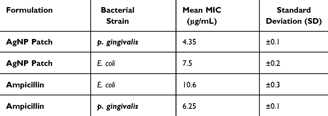 |
Table 1 Antibacterial Activity of Silver Nanoparticle Loaded Patch (AgNP Patch) Against P. Gingivalis and S. Aureus (Mean ± Standard Deviation) |
Behavioural Reflex
The behavioural parameters observed across the study groups included respiration, piloerection, gait, catalepsy, righting reflex, pinna reflex, arousal, sedation, spontaneous activity, redness, swelling, and abnormal secretions as shown in (Table 2). Respiration, piloerection, gait, catalepsy, arousal, sedation, spontaneous activity, redness, swelling, and abnormal secretion were not observed in any of the experimental groups (Group I–V) at any of the time intervals assessed, including 12 h, 24 h, 2 days, 8 days, and 14 days post-treatment as shown in Figure 8. The absence of these parameters (NA) suggests that there were no apparent adverse effects or severe stress responses induced by either the ligature placement or the treatment interventions in the animals. The righting reflex was consistently present (P) in all groups (Group I–V) across all time intervals. This indicates that the animals retained their ability to regain an upright position when placed on their backs, reflecting an absence of neurological impairments or significant physical weakness during the experimental period. Similarly, the pinna reflex was observed to be consistently present (P) in all groups at all time intervals. This indicates retention of normal sensory and reflexive responses throughout the study, suggesting that the treatments and procedures did not adversely affect the animals’ responsiveness to light touch or auditory stimuli near the ears. In summary, the behavioural assessment indicated no adverse effects or abnormalities in the animals across all groups and time intervals. The consistent presence of the righting and pinna reflexes, along with the absence of other behavioural alterations, highlights the safety and non-toxic nature of the interventions employed.
 |
Table 2 Behavioural Parameters Observed Across Different Animal Groups During Treatment Period |
 |
Figure 8 (A–C) The yellow colour indicating Steps of induction of periodontitis in experimental Wister rat by ligature. (D) The green colour indicating Detection of inflamed tissue post ligature induction. (E) The blue colour indicatingApplication of AgNP-patch over the infected periodontal area. (F) The Red colour indicatingRecovery of inflamed tissue towards normal post 14 days treatment period. |
Histopathological Examination
The histopathological examination of gingival and periodontal tissues across the control and experimental groups revealed significant differences in tissue integrity, inflammation, and regeneration. In the control group (Group I), the gingival and periodontal tissues exhibited normal histological architecture, with intact epithelium and underlying connective tissue, free of any inflammation or damage. The periodontal ligament fibers were well-organized showing no signs of bone resorption or inflammatory cell infiltration. In contrast, the tissues from the periodontitis group (Group II) displayed extensive histopathological alterations indicative of severe periodontal infection. The gingival epithelium was disrupted, with significant detachment from the underlying connective tissue. A dense infiltration of inflammatory cells, including neutrophils and lymphocytes, was observed in the gingiva and periodontal ligament. The periodontal ligament fibres were disorganized and fragmented.
Treatment with 1% chlorhexidine digluconate gel (Group III) led to notable improvements, with partial restoration of the gingival epithelium and a reduction in inflammatory cell infiltration compared to the untreated periodontitis group. However, the tissue regeneration was less advanced than in the AgNP-treated groups. In the group treated with 500 mg/kg AgNP patches (Group IV), moderate improvements were observed. The gingival epithelium showed partial restoration with reduced detachment, and inflammatory cell infiltration was significantly decreased, with fewer neutrophils and lymphocytes. The periodontal ligament fibres showed partial reorganization. Mild inflammation persisted in localized regions, but overall, the healing process was progressing. However, the group treated with 1000 mg/kg AgNP patches (Group V) demonstrated the most significant histopathological improvement (p ˂ 0.05). The gingival epithelium was almost completely restored, with minimal detachment and a re-established normal architecture. Inflammatory cell infiltration was markedly reduced, with only occasional lymphocytes observed. The periodontal ligament fibres were well-organized. The tissue response in Group V indicated enhanced healing and regeneration compared to the lower-dose treatment group, highlighting the superior efficacy of the higher AgNP dose in promoting tissue regeneration (Figure 9).
 |
Figure 9 Histopathological evaluation of gingival and periodontal tissues across study groups. (A) Control group (Group I): Normal histological architecture with intact gingival epithelium, underlying connective tissue, well-organized periodontal ligament fiber. (B) The arrow indicating Periodontitis group (Group II): Disrupted gingival epithelium with significant detachment from the connective tissue, dense inflammatory cell infiltration, disorganized periodontal ligament fiber. (C) The arrow indicating Periodontitis + 1% chlorhexidine digluconate gel treated group (Group III): Partial restoration of gingival epithelium with reduced inflammatory cell infiltration and early signs of healing in the periodontal ligament . (D) The arrow indicating 500 mg/kg AgNP patch-treated group (Group IV): Moderate improvement with partial restoration of gingival epithelium, decreased inflammatory infiltration, reorganization of periodontal ligament fiber. (E) The arrow indicating 1000 mg/kg AgNP patch-treated group (Group V): Near-complete restoration of gingival epithelium, minimal inflammatory cell infiltration, well-organized periodontal ligament fibers, indicating superior therapeutic efficacy. |
Discussion
This research investigated the potential of AgNP-infused patches as a novel treatment for periodontal infection. The use of neem extract for the green synthesis of AgNPs offers a promising eco-friendly and biocompatible alternative to traditional methods, promoting sustainable biomedical practices.23,24 The synthesis process is in line with the increasing demand for environmentally friendly and non-toxic treatments in modern medicine, ensuring that the AgNPs are safe for biological use.23 The advanced characterization of AgNPs through AFM, FESEM, SEM, and EDAX confirmed their spherical morphology, nanoscale dimensions, and uniform incorporation into PVA based patch matrix.22 These physical properties are critical for ensuring the stability, uniform distribution, and controlled release of the AgNPs, all of which contribute to the therapeutic efficacy of the patches.25 However, while these characterization methods establish the basic material properties, additional in vivo testing could provide further insights into how the nanoparticles interact with the tissue over time and their long-term stability.
Histopathological analysis demonstrated that AgNP-infused patches significantly improved periodontal tissue integrity, reduced inflammation in the ligature-induced rat model. The process of tissue repair was evaluated based on key histopathological criteria, including degree of epithelial restoration, the extent of inflammatory cell infiltration, and the reorganization of periodontal ligament fibres. In the control group, gingival and periodontal tissues exhibited normal architecture, with intact epithelium, well-structured connective tissue, and organized periodontal ligament fibers. In contrast, the untreated periodontitis group displayed extensive histopathological alterations, including epithelial disruption, significant detachment from connective tissue, dense infiltration of neutrophils and lymphocytes, disorganized periodontal ligament fibers. Treatment with 1% chlorhexidine digluconate gel led to partial restoration of epithelial integrity and a reduction in inflammatory cell infiltration; however, bone regeneration remained limited, with only minor improvements in periodontal ligament organization and moderate reductions in alveolar bone resorption.
On the other hand, the 500 mg/kg AgNP-treated group exhibited a more substantial healing response, with partial epithelial restoration, reduced inflammatory cell infiltration, and improved periodontal ligament organization. The epithelial architecture was nearly intact, inflammatory cell presence was minimal, and periodontal ligament fibers were well-organized. These results are consistent with existing literature demonstrating the regenerative and antimicrobial properties of AgNPs.23,26,27 The observed reduction in bacterial load and inflammatory responses aligns with the multifaceted mechanisms through which AgNPs exert their action, including bacterial membrane disruption, the generation of reactive oxygen species (ROS), and interference with bacterial DNA replication and protein synthesis. The mechanistic approach and further insights on the mechanism of AgNPs against bacterial infections and tissue regeneration have been depicted in Figure 10. These combined mechanisms reduce the likelihood of bacterial resistance compared to traditional antibiotics.
 |
Figure 10 Displays the mechanistic approach of silver nanoparticles (AgNPs) against bacterial infections and tissue regeneration. |
In addition to antimicrobial activity, AgNPs have shown promise in promoting tissue repair and regeneration. AgNPs stimulate fibroblast proliferation, collagen synthesis, and angiogenesis, essential processes for effective tissue healing. These regenerative properties were evident in the improvements in periodontal ligament fibre organization in the treated groups.28,29 However, the precise molecular pathways involved in these regenerative effects need further exploration to understand the role of AgNPs in tissue healing fully and to optimize their therapeutic potential.
Compared to standard treatment with 1% chlorhexidine gel, AgNP-infused patches showed superior therapeutic efficacy. While chlorhexidine is a well-established antimicrobial agent, its use is limited by cytotoxicity at higher concentrations and its lack of regenerative properties.30,31 In contrast, AgNP patches offer both antimicrobial and regenerative benefits, addressing the dual challenge of combating infection while promoting tissue repair.32 This dual mechanism of action is a significant advantage over conventional treatments, making AgNP patches a more comprehensive therapeutic option for periodontal disease. Additionally, the localized delivery of AgNPs ensures targeted action at the site of infection, minimizing systemic side effects and potentially improving patient compliance. However, it is important to note that the clinical translation of AgNP-based patches requires further evaluation of factors such as long-term biocompatibility, possible toxicity at higher doses, and the potential for accumulation in systemic circulation. The ideal dosage and formulation must also be tailored to ensure optimal therapeutic outcomes without adverse effects. Furthermore, future studies should consider the potential interactions between AgNPs and other components in the oral microbiome and assess the sustainability of AgNP treatments over time.
Conclusion
The study conclusively demonstrated the therapeutic potential of AgNP-infused patches in managing periodontal disease. AgNPs, synthesized via a green method using neem extract, exhibited superior antimicrobial properties as compared to standard drug therapy. Characterization techniques such as AFM, FESEM, SEM, and EDAX validated the uniform morphology, size, and effective integration of AgNPs into PVA-based patches, creating a microporous and stable delivery system. In vivo efficacy evaluation in ligature-induced periodontitis model highlighted significant improvements in tissue integrity and inflammation in AgNP-treated groups. The high-dose AgNP patch group (1000 mg/kg) demonstrated the most pronounced therapeutic effects, with near-complete restoration of gingival epithelium, minimal inflammatory infiltration. Compared to standard treatment with 1% chlorhexidine gel, AgNP patches offered superior efficacy, emphasizing their dual role in antimicrobial action and tissue repair. This localized therapy approach would minimize systemic side effects and reduce reliance on antibiotics, addressing challenges like antibiotic resistance and recurrence. While the findings underscore the potential of AgNP-infused patches as a transformative solution for periodontal disease management, further studies are warranted to explore their long-term safety, biocompatibility, and clinical efficacy in human trials. These results would lay the foundation towards clinical application of AgNPs as innovative, affordable, and localized treatment modality, offering hope for improved periodontal care, particularly in underserved populations.
Ethical Statement
All animal studies were conducted as per the approval of the Institute Animal Ethical Committee, Centurion University of technology and Management, Odisha, India. (IAEC Protocol number CUTM/IAEC-23, Institutional IAEC number-2024/PO/Re/S/18/CCSEA). All animal-related experiments complied with ARRIVE guidelines.
Acknowledgment
Researchers acknowledge Supporting Project number (RSP2025R140), King Saud University, Riyadh, Saudi Arabia, for the support to execute the work.
Author Contributions
All authors contributed to data analysis, drafting or revising the article, have agreed on the journal to which the article will be submitted, gave final approval of the version to be published, and agree to be accountable for all aspects of the work.
Disclosure
The author(s) report no conflicts of interest with any other third party or organization for the data presented in the work.
References
1. Ray RR. Periodontitis: an oral disease with severe consequences. Appl Biochem Biotechnol. 2023;195(1):17–32. doi:10.1007/s12010-022-04127-9
2. Villoria GE, Fischer RG, Tinoco EM, Meyle J, Loos BG. Periodontal disease: a systemic condition. Periodontology 2000. 2024;96(1):7–19. doi:10.1111/prd.12616
3. Janakiram C, Mehta A, Venkitachalam R. Prevalence of periodontal disease among adults in India: a systematic review and meta-analysis. J Oral Biol Craniofacial Res. 2020;10(4):800–806. doi:10.1016/j.jobcr.2020.10.016
4. Hughes S, Davies L, Monaghan U, Stennett M. Implementation of a CBT-based dental anxiety management pathway for patients with learning disabilities. Br Dent J. 2024;237(1):40–44. doi:10.1038/s41415-024-7557-7
5. Vaismoradi M, LilloCrespo M, Turjamaa R. Nurse-led medication management for older people in home care: a systematic review of evolving nurse responsibilities in technology-assisted care. Home Health Care Manag Pract. 2024;10848223241283415.
6. Barua N, Buragohain AK. Therapeutic potential of silver nanoparticles (agnps) as an antimycobacterial agent: a comprehensive review. Antibiotics. 2024;13(11):1106. doi:10.3390/antibiotics13111106
7. Rodrigues AS, Batista JG, Rodrigues MÁ, et al. Advances in silver nanoparticles: a comprehensive review on their potential as antimicrobial agents and their mechanisms of action elucidated by proteomics. Front Microbiol. 2024;15:1440065. doi:10.3389/fmicb.2024.1440065
8. Zhang K, Li X, Yu C, Wang Y. Promising therapeutic strategies against microbial biofilm challenges. Front Cell Infect Microbiol. 2020;10(359). doi:10.3389/fcimb.2020.00359
9. Lauritano D, Limongelli L, Moreo G, Favia G, Carinci F. Nanomaterials for periodontal tissue engineering: chitosan-based scaffolds. A systematic review. Nanomaterials. 2020;10(4):605. doi:10.3390/nano10040605
10. Craciunescu O, Seciu AM, Zarnescu O. In vitro and in vivo evaluation of a biomimetic scaffold embedding silver nanoparticles for improved treatment of oral lesions. Mater Sci Eng. 2021;123:112015. doi:10.1016/j.msec.2021.112015
11. Chen S, Huang X. Nanomaterials in scaffolds for periodontal tissue engineering: frontiers and prospects. Bioengineering. 2022;9(9):431. doi:10.3390/bioengineering9090431
12. Barik B, Pattnaik G, Satapathy BS, Acharya B, Kumar LA. An ethnobotanical study of medicinal plants used by ethnic communities of Nuapada District, Odisha to treat periodontal disorders. Ethnobot Res App. 2024;28:1–20. doi:10.32859/era.29.3.1-20
13. Cadinoiu AN, Rata DM, Daraba OM, et al. Silver nanoparticles biocomposite films with antimicrobial activity: in vitro and in vivo tests. Int J mol Sci. 2022;23(18):10671. doi:10.3390/ijms231810671
14. Ruffo M, Parisi OI, Dattilo M, et al. Synthesis and evaluation of wound healing properties of hydro-diab hydrogel loaded with green-synthetized AGNPS: in vitro and in ex vivo studies. Drug Delivery Transl Res. 2022;12(8):1881–1894. doi:10.1007/s13346-022-01121-w
15. Barik B, Satapathy BS, Pattnaik G, Bhavrao DV, Shetty KP. Sustainable synthesis of silver nanoparticles from Azadirachta indica: antimicrobial, antioxidant and in silico analysis for periodontal treatment. Front Chem. 2024;12:1489253. doi:10.3389/fchem.2024.1489253
16. Thoms S, Gonsalves RA, Jose J, Zyoud SH, Prasad AR, Garvasis J. Plant-based synthesis, characterization approaches, applications and toxicity of silver nanoparticles: a comprehensive review. J Biotechnol. 2024;394:135–149. doi:10.1016/j.jbiotec.2024.08.009
17. Wani IA, Ahmad T, Khosla A. Recent advances in anticancer and antimicrobial activity of silver nanoparticles synthesized using phytochemicals and organic polymers. Nanotechnology. 2021;32(46):462001.
18. Suksaeree J, Thuengernthong A, Pongpichayasiri K, Maneewattanapinyo P, Settharaksa S, Pichayakorn W. Formulation and evaluation of matrix type transdermal patch containing silver nanoparticles. J Polym Environ. 2018;26:4369–4375. doi:10.1007/s10924-018-1305-5
19. Wang C, Jiang Y, He K, Wāng Y. Eco-friendly synthesis of silver nanoparticles against mosquitoes: pesticidal impact and indispensable biosafety assessment. Scie Total Environ. 2024;953:176006. doi:10.1016/j.scitotenv.2024.176006
20. Vendidandala NR, Yin TP, Nelli G, Pasupuleti VR, Nyamathulla S, Mokhtar SI. Gallocatechin-silver nanoparticle impregnated cotton gauze patches enhance wound healing in diabetic rats by suppressing oxidative stress and inflammation via modulating the Nrf2/HO-1 and TLR4/NF-κB pathways. Lifesciences. 2021;286:120019.
21. Suvandee W, Teeranachaideekul V, Jeenduang N, et al. One-pot and green preparation of phyllanthus emblica extract/silver nanoparticles/polyvinylpyrrolidone spray-on dressing. Polymers. 2022;14(11):2205. doi:10.3390/polym14112205
22. Mahmood S, Mei TS, Yee WX, Hilles AR, Alelwani W, Bannunah AM. Synthesis of capsaicin loaded silver nanoparticles using green approach and its anti-bacterial activity against human pathogens. J Biomed Nanotechnol. 2021;17(8):1612–1626. doi:10.1166/jbn.2021.3122
23. Khamrai M, Banerjee SL, Paul S, Ghosh AK, Sarkar P, Kundu PP. AgNPs ornamented modified bacterial cellulose based self-healable LBL assembly via a Schiff Base reaction: a potential wound healing patch. ACS Appl Bio Mater. 2020;4(1):428–440. doi:10.1021/acsabm.0c00915
24. Dahab M, Taha R, Ghali LS. Effect of Local Nanosilver Loaded with Tetracycline in Treatment of Induced Infection of Rat Buccal Mucosa with Porphyromonas Gingivalis (Histological and Immunohistochemical Study). Dental Science Updates. 2022;3(1):65–72. doi:10.21608/dsu.2022.97155.1081
25. Kumar M, Kurup M, Jayaprakash J (2024, July). Design and optimization of herbal formulations in silver nanoparticles to enhance wound healing activity. In
26. Takallu S, Mirzaei E, ZakeriBazmandeh A, GhaderiJafarbeigloo HR, Khorshidi H. Addressing antimicrobial properties in guided tissue/bone regeneration membrane: enhancing effectiveness in periodontitis treatment. ACS Infect Dis. 2024;10(3):779–807. doi:10.1021/acsinfecdis.3c00568
27. Nandhini J, Karthikeyan E, Rani EE, et al. Advancing engineered approaches for sustainable wound regeneration and repair: harnessing the potential of green synthesized silver nanoparticles. Engineered Regeneration. 2024;5(3):306–325. doi:10.1016/j.engreg.2024.06.004
28. Noga M, Milan J, Frydrych A, Jurowski K. Toxicological aspects, safety assessment, and green toxicology of silver nanoparticles (AgNPs)—critical review: state of the art. Int J mol Sci. 2023;24(6):5133. doi:10.3390/ijms24065133
29. Jagadeesh P, Rangappa SM, Siengchin S. Advanced characterization techniques for nanostructured materials in biomedical applications. Adv Ind Eng Polym Res. 2024;7(1):122–143. doi:10.1016/j.aiepr.2023.03.002
30. Sabarees G, Velmurugan V, Tamilarasi GP, Alagarsamy V, Raja Solomon V. Recent advances in silver nanoparticles containing nanofibers for chronic wound management. Polymers. 2022;14(19):3994. doi:10.3390/polym14193994
31. Nqakala ZB, Sibuyi NR, Fadaka AO, Meyer M, Onani MO, Madiehe AM. Advances in nanotechnology towards development of silver nanoparticle-based wound-healing agents. Int J mol Sci. 2021;22(20):11272. doi:10.3390/ijms222011272
32. Xin H, Liu Y, Xiao Y, Wen M, Sheng L, Jia Z. Design and nanoengineering of photoactive antimicrobials for bioapplications: from fundamentals to advanced strategies. Adv Funct Mater. 2024;34:2402607. doi:10.1002/adfm.202402607
 © 2025 The Author(s). This work is published and licensed by Dove Medical Press Limited. The
full terms of this license are available at https://www.dovepress.com/terms.php
and incorporate the Creative Commons Attribution
- Non Commercial (unported, 4.0) License.
By accessing the work you hereby accept the Terms. Non-commercial uses of the work are permitted
without any further permission from Dove Medical Press Limited, provided the work is properly
attributed. For permission for commercial use of this work, please see paragraphs 4.2 and 5 of our Terms.
© 2025 The Author(s). This work is published and licensed by Dove Medical Press Limited. The
full terms of this license are available at https://www.dovepress.com/terms.php
and incorporate the Creative Commons Attribution
- Non Commercial (unported, 4.0) License.
By accessing the work you hereby accept the Terms. Non-commercial uses of the work are permitted
without any further permission from Dove Medical Press Limited, provided the work is properly
attributed. For permission for commercial use of this work, please see paragraphs 4.2 and 5 of our Terms.