")
Back to Journals » International Journal of Nanomedicine » Volume 20
Zinc Doped Synthetic Polymer Composites for Bone Regeneration: A Promising Strategy to Repair Bone Defects
Authors Chen L, Zhou C, Xie Q, Xia L, Liu L, Bao W, Lin H, Xiong X, Zhang H, Zheng Z, Zhao J, Liang W
Received 20 January 2025
Accepted for publication 22 May 2025
Published 1 July 2025 Volume 2025:20 Pages 8567—8586
DOI https://doi.org/10.2147/IJN.S512994
Checked for plagiarism Yes
Review by Single anonymous peer review
Peer reviewer comments 4
Editor who approved publication: Dr Krishna Nune
Long Chen,1,* Chao Zhou,2,* Qiong Xie,3 Linying Xia,3 Lu Liu,3 Wenwen Bao,3 Hongming Lin,4 Xiaochun Xiong,4 Hao Zhang,4 Zeping Zheng,4 Jiayi Zhao,4 Wenqing Liang4
1Department of Orthopedics, Affiliated Hospital of Shaoxing University, Shaoxing, 312000, People’s Republic of China; 2Department of Orthopedics, Zhoushan Guanghua Hospital, Zhoushan, 316000, People’s Republic of China; 3Medical Research Center, Zhoushan Hospital of Traditional Chinese Medicine Affiliated to Zhejiang Chinese Medical University, Zhoushan, 316000, People’s Republic of China; 4Department of Orthopedics, Zhoushan Hospital of Traditional Chinese Medicine Affiliated to Zhejiang Chinese Medical University, Zhoushan, 316000, People’s Republic of China
*These authors contributed equally to this work
Correspondence: Wenqing Liang, Department of Orthopaedics, Zhoushan Hospital of Traditional Chinese Medicine Affiliated to Zhejiang Chinese Medical University, 355 Xinqiao Road, Dinghai District, Zhoushan, Zhejiang Province, 316000, People’s Republic of China, Tel +86-580-2615027, Fax +86-580-2616545, Email [email protected] Jiayi Zhao, Department of Orthopaedics, Zhoushan Hospital of Traditional Chinese Medicine Affiliated to Zhejiang Chinese Medical University, 355 Xinqiao Road, Dinghai District, Zhoushan, Zhejiang Province, 316000, People’s Republic of China, Tel +86-580-2615024, Email [email protected]
Abstract: Zinc-doped synthetic polymer composites are a potential approach for bone regeneration, effectively meeting the urgent requirement for efficient bone repair materials. This review critically examines recent advancements in zinc-doped synthetic polymer composites for bone regeneration, with a focus on their synthesis methods, material properties, biological interactions, and potential clinical applications. The primary findings indicate that adding zinc to synthetic polymers, such as poly (lactic-co-glycolic acid), polycaprolactone, and polyethylene glycol, enhances their mechanical strength, bioactivity, and biocompatibility. Those composite materials enable the regulated release of zinc, which stimulates cellular responses crucial for bone repair. The importance of this technique lies in its ability to give a versatile framework that promotes the regeneration of bone tissue, presenting a new alternative for patients with bone abnormalities and enhancing clinical results. Despite these advantages, Zn-doped polymer composites need to overcome challenges including high Zn concentration toxicity and altered degradation kinetics and changes in mechanical properties for optimized clinical success. Moreover, homogeneous Zn distribution within polymer matrices stands as an essential requirement to accomplish both stable bioactivity properties and uniform mechanical characteristics. The use of advanced fabrication methods will help overcome pelletization issues to improve the therapeutic application of Zn-doped polymer composites in bone tissue regeneration. This review is intended to provide a valuable resource for future research, contributing to the advancement of innovative therapies for bone regeneration. It facilitates the development of innovative solutions in regenerative medicines and bone repair by emphasizing the prospective role of zinc-doped polymers.
Keywords: zinc doping, synthetic polymer composites, bone regeneration, bone defects, biomaterials
Graphical Abstract:

Introduction
Bone cancer and osteoporosis, lead to a growing demand for sophisticated bone healing materials appropriate for skeletal reconstruction. Bone is widely recognized as the second most regenerative tissue in the body after blood. The body depends on bones for several essential functions, including the protection of vital organs, support for the body’s structure, and enabling movement by serving as levers for muscles. Additionally, bones are crucial for storing minerals like calcium and phosphorus and house the bone marrow, where blood cells are produced.1 Several causes can lead to bone defects, including congenital deformity, trauma, and infection. These defects cause severe pain and place a considerable strain on the healthcare system.2 Each year, around $45 billion is spent on the healthcare of 15 million individuals with bone-related issues, including 1.6 million trauma-induced fractures and 2 million osteoporosis-related abnormalities.3 There are almost 2.2 million bone transplant surgeries conducted globally every year, with an approximate annual cost of 2.5 billion dollars.4
Bone deficiencies remain a major clinical challenge, with the demand for bone substitutes outpacing the availability of autogenous, allogeneic, and xenogeneic grafts. Autogenous grafts, harvested from the patient’s own body, are considered the gold standard due to their low risk of immune rejection and their ability to promote bone formation. However, they are limited by donor site morbidity and the volume of graftable tissue available. Allografts, derived from human donors, offer an alternative with sufficient supply and the ability to support bone regeneration. While they share osteoinductive and osteoconductive properties, their effectiveness is constrained by donor availability, immune rejection risks, and slower integration. Advances in processing techniques are improving their immunocompatibility. Xenografts, sourced from animals such as cows or pigs, can also support bone regeneration but face challenges related to immune response and disease transmission. Recent developments in graft sterilization are enhancing their safety and efficacy for bone repair. While all three types of grafts provide valuable solutions for bone repair, each has limitations that ongoing research aims to address, aiming to improve clinical outcomes and meet the growing demand for bone substitutes.5
Consequently, the development of techniques for tissue engineering has met the requirements for new and creative methods to restore and regenerate bone abnormalities. Bone tissue engineering (BTE) seeks to create a bone replacement using cell biology and architecture principles. Bone defects arise from congenital abnormalities, trauma, tumor resection, and infections, which not only result in significant pain but also impose considerable strain on healthcare systems. In recent decades, bone tissue engineering (BTE) has emerged as a promising approach for addressing these defects. BTE involves the combination of biomaterials and stem cells to promote the regeneration of damaged bone. A diverse range of biomaterials, including ceramics, metals, natural and synthetic polymers, and their composites, have been utilized in BTE to repair and replace compromised bone tissue.6 Recent advancements in bone tissue engineering (BTE) have leveraged various processing techniques, including solvent casting, freeze-drying, and three-dimensional (3D) printing, to develop synthetic polymer scaffolds for bone regeneration. Solvent casting involves dissolving a polymer in a solvent, which is then cast into a mold to form a scaffold. This method allows for precise control over the scaffold’s porosity and surface characteristics, which are critical for cell attachment and osteointegration. However, challenges such as solvent toxicity and the potential limitations in mechanical strength can affect the functionality of the resulting scaffold. Freeze-drying, or lyophilization, creates highly porous scaffolds by freezing a polymer solution and subsequently removing water under vacuum. This technique enhances cellular infiltration and nutrient diffusion, promoting bone regeneration. However, the mechanical properties of freeze-dried scaffolds may not always meet the demands of load-bearing applications. Three-dimensional (3D) printing enables the precise fabrication of scaffolds with complex geometries, offering the ability to tailor porosity, mechanical strength, and bioactivity. This method allows for the creation of patient-specific scaffolds that closely match the defect site, potentially improving the efficiency of bone regeneration. 3D printing also facilitates the integration of multiple materials, further enhancing scaffold functionality. Each of these techniques offers distinct advantages, and ongoing research aims to optimize their use in the development of more effective bone repair scaffolds.7
Synthetic polymers used in bone tissue engineering can be tailored in terms of their composition, architecture, and physical properties, providing flexibility in their design and function. These materials are also highly reproducible, allowing for consistent manufacturing of scaffolds with desired characteristics. Additionally, synthetic polymers possess the unique ability to degrade over time, offering initial mechanical support to the defect site while gradually breaking down to accommodate the growth of newly formed tissue. However, while these polymers may exhibit osteoconductive properties, they often fail to achieve complete bone regeneration when used alone. To overcome this limitation, the incorporation of zinc (Zn) is emerging as a promising strategy. The release of Zn²⁺ ions from these polymers has been shown to stimulate the proliferation of bone-forming cells, thereby accelerating the bone regeneration process and enhancing the overall efficacy of the scaffold.8 Zn-doped synthetic polymer composites receive attention because Zn²⁺ ions serve dual functions in the bone metabolic process. During osteogenesis Zn functions as a vital stimulant for osteoblasts to multiply and differentiate and simultaneously suppresses osteoclast activity. Zn-doped synthetic polymers demonstrate dual antibacterial properties together with angiogenesis promotion which increases their potential application as bioactive components for bone scaffolds.9–12 Nanocomposite biomaterials are advanced materials designed for biomedical applications, combining a matrix made of biopolymers and biodegradable materials with small-sized, bioactive fillers. Biopolymers, such as collagen or chitosan, are naturally derived and biocompatible, offering a supportive scaffold for tissue regeneration. Biodegradable materials, like polylactic acid (PLA) or polycaprolactone (PCL), gradually break down within the body, eliminating the need for surgical removal as new tissue forms. Bioactive fillers, often at the nanometer scale, include substances like hydroxyapatite or growth factors that enhance the material’s biological activity, promoting cell proliferation and tissue regeneration. The integration of these components creates a synergistic effect, where the matrix provides structural support, the fillers stimulate biological responses, and the biodegradability ensures the material gradually disappears, making nanocomposite biomaterials ideal for applications in bone regeneration and other areas of tissue engineering.13 The incorporation of nano-fillers into the polymer matrix enhances both the mechanical and biological properties of the scaffold, making it more suitable for bone regeneration applications. This is crucial for ensuring compatibility with tissue regeneration.14
Zinc ions integrated into bone tissue engineering scaffolds has gained significant attention because of their fundamental importance in osteogenic processes.15
Scientific interest in zinc-based materials has witnessed dramatic changes as research activity and focus developed from 2000 through 2025. A bibliometric analysis of zinc-based biodegradable metallic materials for publication research between 2000 and 2022 has revealed 632 results including 553 research articles and 50 reviews.16 Throughout 2007 to 2013 the number of publications remained rare. Research interest in this field experienced a dramatic yearly rise starting from 2014 according to publication records. The increased attention toward zinc applications in biomedical research corresponds to general scientific acknowledgment of its biomedical potential. The research community investigates zinc-based biomaterials since they provide multiple advantages that include their biological relevance and both compatibility and degradability features alongside tissue regeneration properties. The breakdown of metallic zinc implants supports healing processes through a rate that matches tissue regeneration rates while triggering tissue rearrangement mechanisms.17
The method through which Zn²⁺ leaves scaffolds determines both bone tissue growth outcomes and influences cellular behavior and degradation.17 The controlled and extended release of Zn²⁺ activates osteoblasts along with promoting angiogenesis and enabling extracellular matrix formation which are necessary elements for successful bone healing.18 The rapid release of Zn²⁺ will generate toxicity that can harm cellular functions and cause damage to tissue regenerative processes. Different initiatives have been used to maximize Zn²⁺ release patterns through material structure modification and surface treatment developments.15 Zn-based biodegradable metals along with Zn-Mg and Zn-Cu alloys provide the ability to customize degradation speeds and regulate Zn²⁺ ion discharge thus enhancing mechanical performance while improving biocompatibility.19,20 Understanding and regulating Zn²⁺ release kinetics is crucial for maximizing the benefits of zinc in bone tissue engineering while minimizing potential cytotoxic risks associated with uncontrolled ion release. The tuning of these specific parameters allows Zn²⁺-incorporated biomaterials to become highly effective for improving bone healing together with scaffold integration.
This review examines zinc-doped synthetic polymer composites used in bone tissue engineering by investigating their potential upshots on mechanical properties and bioactivity and biocompatibility. Moreover, it examines the methods of synthesis and structural features and biological interactions of materials to understand their capability in bone regeneration. Studies were selected based on their relevance to the topic, methodological rigor, and the significance of their experimental or clinical findings. Preference was given to more recent studies that contribute to understanding key developments in the field. This review starts with a section that explains synthesis techniques of zinc-doped polymer composites before detailing their physicochemical properties, biological interactions and their function in bone regeneration. The review evaluates current challenges alongside prospects in this field after investigating their utilization challenges. Moreover, it focuses on building knowledge that promotes the development of innovative materials to enhance bone repair and regeneration.
Zinc Oxide (ZnO)-Doped Polymers in Bone Tissue Engineering
The combination of zinc oxide (ZnO) with polymer-based scaffolds and nanofibers and nanoparticles has become prominent because ZnO serves simultaneously as a structural support material and biological functional component. The core advantage of ZnO stems from its antimicrobial effects alongside its biological compatibility together with its capability to support osteogenesis by steady Zn²⁺ ion release.21 ZnO induces the gradual release of Zn²⁺ ions which modulates cellular pathways towards differentiation of osteoblasts and stimulates new blood vessel formation.22 The controlled release mechanism of ZnO-functionalized scaffolds permits the scaffolds to sustain a proper equilibrium between their bioactive properties and their mechanical support capabilities. The inclusion of Zn²⁺ directly into matrix polymers leads to rapid cellular responses yet demands exact control of the release rates to prevent toxicity.23,24
Doping involves incorporating small quantities of filler materials, or dopants, into a host polymer to modify its physical properties. However, large filler particles that do not chemically interact with the polymer are not considered true dopants. Nanomaterials, due to their high surface-to-volume ratio and enhanced surface reactivity, are particularly effective as polymer dopants. These materials, often exhibiting quantum confinement effects, interact uniquely with polymers, offering significant potential for performance enhancement. When a non-polymeric nanomaterial is incorporated into a polymer matrix, it forms a nanomaterial-doped polymer, and when the dopant is inorganic, the resulting structure is referred to as a “hybrid nanocomposite”, which combines both inorganic and organic components (Figure 1).10 This section focuses on the impact of doping polymers with zinc oxide (ZnO) in the form of scaffolds, nanofibers, and nanoparticles. The doping of ZnO into polymers can achieve three primary objectives: (1) modification of the ZnO itself, (2) modification of the polymer matrix, or (3) creation of a hybrid nanocomposite with enhanced or novel properties,25 as illustrated in Figure 1.
 |
Figure 1 The flowchart illustrates six major techniques used to incorporate zinc into polymeric scaffolds for biomedical applications. Each approach enhances the biological, mechanical, and osteogenic properties of the scaffolds, making them suitable for bone tissue engineering and regenerative medicine. |
Electrospinning Method
Polyethylene glycol (PES-PEG) scaffolds modified with NH3 plasma to improve hydrophilicity and promote cell adhesion, proliferation, and osteogenic differentiation.26 Incorporating Willemite (Zn2SiO4) bioceramic nanoparticles into electrospun fibers significantly enhanced the proliferation and osteogenic differentiation of human mesenchymal stem cells (hMSCs), as evidenced by increased alkaline phosphatase (ALP) activity, calcium deposition, and upregulation of osteogenic genes and proteins. This Zn2SiO4-loaded PES-PEG scaffold offered a promising matrix for bone tissue engineering.27
Another study involved the incorporation of metal-organic framework (MOF) nanoparticles (using zinc and copper as metal ions) into electrospun PLLA nanofibers, with air plasma treatment improving scaffold hydrophilicity. This modification enhanced osteogenic activity, reducing the water contact angle and promoting better cell proliferation. While the tensile strength of the scaffold was not significantly improved by the MOF addition, the improved osteogenic properties made it a valuable candidate for bone regeneration.28 Zinc chloride/polycaprolactone (PCL) electrospun scaffolds with antibacterial microgrooves were shown to enhance mesenchymal stem cell proliferation, highlighting their potential for nerve regeneration.29
Sol-Gel Method
Nano-bioglass ceramic nanoparticles were synthesized using the sol-gel method with a SiO2:CaO:P2O5 ratio of 55:40:5 (Mol %) and doped with zinc sulfate.30 These nanoparticles were evaluated for their chemical, physical, and biological properties, showing potential for reducing post-surgical infections and enhancing bone tissue engineering applications.30 Water-based sol-gel method to create strontium- and zinc-doped nano-hydroxyapatite (n-HAp), which was incorporated into composite scaffolds with PLGA using supercritical carbon dioxide. These Sr/Zn n-HAp-PLGA scaffolds exhibited compressive properties similar to cancellous bone, suggesting their potential as a bone substitute in tissue engineering.31
In-Situ Polymerization
In-situ polymerization is a technique used to create polymer nanocomposites by chemically reacting nanoparticles within the polymerization medium, ensuring a homogeneous dispersion of nanoparticles within the matrix. This method has been applied to enhance poly (L-lactic acid) (PLLA), which lacks inherent osteogenic activity, by incorporating zinc (Zn) to stimulate bone regeneration. Zinc-doped mesoporous silica (Zn-MS) particles were synthesized and integrated into PLLA scaffolds, promoting osteoblast differentiation and improving compressive strength through micromechanical coupling with the PLLA matrix.32 Additionally, polyaniline was used to create a polyelectrolyte shell on mesoporous silica nanoparticles, with zinc cations incorporated during in-situ polymerization to improve surface properties and enhance osteogenic potential.33 These approaches demonstrate the efficacy of in-situ polymerization for improving the biological and mechanical properties of polymer-based scaffolds for bone tissue engineering.
3D Printing
Composite filaments were developed by incorporating hydroxyapatite (HA) and zinc oxide (ZnO) nanoparticles into poly(caprolactone) (PCL), enabling the fabrication of scaffolds with enhanced osteoconductive properties. The use of PCL allowed for low-temperature printing, preventing thermal damage to surrounding tissues during scaffold fabrication.34 The in situ-printed scaffolds demonstrated good adhesion to moist bone tissue, reducing the risk of scaffold displacement. These scaffolds promoted osteodifferentiation of mesenchymal stem cells, showing potential for bone regeneration. Further studies have explored zinc-infused bioactive glass/PCL composite scaffolds for bone regeneration, produced through both direct and indirect 3D printing techniques. These scaffolds released Zn²⁺ ions, which not only supported bone formation but also helped reduce inflammation, enhancing their overall effectiveness for bone defect healing.35 Additionally, research is ongoing into the combination of freeze-drying and 3D-printing methods to create microporous PLA/PCL/HA scaffolds, which are expected to improve the mechanical and biological properties for bone tissue engineering applications.36
Chemical Vapor Deposition (CVD)
Chemical Vapor Deposition (CVD) techniques have been employed to enhance the properties of biomaterial coatings for tissue engineering. A study involving ZnSrMg-HAp coatings on implants demonstrated significant bone tissue production and growth, suggesting their potential for bone regeneration. The use of Vapor-Induced Pore-Forming Atmospheric Plasma Spraying (VIPF-APS) on titanium implants also showed promise in preventing bacterial infection due to its porous nature.37 In another study, zinc oxide (ZnO) was deposited on 3D porous iron scaffolds using Atomic Layer Deposition (ALD).38 The ZnO coating improved cell adhesion, exhibited antibacterial activity, and maintained scaffold degradability, making it a promising strategy for enhancing the performance of metal scaffolds in bone tissue engineering.39
Precipitation Method
The precipitation method was used to create PCL-nZnO scaffolds by incorporating zinc oxide nanoparticles into PCL via electrospinning.40 The resulting scaffolds were nanoporous, with fibers that exhibited random alignment and expanded diameter. Characterization through SEM, EDX, ATR-FTIR, XRD, and contact angle measurements confirmed their composition.41 Cell culture studies with MG63 cells showed enhanced cell growth and improved cell adhesion on the PCL-nZnO scaffolds. The presence of ZnO particles promoted bone growth, indicating their potential for bone tissue regeneration.42
Significance of Zinc in Bone Regeneration
Zinc (Zn) is a vital micronutrient with a significant concentration in bone tissue, accounting for about 30% of the body’s total zinc. It plays an essential role in bone formation, mineralization, and maintenance, as zinc is crucial for the function and differentiation of osteoblasts, which are responsible for bone synthesis.43 Zinc also influences collagen production, a key component of the bone matrix. Zinc deficiency can lead to skeletal disorders, such as osteoporosis and impaired bone growth.44 Recent studies suggest that incorporating zinc into synthetic polymer scaffolds for bone tissue engineering can enhance osteoblast differentiation and accelerate bone regeneration by releasing Zn²⁺ ions.45 This approach holds promise for improving the efficacy of biomaterials in bone repair and regeneration.8 In addition to hydroxyapatite (HAp)-based scaffolds, studies have demonstrated that Zn²⁺ ions contribute to osteogenesis through their interactions with polymeric scaffolds, facilitating enhanced mineralization, stem cell differentiation, and mechanical reinforcement of biomaterials.46,47
Numerous studies have demonstrated that Zn²⁺ ions play a crucial role in enhancing stem cell osteogenesis and promoting mineral deposition in bone tissue. Zinc is known to stimulate the differentiation of mesenchymal stem cells (MSCs) into osteoblasts, the cells responsible for bone formation (Figure 2).48 This process is essential for the regeneration of bone tissue, as zinc supports the synthesis of osteogenic proteins and the deposition of mineralized matrix components, such as hydroxyapatite.49 Moreover, zinc’s influence on key signaling pathways, such as the Wnt/β-catenin (for example, the C17.2 neural stem cells were used as a subject, low zinc concentration produced both reduced GSK-3β phosphorylation and diminished β-catenin levels. The data shows that appropriate levels of zinc must exist for Wnt/β-catenin pathway maintenance since it controls GSK-3β phosphorylation states that stabilize β-catenin and promote its transport into the nucleus for activating proliferation and differentiation control genes.50 Research on osteosarcoma cells demonstrates that zinc treatment turns on the Wnt-3a/β-catenin signaling pathway which results in elevated Wnt-3a and β-catenin protein levels.51 The pathway activation resulted in restrained cell proliferation together with cell invasion suppression and cellular apoptosis promotion of osteosarcoma cells. The results suggest zinc works through Wnt/β-catenin pathway components but researchers have not yet determined the complete molecular actions of zinc51), Mitogen-Activated Protein Kinase (MAPK pathway (Several studies show modified titanium surfaces release zinc ions that activate MAPK/ERK pathway to enhance osteogenesis and angiogenesis through osteoblast behavior activation52) and Bone Morphogenetic Protein-2 (BMP-2) signaling pathway (The activation of BMP-2 pathway causes transcription factors Runx2 and Osterix (Sp7) to become active for osteoblast generation53,54), further enhances osteogenic differentiation and mineralization.55,56 As a result, the incorporation of zinc into biomaterials and scaffolds used in bone tissue engineering has been shown to significantly accelerate bone healing and improve the mechanical strength of the regenerated bone, making it a promising strategy for enhancing bone regeneration therapies. Several experimental studies prove that adding zinc to biomaterials leads to rapid bone healing and it enhances the mechanical properties of repaired tissue.57,58 The discussion of Zn’s cell adhesion functions and proliferative effects on osteogenesis expands in detail in Role of Zinc Ions in Cell Adhesion, Proliferation, and Osteogenesis where the scaffold-polymer relationships and material composition interactions are presented.
 |
Figure 2 Role of zinc in bone regeneration. Adapted from O’Connor JP, Kanjilal D, Teitelbaum M, et al. Zinc as a therapeutic agent in bone regeneration. Materials. 2020;13(10):2211. © 2020 by the authors. Licensee MDPI, Basel, Switzerland. This article is an open access article distributed under the terms and conditions of the Creative Commons Attribution (CC BY) license (http://creativecommons.org/licenses/by/4.0/).11 |
The Impact of Zn Ions on Inflammation Behaviors and Bone Scaffold Degradation
Recent studies have shown that incorporating Zn²⁺ ions into polymeric scaffolds can significantly influence their swelling behavior, which is essential for facilitating nutrient and oxygen transport to encapsulated cells during the early stages of bone healing.59 Zinc ions enhance the hydrophilicity of scaffolds, promoting water absorption and improving the scaffold’s capacity to support cellular infiltration and growth, crucial for osteoblast proliferation and extracellular matrix (ECM) formation.60
In addition to modulating swelling, Zn²⁺ ions play a pivotal role in the degradation of bone scaffolds. Zinc accelerates the hydrolytic breakdown of biodegradable polymers such as polylactic acid (PLA) and polycaprolactone (PCL), thereby regulating the scaffold’s degradation rate.45 Zn²⁺ incorporation facilitated the controlled degradation of these materials, allowing scaffolds to provide temporary mechanical support while being gradually replaced by newly formed bone tissue.61 Furthermore, studies have shown that the release kinetics of Zn²⁺ ions from scaffolds significantly influence the rate of bone regeneration. Zn²⁺ ions stimulate osteoblast differentiation and mineralization, promoting more efficient bone formation.45 However, research emphasized that an imbalanced release rate—either too rapid or too slow—can hinder bone healing and scaffold performance.62 Thus, understanding and optimizing the release profile of Zn²⁺ ions is crucial for enhancing both the degradation behavior and therapeutic efficacy of bone scaffolds.
Impact of Zn Ions on the Physical Characteristics of Bone Scaffold
Zinc (Zn) ions have been shown to significantly influence the physical and mechanical properties of bone scaffolds, primarily by enhancing their compressive strength, porosity, and degradation rate.63 Zinc incorporation into bone scaffold materials, such as hydroxyapatite (HA), β-tricalcium phosphate (β-TCP), and polymer-based composites, alters their microstructure, leading to an increase in crystallinity and stability, which in turn improves mechanical properties.64 Recent studies have demonstrated that the optimal concentration of zinc (typically in the range of 1–5 wt%) results in enhanced compressive strength and elasticity without compromising the scaffold’s biodegradability.65 Zinc incorporation also modulates scaffold porosity, enhancing pore connectivity, which facilitates better cell infiltration and nutrient exchange—key factors for tissue regeneration.66 Furthermore, zinc ions promote accelerated scaffold degradation by increasing the dissolution rate of calcium phosphate materials, facilitating the timely replacement of the scaffold by newly formed bone tissue.67 The ion release rate, however, must be carefully controlled, as excessive zinc may induce cytotoxicity, while inadequate release could impair osteogenesis. Advanced release systems, such as mesoporous silica or polymeric carriers, are being explored to regulate the controlled release of zinc ions, ensuring sustained biological effects and minimizing adverse reactions.68 Moreover, zinc ions are known to promote osteoblast differentiation via the Wnt/β-catenin and MAPK signaling pathways, while also enhancing angiogenesis, which is critical for bone regeneration in vivo.18
Role of Zinc Ions in Cell Adhesion, Proliferation, and Osteogenesis
Cell adhesion and proliferation can be aided by zinc ions as Zinc could improve MBG scaffold colonization by mesenchymal stem cells of human bone marrow.69 According to Mangaraj et al, MG-63 cells showed enhanced adhesion and proliferation when grown on β-TCP scaffolds that contained 2.5 wt % ZnO.70 Ghorbani et al discovered that incorporating 5% of nano zinc hydroxyapatite (nZnHA) into nanocomposites made of polycaprolactone (PCL), chitosan, and nZnHA can greatly improve and speed up their proliferation and development; stem cells derived from human adipose tissue.71 Xiong et al discovered that bone marrow stem cells (BMSCs) from mice proliferated faster when zinc was released from calcium phosphate cement (CPC) at concentrations varying from 10.91 μM to 27.15 μM. In addition, they found that the effect was dose-dependent, with the opposite effect of decreased proliferation occurring when the dosage of zinc exceeded 128.58 μM.72
Recent research has shown that Zn ions play a role in osteogenesis, the process by which new bone tissue is produced. Researchers found that nano bioglass ceramic particles with zinc ions added had better osteoconductive properties than those without the metal.73 Research also suggests that zinc, an important ALP cofactor, can modulate ALP activity to a considerable degree.74 The discipline of bone tissue engineering is currently very interested in immunomodulated osteogenesis. Zinc enhances the process of osteogenic differentiation by controlling the polarisation of macrophages.23 During the initial stage (1–3 days) of bone regeneration, Bai et al found that macrophages were polarised into an inflammation-related phenotype (M1 subtype) when exposed to a dose of 1.39 ppm of zinc.75 Another hypothesis is that zinc has insulin-like properties that aid in osteochondral tissue regeneration.74 It reduces the development of bone-destroying cells called osteoclasts by blocking the production of RANKL in bone-forming cells known as osteoblasts.
Impact of Zinc Ions on Antibacterial Properties
Surgical infection is a complication that often arises after surgery and has significant detrimental effects on bone healing, including the need for additional surgical procedures, prolonged hospitalization, increased mortality, and other related consequences.76 Zn ions possess notable antibacterial properties,77 as depicted in Figure 3 and Table 1. The antibacterial efficacy of hydrogels was improved by including zinc ions, leading to increased efficiency against both Gram-negative and Gram-positive bacteria.78 Furthermore, Heras et al identified that the concurrent use of zinc and antibiotics can provide synergistic effects, resulting in comparable therapeutic effectiveness with a lower antibiotic dosage.76
 |
Figure 3 Schematic representation of the antibacterial mechanism of zinc ions. The schematic shows how zinc-based nanomaterials trigger the release of Zn²⁺ ions and ROS reactive oxygen species. Bacterial cell membranes become more permeable when Zn²⁺ encounters them. The penetration of Zn²⁺ inside bacterial cells cause enzymatic failure and creates oxidative stress through ROS production. The bacterial cell death occurs because of DNA damage along with protein dysfunction while lipid peroxidation happens simultaneously. These antimicrobial effects result in bacterial lysis and prevent bacterial proliferation while being captured through the image representation. |
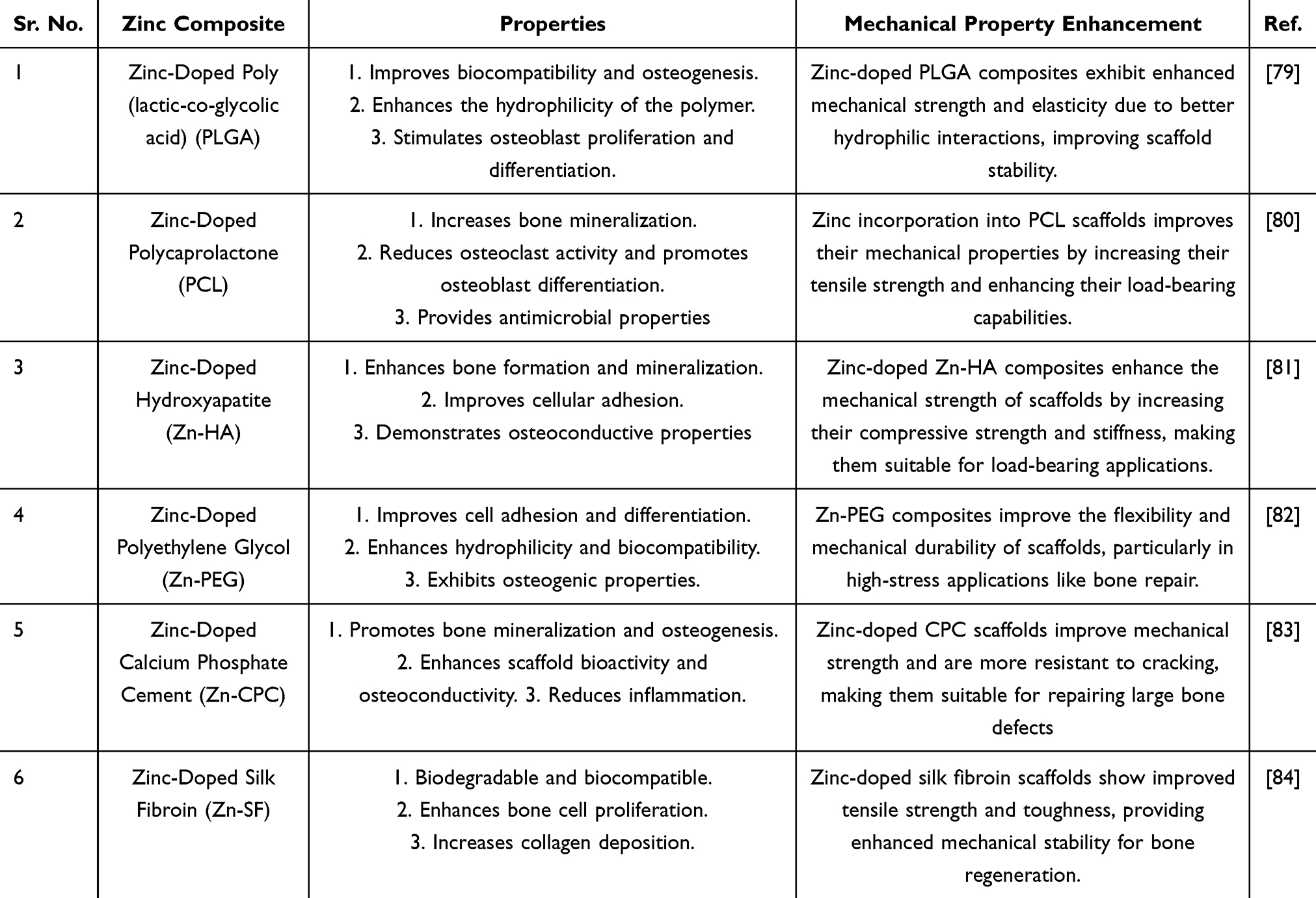 |
Table 1 Zinc-Doped Composites, Their Properties, and Mechanical Property Enhancements |
Synthetic Polymer Composites for the Regeneration of Bone
Synthetic polymers (have been widely employed as scaffolding materials in bone tissue engineering. The functionality of these materials receives added enhancement through Zn²⁺ incorporation due to its ability to stimulate osteogenesis and angiogenesis processes as well as promote scaffold breakdown. Polymeric composites that contain Zn²⁺ ions deliver specific ion levels which enable long-term bioactivity without producing harmful effects to cells. The results from various studies showed that Zn²⁺ functionalized polymer scaffolds improve cellular response outcomes and enhance mechanical strength and integration potential in host tissue10 (Table 2).
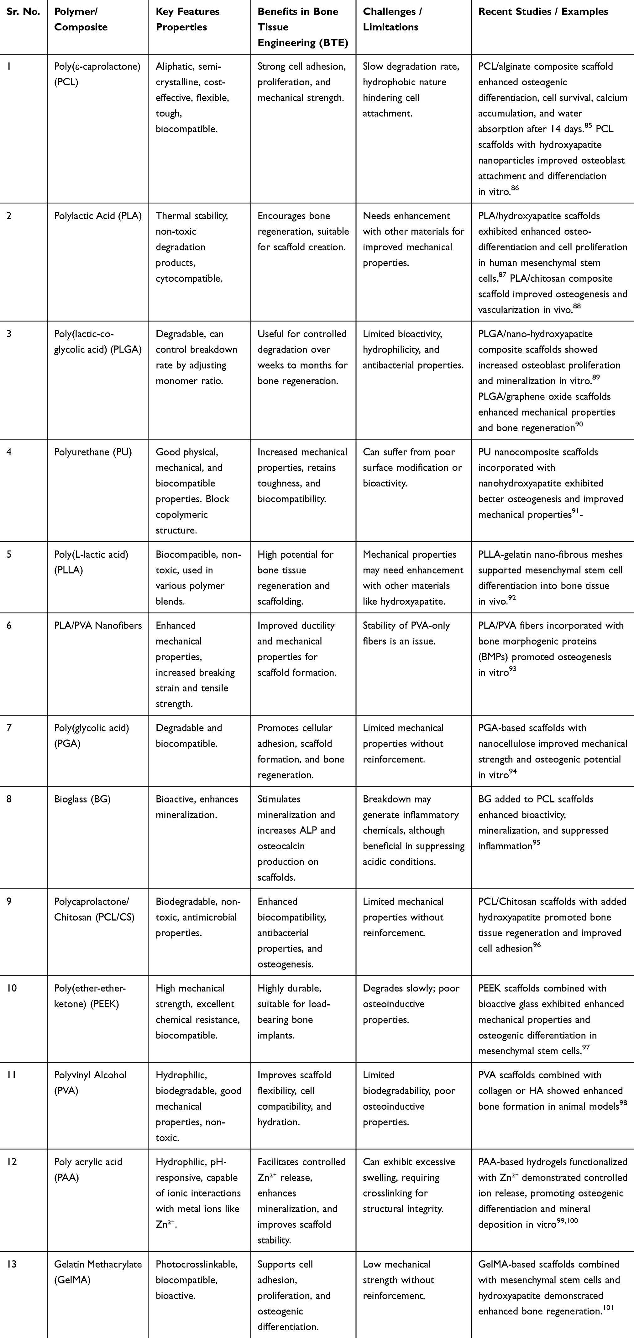 |
Table 2 Summary of Synthetic Polymer Composites for Bone Tissue Engineering (BTE) |
Zinc-Doped Synthetic Polymer Composites
Zinc-doped synthetic polymer composites are gaining attention for their ability to enhance bone tissue regeneration. Zinc (Zn) plays a vital role in osteogenesis by promoting osteoblast differentiation and mineralization. Incorporating Zn into polymers such as PCL, PLGA, and PLA improves mechanical properties, degradation rates, and cell interactions, facilitating better bone integration.102 Recent studies have shown that Zn-doped scaffolds improve cell adhesion, proliferation, and osteogenic differentiation of mesenchymal stem cells, while also enhancing the swelling behavior and hydrophilicity of the scaffolds.103 However, challenges remain in optimizing Zn ion concentrations, as higher doses can lead to cytotoxicity and reduce biocompatibility. Therefore, careful control of Zn doping levels is essential for balancing osteogenic benefits with scaffold safety.104 Additionally, variability in study results highlights the need for standardized fabrication methods and long-term in vivo studies to assess the reproducibility and effectiveness of Zn-doped scaffolds in clinical applications.105 Despite these challenges, Zn-doped composites hold significant potential for advancing bone tissue engineering.
In vitro and in vivo Experiments of Zinc-Doped Synthetic Polymer
The incorporation of degradable polymers combined with bioactive inorganic glasses, such as zinc-doped bioactive glass (ZnBG), offers a promising strategy for enhancing bone regeneration, as illustrated in Figure 4. In a laboratory study, rat bone marrow mesenchymal stem cells (rBMSCs) were cultured on various membrane substrates, including pure PLA (polylactic acid), PLA–BG (bioactive glass), and PLA–ZnBG (zinc-doped bioactive glass), to investigate the synthesis of osteogenic phenotypes and cellular mineralization.106 There was significant increase in the synthesis of osteocalcin and alkaline phosphatase at days 7 and 21 in the presence of bioactive glass and ZnBG, with the highest levels observed in the ZnBG-incorporated samples. Immunostaining analysis revealed that ZnBG significantly upregulated the expression of bone sialoprotein in rBMSCs.107
 |
Figure 4 Schematic representation of the fabrication and biological response of zinc-doped bioactive scaffolds for bone regeneration. |
In addition to bioactive glass, pro-osteogenic components, such as those found in bone graft replacements, play a crucial role in accelerating bone healing. In one study, scaffolds made from calcium phosphate cement were enhanced with PLGA microspheres and a Si-Zn dual component to promote bone regeneration.67 The incorporation of Si/Zn PLGA microspheres significantly improved the assembly and compressive strength of the CPC scaffolds. These scaffolds exhibited a gradual release of Si and Zn ions, which, in turn, facilitated the production of anti-inflammatory cytokines by M2 RAW 264.7 cells in vitro.108 Furthermore, when grown on PLGA/CPC-Si/Zn scaffolds, rat bone marrow stem cells (rBMSCs) demonstrated improved bone formation.108 In a femur defect model, rats treated with these scaffolds exhibited new bone formation after 4 weeks, with the PLGA microspheres promoting further bone development over 12 weeks.109 By 24 weeks, the microspheres had degraded, and the defect was almost completely filled with newly formed bone. The synergistic effects of PLGA/CPC-Si/Zn scaffolds, enhancing both immunological responses and biodegradability, significantly promoted bone formation and repair.109
Zinc, widely known for its anti-inflammatory properties, plays an essential role in the regulation of the immune response during bone healing. Zinc’s anti-inflammatory mechanism involves the suppression of the NF-kB pathway, inhibition of COX-2 enzyme activity, and downregulation of pro-inflammatory cytokines such as IL-1β, IL-6, and TNF-α. As a result, zinc reduces inflammation, creating a favorable immunological environment for early bone defect repair.110 Moreover, the regulated release of zinc and silicon ions from scaffolds can accelerate bone remodeling in the intermediate and advanced stages of healing. In a study by Qian et al, CPC, when combined with zinc silicate (ZS) and PLGA microspheres, demonstrated improved osteogenic and anti-inflammatory properties.111 The presence of zinc ions upregulated the anti-inflammatory cytokine IL-10 while suppressing pro-inflammatory cytokines, facilitating an optimal immune response conducive to bone regeneration.55
Proliferation and Viability of Cells
Recent studies on zinc-doped synthetic polymer composites for bone regeneration have shown promising results in enhancing cell proliferation and osteogenesis (Figure 5). Zinc-containing polymers, such as polyacrylic acid (PAA), exhibit low cytotoxicity at moderate concentrations, promoting cell viability and supporting bone regeneration.18 Specifically, PAA at concentrations of 0.025 and 0.05 mol/L has been found to enhance cell proliferation, while higher concentrations lead to toxicity and reduced cell viability.112 The addition of zinc further improves the osteogenic properties of these polymers, stimulating osteoblast differentiation and enhancing scaffold integration. However, excessive zinc concentrations can disrupt cellular functions and impair tissue repair. Therefore, optimizing the concentration of both zinc and the polymer is crucial to achieving a balance between promoting bone regeneration and ensuring biocompatibility, making zinc-doped polymer composites a promising strategy for repairing bone defects.97
 |
Figure 5 Schematic representation of the applications and biological roles of zinc-doped bioactive glass. Zinc incorporation enhances the bioactivity of bioactive glass, making it a promising material for bone tissue engineering. |
Prior research has examined the potential cytotoxic impact of Zn on cellular structures. Several researchers have noted that cytotoxicity occurs when the concentration of Zn doping in BGs exceeds approximately 5 mol%. In addition, it was shown that only the elution extracts with the greatest concentration (5 mg/L) were able to suppress the proliferation of fibroblasts of mouse embryonic (MEFs). Neščáková et al113 demonstrated that Zn-doped bioactive glasses (containing approximately 8% ZnO) did not exhibit harmful effects on MEF or MG-63 cells. Utilizing a solution provides a more precise measure than the amount of zinc in materials.114 Studies have demonstrated that Zn²⁺ enhances the attachment, expansion, growth, and movement of vascular smooth muscle cells. This effect is observed at concentrations ranging from 60–80 μM. However, at higher concentrations of Zn²⁺ (80–120 μM), the reversible are observed.115 Aina et al116 found that a dosage of 1.1 mg/L of Zn²⁺ stimulated endothelial cell proliferation, whereas a dose of 2.7 mg/L led to cell death. Yamaguchi et al discovered that MC3T3-E1 osteoblasts synthesized more regucalcin mRNA, Runx-2, and osteoprotegerin when Zn was present at quantities ranging from 10−6 to 65 μg/L–6.5 mg/L.117 The author’s results showed that the concentration of Zn²⁺ ranging from 10 to 250 μM (equal to 0.65 and 16.25 mg/L) hindered the production of osteoclasts in RAW264.7 cells. The zinc amount had an impact on the activity of SaOS-2, a cell line that resembles human osteoblasts.
Overextended stimulation, this impact was shown without affecting cell growth.74 Previous studies have shown that, in laboratory conditions, the released Zn²⁺ may stimulate osteoblast growth, osteogenic mesenchymal cell (MSC) differentiation, and extracellular matrix (ECM) mineralization when present in tiny concentrations,17 Zn²⁺ may be harmful to cells because it prevents the extracellular matrix (ECM) from mineralizing at high amounts. The research that is now available suggests that adding zinc to a bone scaffold at optimum concentrations and with regulated release characteristics could improve the procedure of bone regeneration.
Cellular Osteogenesis Differentiation
Recent studies investigating the role of zinc in polymeric scaffolds for bone regeneration have consistently shown that the incorporation of zinc enhances osteogenic differentiation and cell proliferation. Additionally, PAA scaffolds with lower zinc concentrations promoted greater calcium deposition and bone differentiation than those with higher zinc levels.10 Similar findings were observed with zinc-doped polycaprolactone (PCL) scaffolds, which exhibited increased alkaline phosphatase (ALP) expression and apatite formation.118 Furthermore, the combination of silver and zinc ions in SPEEK polymers resulted in greater cell proliferation and bone differentiation than zinc alone.119 Other studies, such as those involving PLGA/CPC -Si/Zn composites, also reported improved cell proliferation and bone formation.120 Zinc-silibinin complexes have additionally been shown to regulate key signaling pathways, promoting osteoblast growth.121 These findings underscore the significant potential of zinc-doped polymeric scaffolds in enhancing bone regeneration, suggesting that the incorporation of zinc, especially in combination with other metal ions, can optimize the osteogenic properties of biomaterials for bone tissue engineering.122
Potential Drawbacks of Zinc-Doped Synthetic Polymer Composites in Bone Regeneration
Synthetic polymer composites containing zinc ions have gained popularity in bone tissue engineering because zinc shows both improved bone cell growth and antimicrobial properties. Such an approach brings certain unfavorable aspects that need thorough evaluation (Table 3).
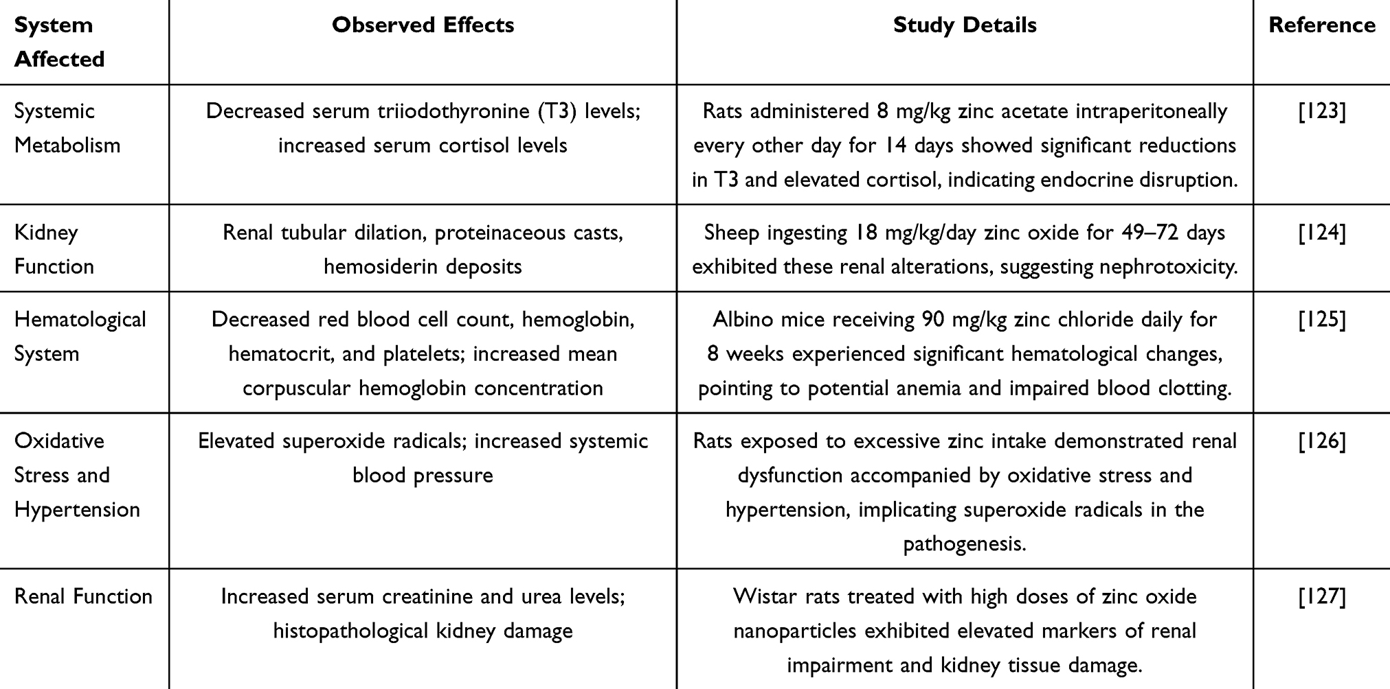 |
Table 3 This Table Summarizes the Observed Toxic Effects of High-Dose Zinc (Zn²⁺) Exposure on Systemic Metabolism, Kidney Function, and the Hematological System in Various Animal Models |
The main drawback of elevated Zn²⁺ concentrations emerge as toxic effects on cells. The presence of zinc ions at lower levels stimulates cell growth along with bone-forming cells but excessive amounts result in negative actions against these processes while simultaneously producing toxic effects to cells. For instance, a systematic review highlighted that high-dose Zn²⁺ resulted in cytotoxicity and inhibition of osteogenic differentiation in vitro.10 The integration of Zn²⁺ ions may modify the apatite formation which represents an essential mineral for bone structure development. Some studies have reported that Zn²⁺ can reduce apatite formation in simulated body fluid, potentially hindering the nucleation process essential for effective bone regeneration.128 Scaffold-based Zn²⁺ release must be controlled for optimal clinical outcomes. Without proper regulation of Zn²⁺ release an undesirable discrepancy will develop between oxide strength and biological function thus affecting healing success of repaired bones. The studies shows Zn²⁺ has positive effects on bone formation yet high rates of its release could damage the complex mechanisms vital for performing successful bone regeneration.10
Zn²⁺ addition during the fabrication of polymer matrices leads to changes in scaffold degradation speed. Prefabricated modifications in scaffold materials can cause early failure of mechanical properties or excessive time delay of scaffold retention and this affects healing negatively.
Moreover, limited research exists on zinc ion doped synthetic polymer composites for human subject use due to their absence in clinical trials. A comprehensive review published in June 2020 revealed 13 suitable studies regarding this subject, but these studies used only in vitro methods and animal experiments without reporting any human clinical trials.10 Research translation becomes crucial to establish safety parameters and clinical value of Zn²⁺-doped synthetic polymer composites for human bone regeneration.
Conclusion and Future Perspectives
Zinc-doped synthetic polymer composites have gained attention as a promising solution for bone regeneration, offering potential advancements in the treatment of bone defects. Zinc, an essential element in bone metabolism and healing, plays a crucial role in enhancing both the mechanical and biological properties of bone grafts. Research has demonstrated that zinc stimulates osteoblast activity, improves bone mineral density, and promotes the proliferation and differentiation of osteogenic cells. Additionally, the incorporation of synthetic polymers strengthens the composite, supporting bone regeneration and enhancing the integration of the scaffold with the surrounding bone tissue. Zn composites also exhibits antimicrobial properties which contribute to reducing infection risks during bone repair, aiding in post-operative recovery. A key advantage of zinc-doped composites is their controlled degradation, which ensures the scaffold remains structurally stable as natural bone replaces it. This process promotes seamless integration and minimizes the need for additional surgeries. However, challenges remain, particularly in achieving a uniform distribution of zinc within the polymer matrix to maintain consistent scaffold performance. Furthermore, understanding the interactions between zinc and other biological factors is essential to improve the biocompatibility of these materials and reduce potential negative effects. Future research should focus on optimizing manufacturing techniques to enhance the consistency and mechanical strength of these composites. Additionally, in vivo studies are necessary to validate the safety and effectiveness of these materials in clinical applications. Combining zinc with other bioactive compounds may also enhance the healing process and improve bone regeneration. To facilitate the successful clinical application of zinc-doped composites in bone regeneration, several critical factors must be addressed. Regulatory challenges represent a significant barrier, as these materials must undergo comprehensive preclinical and clinical testing to satisfy stringent safety and efficacy standards. Furthermore, the scalability of production processes is essential for ensuring that zinc-doped composites can be manufactured at a commercial scale without compromising quality or consistency. Recent studies underscore the importance of developing cost-effective, large-scale manufacturing techniques to support the widespread adoption of these materials in clinical practice. In addition, long-term biocompatibility must be thoroughly assessed to ensure that the materials do not provoke adverse immune responses or degrade in a manner that would impede the desired regenerative outcomes over time. Addressing these regulatory, production, and biocompatibility challenges will be crucial for the successful integration of zinc-doped composites into bone regeneration therapies.
Data Sharing Statement
Not Applicable. This is a review article and all relevant information is provided in the article.
Ethical Approval and Consent to Participate
Not Applicable. This is a review paper and do not involve direct research on humans or animals.
Consent for Publication
“Not applicable” as this manuscript does not contain data from any individual person.
Funding
This work was supported by Public Technology Applied Research Projects of Zhejiang Province (LGF22H060023 to WQL), Medical and Health Research Project of Zhejiang Province (2022KY433 to WQL), Traditional Chinese Medicine Science and Technology Projects of Zhejiang Province (2022ZB380 to JYZ,2022ZB382 to WQL), Science and Technology Project of Zhoushan (2024C31017 to QX, 2024C31018 to XCX, 2024C31020 to HML), Research Fund Projects of The Affiliated Hospital of Zhejiang Chinese Medicine University (2023FSYYZQ23 to LYX).
Disclosure
The Authors declare that they have no competing interests financial or non-financial or any other interests that might be perceived to influence the results and/or discussion reported in this paper.
References
1. Florencio-Silva R, Sasso GRDS, Sasso-Cerri E, et al. Biology of bone tissue: structure, function, and factors that influence bone cells. Biomed Res Int. 2015;2015(1):421746. doi:10.1155/2015/421746
2. Wang X, Zhang A, Huang M, et al. Genetic and clinical characteristics of patients with hereditary spherocytosis in Hubei province of China. Front Genetics. 2020;11:953. doi:10.3389/fgene.2020.00953
3. Bao CLM, Teo EY, Chong MS, et al. Advances in Bone Tissue Engineering, in Regenerative Medicine and Tissue Engineering. IntechOpen; 2013.
4. Archunan MW, Petronis S. Bone grafts in trauma and orthopaedics. Cureus. 2021;13(9).
5. Oryan A, Alidadi S, Moshiri A, et al. Bone regenerative medicine: classic options, novel strategies, and future directions. J Orthopaedic Surg Res. 2014;9:1–27. doi:10.1186/1749-799X-9-18
6. Kashte S, Jaiswal AK, Kadam S. Artificial bone via bone tissue engineering: current scenario and challenges. Tissue Eng Regener Med. 2017;14:1–14. doi:10.1007/s13770-016-0001-6
7. Babilotte J, Guduric V, Le Nihouannen D, et al. 3D printed polymer–mineral composite biomaterials for bone tissue engineering: fabrication and characterization. J Biomed Mater Res Part B. 2019;107(8):2579–2595. doi:10.1002/jbm.b.34348
8. Luo X, Barbieri D, Davison N, et al. Zinc in calcium phosphate mediates bone induction: in vitro and in vivo model. Acta Biomater. 2014;10(1):477–485. doi:10.1016/j.actbio.2013.10.011
9. Kothandam S, Selvatharani V, Vijayakumar N, et al. Zinc Doped Akermanite: A Promising Biomaterial for Orthopedic Application with Enhanced Bioactivity, Mechanical Strength, and Bacterial Study. ACS omega; 2025.
10. Wang S, Li R, Xia D, et al. The impact of Zn-doped synthetic polymer materials on bone regeneration: a systematic review. Stem Cell Res Ther. 2021;12:1–13.
11. O’Connor JP, Kanjilal D, Teitelbaum M, et al. Zinc as a therapeutic agent in bone regeneration. Materials. 2020;13(10):2211. doi:10.3390/ma13102211
12. Jiang S, Zhang Y, Alsaikhan F, et al. A meta-analysis review of the effect of Zn-doped synthetic polymer materials on bone regeneration. J Drug Delivery Sci Technol. 2022;76:103792. doi:10.1016/j.jddst.2022.103792
13. Pina S, Oliveira JM, Reis RL. Natural‐based nanocomposites for bone tissue engineering and regenerative medicine: a review. Adv Mater. 2015;27(7):1143–1169. doi:10.1002/adma.201403354
14. Bonfield W, Grynpas MD, Tully AE, et al. Hydroxyapatite reinforced polyethylene--a mechanically compatible implant material for bone replacement. Biomaterials. 1981;2(3):185–186. doi:10.1016/0142-9612(81)90050-8
15. Li P, Dai J, Li Y, et al. Zinc based biodegradable metals for bone repair and regeneration: bioactivity and molecular mechanisms. Mater Today Bio. 2024;25:100932. doi:10.1016/j.mtbio.2023.100932
16. Yuan K, Deng C, Tan L, et al. Structural and temporal dynamics analysis of zinc-based biomaterials: history, research hotspots and emerging trends. Bioact Mater. 2024;35:306–329. doi:10.1016/j.bioactmat.2024.01.017
17. Su Y, Cockerill I, Wang Y, et al. Zinc-based biomaterials for regeneration and therapy. Trends Biotechnol. 2019;37(4):428–441. doi:10.1016/j.tibtech.2018.10.009
18. Wen X, Wang J, Pei X, et al. Zinc-based biomaterials for bone repair and regeneration: mechanism and applications. J Mat Chem B. 2023;11(48):11405–11425. doi:10.1039/D3TB01874A
19. Vojtěch D, Kubásek J, Šerák J, et al. Mechanical and corrosion properties of newly developed biodegradable Zn-based alloys for bone fixation. Acta Biomater. 2011;7(9):3515–3522. doi:10.1016/j.actbio.2011.05.008
20. García-Mintegui C, Córdoba LC, Buxadera-Palomero J, et al. Zn-Mg and Zn-Cu alloys for stenting applications: from nanoscale mechanical characterization to in vitro degradation and biocompatibility. Bioact Mater. 2021;6(12):4430–4446. doi:10.1016/j.bioactmat.2021.04.015
21. Li Y, Yang Y, Qing Y, et al. Enhancing ZnO-NP antibacterial and osteogenesis properties in orthopedic applications: a review. Int J Nanomed. 2020;15:6247–6262. doi:10.2147/IJN.S262876
22. He T, Chen H, Liu P, et al. One-step co-doping of ZnO and Zn2+ in osteoinductive calcium phosphate ceramics with synergistic antibacterial activity for regenerative repair of infected bone defect. J Mater Sci Technol. 2023;163:168–181. doi:10.1016/j.jmst.2023.04.032
23. Rakhshaei R, Namazi H, Hamishehkar H, et al. In situ synthesized chitosan–gelatin/ZnO nanocomposite scaffold with drug delivery properties: higher antibacterial and lower cytotoxicity effects. J Appl Polym Sci. 2019;136(22):47590. doi:10.1002/app.47590
24. Laurenti M, Cauda V. ZnO nanostructures for tissue engineering applications. Nanomaterials. 2017;7(11):374. doi:10.3390/nano7110374
25. Zadehnazari A. Metal oxide/polymer nanocomposites: a review on recent advances in fabrication and applications. Polym Plast Technol Eng. 2023;62(5):655–700. doi:10.1080/25740881.2022.2129387
26. Petre DG, Leeuwenburgh SC. The use of fibers in bone tissue engineering. Tissue Engineering Part B. Reviews. 2022;28(1):141–159.
27. Karimzadeh Bardeei L, Seyedjafari E, Hossein G, et al. Regeneration of bone defects in a rabbit femoral osteonecrosis model using 3D-printed poly (epsilon-caprolactone)/nanoparticulate willemite composite scaffolds. Int J Mol Sci. 2021;22(19):10332. doi:10.3390/ijms221910332
28. Telgerd MD, Sadeghinia M, Birhanu G, et al. Enhanced osteogenic differentiation of mesenchymal stem cells on metal–organic framework based on copper, zinc, and imidazole coated poly-l -lactic acid nanofiber scaffolds. J Biomed Mater Res Part A. 2019;107(8):1841–1848. doi:10.1002/jbm.a.36707
29. Paterson TE, Beal SN, Santocildes-Romero ME, et al. Selective laser melting–enabled electrospinning: introducing complexity within electrospun membranes. Proc Instit Mech Eng Part HJ Eng Med. 2017;231(6):565–574. doi:10.1177/0954411917690182
30. Arcos Navarrete D, Portolés Pérez MT. Mesoporous bioactive nanoparticles for bone tissue applications. Int J Mol Sci. 2023;24(4):3249. doi:10.3390/ijms24043249
31. Hassan M, Sulaiman M, Yuvaraju PD, et al. Biomimetic PLGA/strontium-zinc nano hydroxyapatite composite scaffolds for bone regeneration. J Funct Biomater. 2022;13(1):13. doi:10.3390/jfb13010013
32. Qian G, Zhang L, Wang G, et al. 3D printed Zn-doped mesoporous silica-incorporated poly-L-lactic acid scaffolds for bone repair. Int J Bioprinting. 2021;7(2). doi:10.18063/ijb.v7i2.346
33. Liao J, Wang H, Liu N, et al. Functionally modified halloysite nanotubes for personalized bioapplications. Adv Colloid Interface Sci. 2023;311:102812. doi:10.1016/j.cis.2022.102812
34. Kumar A, Mandal S, Barui S, et al. Low temperature additive manufacturing of three dimensional scaffolds for bone-tissue engineering applications: processing related challenges and property assessment. Mater Sci Eng R Rep. 2016;103:1–39.
35. Ge Y, Yu Y, Feng C, et al. Enhanced bone healing of photothermal hydrogel via mild heat stimulation and Zn2+ release. Mater Today Commun. 2023;37:107392. doi:10.1016/j.mtcomm.2023.107392
36. Stafin K, Śliwa P, Piątkowski M. Towards polycaprolactone-based scaffolds for alveolar bone tissue engineering: a biomimetic approach in a 3D printing technique. Int J Mol Sci. 2023;24(22):16180. doi:10.3390/ijms242216180
37. Hou -H-H, Lee B-S, Liu Y-C, et al. Vapor-induced pore-forming atmospheric-plasma-sprayed zinc-, strontium-, and magnesium-doped hydroxyapatite coatings on titanium implants enhance new bone formation—an in vivo and in vitro investigation. Int J Mol Sci. 2023;24(5):4933. doi:10.3390/ijms24054933
38. Li Y-L, He J, Ye H-X, et al. Atomic layer deposition of zinc oxide onto 3D porous iron scaffolds for bone repair: in vitro degradation, antibacterial activity and cytocompatibility evaluation. Rare Met. 2022;41:546–558. doi:10.1007/s12598-021-01852-8
39. Chong WJ, Shen S, Li Y, et al. Biodegradable PLA-ZnO nanocomposite biomaterials with antibacterial properties, tissue engineering viability, and enhanced biocompatibility. Smart Mater Manuf. 2023;1:100004. doi:10.1016/j.smmf.2022.100004
40. Singh TA, Sharma A, Tejwan N, et al. A state of the art review on the synthesis, antibacterial, antioxidant, antidiabetic and tissue regeneration activities of zinc oxide nanoparticles. Adv Colloid Interface Sci. 2021;295:102495. doi:10.1016/j.cis.2021.102495
41. Harikrishnan P, Sivasamy A. Preparation, Characterization of Electrospun Polycaprolactone-Nano Zinc Oxide Composite Scaffolds for Osteogenic Applications. Vol. 23. Nano-Structures & Nano-Objects; 2020:100518.
42. Siddiqui N, Kishori B, Rao S, et al. Electropsun polycaprolactone fibres in bone tissue engineering: a review. Mol Biotechnol. 2021;63:363–388. doi:10.1007/s12033-021-00311-0
43. Kumar M, Kumar D, Sharma A, et al. Micronutrients throughout the Life Cycle: needs and Functions in Health and Disease. Curr Nutr Food Sci. 2024;20(1):62–84. doi:10.2174/1573401319666230420094603
44. Stiles LI, Ferrao K, Mehta KJ. Role of zinc in health and disease. Clin Exp Med. 2024;24(1):38.
45. Zarei A, Farazin A. Synergizing additive manufacturing and machine learning for advanced hydroxyapatite scaffold design in bone regeneration. J Aust Ceram Soc. 2024;1–17.
46. Ofudje EA, Adeogun AI, Idowu MA, et al. Synthesis and characterization of Zn-Doped hydroxyapatite: scaffold application, antibacterial and bioactivity studies. Heliyon. 2019;5(5):e01716. doi:10.1016/j.heliyon.2019.e01716
47. Chopra V, Thomas J, Chauhan G, et al. Gelatin nanofibers loaded with zinc-doped hydroxyapatite for osteogenic differentiation of mesenchymal stem cells. ACS Appl Nano Mater. 2022;5(2):2414–2428. doi:10.1021/acsanm.1c04126
48. Li H, Li M, Ran X, et al. The role of zinc in bone mesenchymal stem cell differentiation. Cell Reprogramming. 2022;24(2):80–94. doi:10.1089/cell.2021.0137
49. Dornelas J, Dornelas G, Rossi A, et al. The incorporation of zinc into hydroxyapatite and its influence on the cellular response to biomaterials: a systematic review. J Funct Biomater. 2024;15(7):178. doi:10.3390/jfb15070178
50. Zhao J, Han J, Jiang J, et al. The downregulation of Wnt/β-catenin signaling pathway is associated with zinc deficiency-induced proliferative deficit of C17. 2 neural stem cells. Brain Res. 2015;1615:61–70. doi:10.1016/j.brainres.2015.04.028
51. Gao K, Zhang Y, Niu J, et al. Zinc promotes cell apoptosis via activating the Wnt-3a/β-catenin signaling pathway in osteosarcoma. J Orthopaedic Surg Res. 2020;15:1–8. doi:10.1186/s13018-020-01585-x
52. Zhu W-Q, Li K, Su S, et al. Effects of zinc ions released from Ti-nw-Zn surface on osteogenesis and angiogenesis in vitro and in an in vivo zebrafish model. Front Bioeng Biotechnol. 2022;10:848769. doi:10.3389/fbioe.2022.848769
53. Cho Y-E, Kwun I-S. Zinc upregulates bone-specific transcription factor Runx2 expression via BMP-2 signaling and Smad-1 phosphorylation in osteoblasts. J Nutr Health. 2018;51(1):23–30. doi:10.4163/jnh.2018.51.1.23
54. Molenda M, Kolmas J. The role of zinc in bone tissue health and regeneration—a review. Biol Trace Elem Res. 2023;201(12):5640–5651. doi:10.1007/s12011-023-03631-1
55. Bai L, Song P, Su J. Bioactive elements manipulate bone regeneration. Biomater Transl. 2023;4(4):248. doi:10.12336/biomatertransl.2023.04.005
56. Song Y, Wu H, Gao Y, et al. Zinc silicate/nano-hydroxyapatite/collagen scaffolds promote angiogenesis and bone regeneration via the p38 MAPK pathway in activated monocytes. ACS Appl Mater Interfaces. 2020;12(14):16058–16075. doi:10.1021/acsami.0c00470
57. Feng P, Wei P, Shuai C, et al. Characterization of mechanical and biological properties of 3-D scaffolds reinforced with zinc oxide for bone tissue engineering. PLoS One. 2014;9(1):e87755. doi:10.1371/journal.pone.0087755
58. Liu Y, Yu L, Chen J, et al. Exploring the osteogenic potential of zinc-doped magnesium phosphate cement (zmpc): a novel material for orthopedic bone defect repair. Biomedicines. 2024;12(2):344. doi:10.3390/biomedicines12020344
59. Fan L, Chen S, Yang M, et al. Metallic materials for bone repair. Adv Healthcare Mater. 2024;13(3):2302132. doi:10.1002/adhm.202302132
60. Jin X, Xie D, Zhang Z, et al. In vitro and in vivo studies on biodegradable Zn porous scaffolds with a drug-loaded coating for the treatment of infected bone defect. Mater Today Bio. 2024;24:100885. doi:10.1016/j.mtbio.2023.100885
61. Nedovic V, Willaert R. Fundamentals of Cell Immobilisation Biotechnology. Vol. 8. Springer Science & Business Media; 2013.
62. Liu X, Zhou C, Xie Q, et al. Recent advances in layer-by-layer assembly scaffolds for co-delivery of bioactive molecules for bone regeneration: an updated review. J Transl Med. 2024;22(1):1001.
63. Md yusop AH, Ulum MF, Al Sakkaf A, et al. Current status and outlook of porous Zn-based scaffolds for bone applications: a review. J Bionic Eng. 2022;19(3):737–751. doi:10.1007/s42235-022-00152-w
64. Ansari MAA, Golebiowska AA, Dash M, et al. Engineering biomaterials to 3D-print scaffolds for bone regeneration: practical and theoretical consideration. Biomater Sci. 2022;10(11):2789–2816.
65. Zhao L, Yuan P, Zhang M, et al. Preparation and properties of porous Zn-based scaffolds as biodegradable implants: a review. J Mater Sci. 2023;58(20):8275–8316. doi:10.1007/s10853-023-08561-w
66. Cockerill I, Su Y, Sinha S, et al. Porous zinc scaffolds for bone tissue engineering applications: a novel additive manufacturing and casting approach. Mater Sci Eng C. 2020;110:110738. doi:10.1016/j.msec.2020.110738
67. Yuan X, Wu T, Lu T, et al. Effects of zinc and strontium doping on in vitro osteogenesis and angiogenesis of calcium silicate/calcium phosphate cement. ACS Biomater Sci Eng. 2023;9(10):5761–5771. doi:10.1021/acsbiomaterials.3c00193
68. Huang P, Lian D, Ma H, et al. New advances in gated materials of mesoporous silica for drug controlled release. Chin Chem Lett. 2021;32(12):3696–3704. doi:10.1016/j.cclet.2021.06.034
69. García A, Cabañas MV, Peña J, et al. Design of 3d scaffolds for hard tissue engineering: from apatites to silicon mesoporous materials. Pharmaceutics. 2021;13(11):1981. doi:10.3390/pharmaceutics13111981
70. Mangaraj S, Priyadarshini I, Swain S, et al. ZnO-doped β-TCP ceramic-based scaffold promotes osteogenic and antibacterial activity. Bioinspired. Biomimetic Nanobiomater. 2024;13(2):37–44. doi:10.1680/jbibn.23.00065
71. Ghorbani FM, Kaffashi B, Shokrollahi P, et al. PCL/chitosan/Zn-doped nHA electrospun nanocomposite scaffold promotes adipose derived stem cells adhesion and proliferation. Carbohydr Polym. 2015;118:133–142. doi:10.1016/j.carbpol.2014.10.071
72. Xiong K, Zhang J, Zhu Y, et al. Zinc doping induced differences in the surface composition, surface morphology and osteogenesis performance of the calcium phosphate cement hydration products. Mater Sci Eng C. 2019;105:110065. doi:10.1016/j.msec.2019.110065
73. Paramita P, Curtarelli RB, da Silva IT, et al. Sol–gel based synthesis and biological properties of zinc integrated nano bioglass ceramics for bone tissue regeneration. J Mater Sci Mater Med. 2021;32:1–11. doi:10.1007/s10856-020-06475-6
74. Wang B, Yang M, Liu L, et al. Osteogenic potential of Zn2+ -passivated carbon dots for bone regeneration in vivo. Biomater Sci. 2019;7(12):5414–5423. doi:10.1039/C9BM01181A
75. Bai X, Liu W, Xu L, et al. Sequential macrophage transition facilitates endogenous bone regeneration induced by Zn-doped porous microcrystalline bioactive glass. J Mat Chem B. 2021;9(12):2885–2898. doi:10.1039/D0TB02884C
76. Heras C, Jiménez-Holguín J, Doadrio AL, et al. Multifunctional antibiotic-and zinc-containing mesoporous bioactive glass scaffolds to fight bone infection. Acta Biomater. 2020;114:395–406. doi:10.1016/j.actbio.2020.07.044
77. Edmund C, Rinti B. Mechanically stiff, zinc cross-linked nanocomposite Scaffolds with improved osteostimulation and antibacterial properties; 2016.
78. Hu Y, Zeng Q, Hu Y, et al. Mxene/Zinc Ion Embedded Agar/Sodium Alginate Hydrogel for Rapid and Efficient Sterilization with Photothermal and Chemical Synergetic Therapy. Vol. 266. Talanta; 2024:125101.
79. Li C, Sun F, Tian J, et al. Continuously released Zn2+ in 3D-printed PLGA/β-TCP/Zn scaffolds for bone defect repair by improving osteoinductive and anti-inflammatory properties. Bioact Mater. 2023;24:361–375. doi:10.1016/j.bioactmat.2022.12.015
80. Elahpour N, Niesner I, Bossard C, et al. Zinc-Doped Bioactive Glass/Polycaprolactone Hybrid Scaffolds Manufactured by Direct and Indirect 3D Printing Methods for Bone Regeneration. Cells. 2023;12(13):1759. doi:10.3390/cells12131759
81. Li Y, Liu X, Gaihre B, et al. Zinc-doped hydroxyapatite and poly (propylene fumarate) nanocomposite scaffold for bone tissue engineering. J Mater Sci. 2022;57(10):5998–6012. doi:10.1007/s10853-022-06966-7
82. Oriňaková R, Gorejová R, Čákyová V, et al. Biodegradable zinc‐based materials with a polymer coating designed for biomedical applications. J Appl Polym Sci. 2024;141(2):e54773. doi:10.1002/app.54773
83. Sedelnikova M, Sharkeev YP, Tolkacheva TV, et al. Additively manufactured porous titanium 3D–scaffolds with antibacterial Zn-, Ag-calcium phosphate biocoatings. Mater Charact. 2022;186:111782. doi:10.1016/j.matchar.2022.111782
84. Singh S, Singh G, Bala N. Electrophoretic Deposition of Hydroxyapatite Incorporated Composite Coatings on Metallic Substrates: A Review of the Fundamentals. Advanced Ceramics; 2023:219–257.
85. Yu J, Lee S, Choi S, et al. Fabrication of a polycaprolactone/alginate bipartite hybrid scaffold for osteochondral tissue using a three-dimensional bioprinting system. Polymers. 2020;12(10):2203. doi:10.3390/polym12102203
86. Ebrahimi Z, Irani S, Ardeshirylajimi A, et al. Enhanced osteogenic differentiation of stem cells by 3D printed PCL scaffolds coated with collagen and hydroxyapatite. Sci Rep. 2022;12(1):12359. doi:10.1038/s41598-022-15602-y
87. Esmaeilzadeh M, Asadi A, Goudarzi F, et al. Evaluation of adhesion, growth and differentiation of human umbilical cord stem cells to osteoblast cells on PLA polymeric scaffolds. Biomed Mater Devices. 2023;1(2):772–788. doi:10.1007/s44174-023-00062-3
88. Donate R, Monzón M, Alemán-Domínguez ME. Additive manufacturing of PLA-based scaffolds intended for bone regeneration and strategies to improve their biological properties. e-Polymers. 2020;20(1):571–599. doi:10.1515/epoly-2020-0046
89. Lu M, Li L, Zheng C, et al. 3D printed porous PLGA/n-HA/MgP composite scaffolds with improved osteogenic and angiogenic properties. Mater Des. 2023;234:112351. doi:10.1016/j.matdes.2023.112351
90. Zhu J, Qi Z, Zheng C, et al. Enhanced cell proliferation and osteogenesis differentiation through a combined treatment of Poly‐L‐Lysine‐Coated PLGA/Graphene oxide hybrid fiber matrices and electrical stimulation. J Nanomater. 2020;2020(1):5892506. doi:10.1155/2020/5892506
91. Zhang T, Li J, Wang Y, et al. Hydroxyapatite/polyurethane scaffolds for bone tissue engineering. Tissue Engineering Part B. Reviews. 2024;30(1):60–73.
92. Capuana E, Lopresti F, Ceraulo M, et al. Poly-l-lactic acid (PLLA)-based biomaterials for regenerative medicine: a review on processing and applications. Polymers. 2022;14(6):1153. doi:10.3390/polym14061153
93. da Silva TN, Gonçalves RP, Rocha CL, et al. Controlling burst effect with PLA/PVA coaxial electrospun scaffolds loaded with BMP-2 for bone guided regeneration. Mater Sci Eng C. 2019;97:602–612. doi:10.1016/j.msec.2018.12.020
94. Yang R, Wang X, Liu S, et al. Bioinspired poly (γ-glutamic acid) hydrogels for enhanced chondrogenesis of bone marrow-derived mesenchymal stem cells. Int J Biol Macromol. 2020;142:332–344. doi:10.1016/j.ijbiomac.2019.09.104
95. He L, Yin J, Gao X. Additive manufacturing of bioactive glass and its polymer composites as bone tissue engineering scaffolds: a review. Bioengineering. 2023;10(6):672. doi:10.3390/bioengineering10060672
96. Ezati M, Safavipour H, Houshmand B, et al. Development of a PCL/gelatin/chitosan/β-TCP electrospun composite for guided bone regeneration. Progress Biomater. 2018;7(3):225–237. doi:10.1007/s40204-018-0098-x
97. Zheng X, Luo H, Li J, et al. Zinc‐doped bioactive glass‐functionalized polyetheretherketone to enhance the biological response in bone regeneration. J Biomed Mater Res Part A. 2024;112:1565–1577. doi:10.1002/jbm.a.37710
98. Dadgar N, Ghiaseddin A, Irani S, et al. Bioartificial injectable cartilage implants from demineralized bone matrix/PVA and related studies in rabbit animal model. J Biomater Appl. 2021;35(10):1315–1326. doi:10.1177/0885328220976552
99. Xie X, Feng X, Hong L, et al. Evaluation of antibacterial and osteogenic properties of a novel Poly (acrylic acid)-calcium-zinc biomineralized hydrogel. Front Mater. 2024;11:1444750. doi:10.3389/fmats.2024.1444750
100. Zhu K, Hong L, Ding Y, et al. Layer-by-layer assembly of chitosan quaternary ammonium salt/polyacrylic acid-Zn2+ as a titanium dental implant coating with prominent antibacterial and osteogenic activities. New J Chem. 2025;49:6578–6586. doi:10.1039/D4NJ05452K
101. Zhou B, Jiang X, Zhou X, et al. GelMA-based bioactive hydrogel scaffolds with multiple bone defect repair functions: therapeutic strategies and recent advances. Biomater Res. 2023;27(1):86. doi:10.1186/s40824-023-00422-6
102. Li F, Li S, Liu Y, et al. Current advances in the roles of doped bioactive metal in biodegradable polymer composite scaffolds for bone repair: a mini review. Adv Eng Mater. 2022;24(8):2101510. doi:10.1002/adem.202101510
103. Liu X, Xia Z, Wang Y, et al. Zinc-doped inorganic bioactive materials: a comprehensive review of properties and their applications in osteogenesis, antibacterial, and hemostasis. Appl Mater Today. 2024;40:102393. doi:10.1016/j.apmt.2024.102393
104. He J, Li K, Wu T, et al. Research progress in degradable metal‐based multifunctional scaffolds for bone tissue engineering. MedComm–Biomater Appl. 2023;2(4):e60. doi:10.1002/mba2.60
105. Khan HM, Liao X, Sheikh BA, et al. Smart biomaterials and their potential applications in tissue engineering. J Mat Chem B. 2022;10(36):6859–6895. doi:10.1039/d2tb01106a
106. Majumdar S, Gupta S, Krishnamurthy S. Multifarious applications of bioactive glasses in soft tissue engineering. Biomater Sci. 2021;9(24):8111–8147. doi:10.1039/D1BM01104A
107. Oh S-A, Won J-E, Kim H-W. Composite membranes of poly (lactic acid) with zinc-added bioactive glass as a guiding matrix for osteogenic differentiation of bone marrow mesenchymal stem cells. J Biomater Appl. 2012;27(4):413–422. doi:10.1177/0885328211408944
108. Liang W, Gao M, Lou J, et al. Integrating silicon/zinc dual elements with PLGA microspheres in calcium phosphate cement scaffolds synergistically enhances bone regeneration. J Mat Chem B. 2020;8(15):3038–3049. doi:10.1039/C9TB02901J
109. Jin S, Xia X, Huang J, et al. Recent advances in PLGA-based biomaterials for bone tissue regeneration. Acta Biomater. 2021;127:56–79. doi:10.1016/j.actbio.2021.03.067
110. Skrajnowska D, Bobrowska-Korczak B. Role of zinc in immune system and anti-cancer defense mechanisms. Nutrients. 2019;11(10):2273. doi:10.3390/nu11102273
111. Qian G, Lu T, Zhang J, et al. Promoting bone regeneration of calcium phosphate cement by addition of PLGA microspheres and zinc silicate via synergistic effect of in-situ pore generation, bioactive ion stimulation and macrophage immunomodulation. Appl Mater Today. 2020;19:100615. doi:10.1016/j.apmt.2020.100615
112. Liu J-H, Bian H, Zhang Y, et al. Formation of Stable Zinc-Rich Amorphous Calcium Phosphate. Crystal Growth & Design; 2024.
113. Neščáková Z, Zheng K, Liverani L, et al. Multifunctional zinc ion doped sol–gel derived mesoporous bioactive glass nanoparticles for biomedical applications. Bioact Mater. 2019;4:312–321. doi:10.1016/j.bioactmat.2019.10.002
114. Haimi S, Gorianc G, Moimas L, et al. Characterization of zinc-releasing three-dimensional bioactive glass scaffolds and their effect on human adipose stem cell proliferation and osteogenic differentiation. Acta Biomater. 2009;5(8):3122–3131. doi:10.1016/j.actbio.2009.04.006
115. Ma J, Zhao N, Zhu D. Bioabsorbable zinc ion induced biphasic cellular responses in vascular smooth muscle cells. Sci Rep. 2016;6(1):26661. doi:10.1038/srep26661
116. Aina V, Malavasi G, Fiorio Pla A, et al. Zinc-containing bioactive glasses: surface reactivity and behaviour towards endothelial cells. Acta Biomater. 2009;5(4):1211–1222. doi:10.1016/j.actbio.2008.10.020
117. Yamaguchi M, Goto M, Uchiyama S, et al. Effect of zinc on gene expression in osteoblastic MC3T3-E1 cells: enhancement of Runx2, OPG, and regucalcin mRNA expressions. Mol Cell Biochem. 2008;312:157–166. doi:10.1007/s11010-008-9731-7
118. Wang S, Gu R, Wang F, et al. 3D-Printed PCL/Zn scaffolds for bone regeneration with a dose-dependent effect on osteogenesis and osteoclastogenesis. Mater Today Bio. 2022;13:100202. doi:10.1016/j.mtbio.2021.100202
119. Chen M, Luo C, Yuan Y, et al. Modification of PEEK for implants: strategies to improve mechanical, antibacterial, and osteogenic properties. Rev Adv Mater Sci. 2024;63(1):20240025. doi:10.1515/rams-2024-0025
120. Guo C, Niu D, Liu J, et al. Application of biodegradable PLGA-PEG-PLGA/CPC composite bone cement in the treatment of osteoporosis. Coatings. 2021;11(7):827. doi:10.3390/coatings11070827
121. Tao Z, Li T-L, Yang M, et al. Silibinin can promote bone regeneration of selenium hydrogel by reducing the oxidative stress pathway in ovariectomized rats. Calcifi Tissue Int. 2022;110(6):723–735. doi:10.1007/s00223-021-00936-y
122. Sharifianjazi F, Sharifianjazi M, Irandoost M, et al. Advances in zinc-containing bioactive glasses: a comprehensive review. J Funct Biomater. 2024;15(9):258. doi:10.3390/jfb15090258
123. Piao F, Yokoyama K, Ma N, et al. Subacute toxic effects of zinc on various tissues and organs of rats. Toxicol Lett. 2003;145(1):28–35. doi:10.1016/S0378-4274(03)00261-3
124. Roney N. Toxicological Profile for Zinc. US Department of Health and Human Services, Public Health Service, Agency; 2005.
125. Van TT, Kieu NHL, Thuong HNT. Effect of zinc on weight gain, hematological parameters and tissue structure of the liver, kidney and spleen in albino mice. Trop J Pharm Res. 2023;22(7):1403–1409. doi:10.4314/tjpr.v22i7.7
126. Yanagisawa H, Miyazaki T, Nodera M, et al. Zinc-excess intake causes the deterioration of renal function accompanied by an elevation in systemic blood pressure primarily through superoxide radical-induced oxidative stress. Int J Toxicol. 2014;33(4):288–296. doi:10.1177/1091581814532958
127. Srivastav AK, Kumar M, Ansari NG, et al. A comprehensive toxicity study of zinc oxide nanoparticles versus their bulk in Wistar rats: toxicity study of zinc oxide nanoparticles. Hum Exp Toxicol. 2016;35(12):1286–1304. doi:10.1177/0960327116629530
128. Garcia MCF. Bacterial nanocellulose biocomposites and hydroxyapatite with cationic substitution by Mg²+, Cu²+, Zn²+ and Sr²+ for bone regeneration; 2022.
 © 2025 The Author(s). This work is published and licensed by Dove Medical Press Limited. The
full terms of this license are available at https://www.dovepress.com/terms.php
and incorporate the Creative Commons Attribution
- Non Commercial (unported, 4.0) License.
By accessing the work you hereby accept the Terms. Non-commercial uses of the work are permitted
without any further permission from Dove Medical Press Limited, provided the work is properly
attributed. For permission for commercial use of this work, please see paragraphs 4.2 and 5 of our Terms.
© 2025 The Author(s). This work is published and licensed by Dove Medical Press Limited. The
full terms of this license are available at https://www.dovepress.com/terms.php
and incorporate the Creative Commons Attribution
- Non Commercial (unported, 4.0) License.
By accessing the work you hereby accept the Terms. Non-commercial uses of the work are permitted
without any further permission from Dove Medical Press Limited, provided the work is properly
attributed. For permission for commercial use of this work, please see paragraphs 4.2 and 5 of our Terms.