")
Back to Journals » International Journal of Nanomedicine » Volume 20
Advances in Ultrasound-Targeted Microbubble Destruction (UTMD) for Breast Cancer Therapy
Authors Wu Y, Liu Y, Wu H, Tong M, Du L, Ren S, Che Y
Received 3 November 2024
Accepted for publication 8 January 2025
Published 3 February 2025 Volume 2025:20 Pages 1425—1442
DOI https://doi.org/10.2147/IJN.S504363
Checked for plagiarism Yes
Review by Single anonymous peer review
Peer reviewer comments 2
Editor who approved publication: Professor Dong Wang
Yunfeng Wu,1 Yuxi Liu,2 Han Wu,1 Mengying Tong,1 Linyao Du,1 Shuangsong Ren,1 Ying Che1
1Department of Ultrasound, The First Affiliated Hospital of Dalian Medical University, Liaoning, Dalian, People’s Republic of China; 2Department of Ultrasound, Shandong Second Medical University Affiliated Hospital, Shan Dong, Weifang, People’s Republic of China
Correspondence: Ying Che, Email [email protected]
Abstract: Breast cancer is one of the most common types of cancer in women worldwide and is a leading cause of cancer deaths among women. As a result, various treatments have been developed to combat this disease. Breast cancer treatment varies based on its stage and type of pathology. Among the therapeutic options, ultrasound has been employed to assist in the treatment of breast cancer, including radiation therapy, chemotherapy, targeted immunotherapy, hormonal therapy, and, more recently, radiofrequency ablation for early-stage and inoperable patients. One notable advancement is ultrasound-targeted microbubble destruction (UTMD), which is gradually becoming a highly effective and non-invasive anti-tumor modality. This technique can enhance chemical, genetic, immune, and anti-vascular therapies through its physical and biological effects. Specifically, UTMD improves drug transfer efficiency and destroys tumor neovascularization while reducing toxic side effects on the body during tumor treatment. Given these developments, the application of ultrasound-assisted therapy to breast cancer has gained significant attention from research scholars. In this review, we will discuss the development of various therapeutic modalities for breast cancer and, importantly, highlight the application of ultrasound microbubble-targeted disruption techniques in breast cancer treatment.
Keywords: ultrasound-targeted microbubble destruction, breast cancer, microbubble, chemotherapy, TME, radiofrequency ablation
Introduction
Breast cancer has emerged as the most prevalent cancer among women globally, accounting for 11.7% of all cancer cases.1 Alarmingly, projections indicate that by 2040, the global incidence of breast cancer could increase by a staggering 40%.2,3 Despite significant advancements in medical care that have improved survival rates, the challenges remain substantial. Approximately 20–30% of patients still face the risk of recurrence, leading to poor prognoses and other burdens. Consequently, breast cancer remains the leading cause of death among middle-aged women worldwide.4,5 In clinical practice, the specific subtype of breast cancer plays a crucial role in determining treatment options and predicting patient prognosis.6 Breast cancer is categorized based on the detection and analysis of certain molecular markers in patients. These markers help divide breast cancer into four main subtypes: Luminal A, Luminal B, HER2-positive, and triple-negative7 (Figure 1). Individualized treatment for Luminal and HER2-positive breast cancer typically involves a combination of surgical procedures, chemotherapy, endocrine therapy, and targeted therapy. Advanced stages of the disease are associated with higher malignancy, earlier recurrence and metastasis, and poorer prognoses, which underscores the importance of early and aggressive intervention.8–10 Triple-negative breast cancer (TNBC) is distinguished by the absence of estrogen receptor (ER), progesterone receptor (PR), and human epidermal growth factor receptor 2 (HER2) expression, making it the most aggressive subtype of invasive breast cancer. Characterized by rapid progression and a pronounced tendency for recurrence and metastasis, TNBC poses significant clinical challenges. Patients with TNBC typically do not benefit from endocrine therapy or HER2-targeted therapies due to the lack of hormone receptor and HER2 gene expression. This lack of therapeutic targets has historically rendered TNBC one of the most difficult to treat.11,12 In cases of highly malignant breast cancers, including TNBC, clinicians often employ neoadjuvant systemic therapies to enhance patient survival and quality of life, and potentially achieve a cure. These therapies typically include cytotoxic chemotherapy, hormone therapy (where applicable), and targeted therapies directed at specific tumor cell characteristics.13 However, recent advancements in immune checkpoint blockade (ICB) offer a novel approach to targeted therapies. Unlike conventional therapies that target tumor cells directly, ICB modulates the tumor microenvironment (TME), thereby influencing tumor growth even in the absence of readily targetable molecular alterations within the tumor cells. To further advance precision oncology in TNBC, a more comprehensive molecular characterization of TNBC subtypes is essential. This molecular typing is crucial for guiding the development and implementation of personalized therapies. The emerging use of immune checkpoint inhibitors (ICIs) and antibody-drug conjugates (ADCs) exemplifies the promise of these new therapeutic strategies, offering hope for improved outcomes in this challenging breast cancer subtype.14,15 For instance, drugs targeting CDK4/6i have significantly inhibited tumor growth in patients with HR+/HER2- breast cancer.16,17 However, despite the development of numerous anti-cancer drugs and therapies, most treatments are limited by drug resistance or relapse, rendering them ineffective.18,19 The underlying mechanism for these therapeutic challenges lies in the tumor microenvironment, which offers a sanctuary and optimal conditions for tumor cell survival and growth. Moreover, it ‘shields’ and ‘promotes’ their malignant biological behaviors.20–22 Consequently, researchers have shifted their focus towards understanding how drugs can penetrate the tumor microenvironment to effectively target and eliminate tumor cells and enhance anti-tumor immunity.
 |
Figure 1 Breast cancer subtypes, their origin and staging. |
Ultrasound-targeted microbubble destruction (UTMD), an emerging non-invasive therapeutic modality, has demonstrated enhanced drug transfer efficiency and tumor neovascularization disruption via its multifaceted physicobiological effects. Consequently, UTMD exerts synergistic effects in tumor-related therapies, encompassing chemical, genetic, immune, and anti-vascular modalities.23–25 Furthermore, UTMD can be integrated with acoustic power therapy and composite nanoparticles to potentiate anti-tumor efficacy, thereby offering a novel avenue for targeted tumor therapy. In a seminal study, the intra-tumoral delivery of STAT3 transcription factor decoys into squamous cell carcinoma (SCC) tumors using UTMD resulted in the significant abrogation of STAT3 signaling, leading to markedly suppressed tumor growth.26 Similarly, in another pivotal investigation, UTMD-mediated delivery of siRNA-loaded nanobubbles (siRNA-NBs) targeting PDLIM5 in human non-small cell lung cancer PC9GR cells effectively silenced PDLIM5 expression, induced autophagy, and promoted both growth inhibition and apoptosis.27 These findings collectively underscore the potential of UTMD as a versatile and effective tool in the armamentarium of anti-cancer therapeutics.27 Moreover, UTMD technology has also been explored in the context of breast cancer therapy, wherein it was utilized to induce reactive oxygen species (ROS) production, thereby modulating the miR-200c/ZEB1 axis and suppressing the epithelial-to-mesenchymal transition (EMT) properties of breast cancer MDA231 cells, as well as inhibiting the migration of breast cancer tumor cells.28 Furthermore, a recent study demonstrated the efficacy of UTMD technology in delivering dual-targeted cationic microbubbles, which augmented gene transfection efficiency, enhanced ultrasound molecular imaging of tumors, and exhibited pronounced tumor growth inhibition in in vivo experiments, with favorable safety and efficacy profiles.29 Collectively, these findings highlight the potential of UTMD technology to surmount the limitations of emerging breast cancer treatments, rendering it a promising adjunctive therapeutic modality for the management of this disease.30
This review commences with an overview of the various subtypes of breast cancer, elucidating the distinct treatment modalities and therapeutic challenges associated with each. We specifically delve into the tumor microenvironmental factors contributing to drug resistance and suboptimal treatment efficacy, as elucidated by recent advancements in ultrasound oncology. Subsequently, we summarize the clinical status and potential of ultrasound-targeted microbubble destruction (UTMD) in combination with emerging therapeutic modalities for breast cancer patients.
Mechanisms of Ultrasound Therapy in Tumor Treatment
Ultrasound-Targeted Microbubble Destruction (UTMD), an innovative non-invasive precision oncology technique, has garnered considerable attention in tumor and tumor microenvironment therapy over recent years.23 This technology capitalizes on the synergy between ultrasound and microbubbles to facilitate targeted drug, gene, and other therapeutic agent delivery, followed by site-specific release. Consequently, UTMD enhances therapeutic efficacy while minimizing collateral damage to healthy tissues24,31(Figure 2).
 |
Figure 2 UTMD combined with nano microbubble therapy for tumor treatment. |
The application of ultrasound in tumor treatment primarily encompasses high-intensity ultrasound and low-frequency, low-intensity ultrasound. High-intensity ultrasound principally eliminates or damages tumor cells directly through thermal and mechanical effects (eg, High Intensity Focused Ultrasound, HIFU).32–34 Clinically, HIFU focuses ultrasound waves to generate localized high temperatures, thereby destroying tumor tissues.35,36 In contrast, low-frequency, low-intensity therapeutic ultrasound, with frequencies ranging from 20 kHz to 1 MHz and intensities between 0.1 to 3.0 W/cm², is characterized by its deep tissue penetration, low tissue attenuation, and minimal damage to normal cells. This modality can enhance the efficacy of antitumor drugs such as curcumin in glioma cells when combined with microbubbles.37,38 Moreover, the targeted and non-invasive nature of low-frequency ultrasound allows it to act as a carrier for gene and drug delivery, inducing apoptosis in tumor cells and blocking tumor microvessels.39,40 In recent years, Ultrasound-Targeted Microbubble Destruction (UTMD) has emerged as a non-invasive treatment method that leverages low-frequency ultrasound and microbubbles to enhance the therapeutic efficacy on tumors by improving drug or gene delivery efficiency.41 The effects of UTMD are multifaceted, encompassing enhanced drug permeability, improved tumor microenvironment, and stimulated immune responses, among other benefits.42,43 Notably, studies have demonstrated that the combination of low-frequency ultrasound and microbubble cavitation can induce apoptosis in various tumor cells, including those associated with breast cancer, bladder cancer, and prostate cancer, ultimately facilitating targeted tumor cell destruction through increased sensitivity to chemotherapeutic agents.44,45 Further research has explored the application of UTMD in miR-34a-mimic delivery to tumor tissues. An in vitro cellular assay revealed that upon localized irradiation of the tumor surface following microbubble injection, ultrasound-triggered cavitation at the target site effectively inhibited tumor growth.46,47 These findings underscore the potential of UTMD in localizing gene or drug delivery to tumors, thereby improving treatment efficacy. Additionally, animal model studies have demonstrated the efficacy of UTMD in enhancing the delivery of therapeutic agents into the interior of solid tumors, thereby reducing the required dose of chemotherapy without compromising treatment efficacy.48,49 This significant reduction in dosage can lead to diminished side effects, making UTMD a promising approach in the treatment of various cancers. Previous research has investigated the silencing of breast cancer-associated genes, such as MTDH, using UTMD technology. The findings of this study indicate that UTMD is more efficacious than liposome transfection alone in reducing MTDH expression levels and consequently inhibiting tumor growth.23 UTMD technology represents an innovative and effective strategy for cancer treatment.50 It not only enhances the accumulation of chemotherapeutic agents or gene therapies within tumor tissues but also minimizes damage to normal tissues, thus exhibiting considerable potential for clinical translation.
Within the tumor microenvironment, the rapid proliferation of tumor cells outpaces the development of an adequate neovascular system, leading to the formation of hypoxic regions. This hypoxic microenvironment not only facilitates the maintenance and proliferation of tumor cells but is also intrinsically linked to enhanced tumor aggressiveness and resistance to chemotherapy and radiotherapy.51,52 To address the challenge of oxygen deprivation, researchers have employed various strategies, such as the development of oxygen-sufficient nanobubbles. These nanobubbles release oxygen at the tumor site upon activation by ultrasound, thereby increasing local oxygen concentration.53 Moreover, the application of UTMD technology has been demonstrated to modulate the tumor microenvironment, including the disruption of tumor tissue barriers and the promotion of drug penetration. For instance, a study employed a paclitaxel prodrug that was activated under hypoxic conditions and combined it with a photosensitizer to form nanoparticles. These nanoparticles exhibited enhanced cytotoxicity towards cancer cells upon exposure to light, highlighting the potential of UTMD-based approaches in cancer therapy.54 Another investigation demonstrated the potential of UTMD in facilitating the delivery of IR780 and oxygen-enriched nanoparticles to tumor sites, thereby promoting the generation of reactive oxygen species (ROS) and inducing apoptosis in cancer cells.53 These findings collectively suggest that UTMD represents a promising strategy to overcome the limitations of tumor resistance and sensitivity. The UTMD approach not only enhances the tumor microenvironment by promoting drug penetration and delivery to tumor cells, but also potentially reduces the systemic toxicity associated with tumor treatment by allowing for targeted drug release.55 Moreover, UTMD may modulate vascular structure and boost the immune response to augment the overall therapeutic effect. However, as an emerging field, further clinical trials are necessary to validate the safety and efficacy of UTMD technology.
Therapeutic Strategies and Challenges Associated with Various Pathological Subtypes of Breast Cancer
Breast cancer, a heterogeneous and complex disease, is driven by a confluence of genetic, hormonal, and environmental factors.56,57 A critical mediator in breast carcinogenesis and progression is the Wnt/β-catenin signaling pathway.58 Clinically, breast cancer subtypes, including estrogen receptor-positive (ER+), progesterone receptor-positive (PR+), human epidermal growth factor receptor 2 overexpressing (HER2+), and triple-negative breast cancer (TNBC), exhibit distinct therapeutic responses.10,59 In HER2-positive breast cancer, targeted therapy with anti-HER2 agents such as trastuzumab (Herceptin), often in combination with chemotherapy, has demonstrated significant clinical benefits.60,61 Conversely, endocrine therapy remains the mainstay for ER+ or PR+ breast cancer.62 The presence of estrogen receptor (ER) and/or progesterone receptor (PR) in hormone receptor-positive (HR+) breast cancer defines a clinically significant subgroup highly responsive to endocrine therapy. This treatment modality leverages the dependence of these tumors on hormonal stimulation for growth, employing agents like tamoxifen and aromatase inhibitors to effectively block estrogen’s actions.63,64 In sharp contrast, triple-negative breast cancer (TNBC) is defined by the absence of ER, PR, and HER2, rendering endocrine and anti-HER2 targeted therapies ineffective. The absence of these established therapeutic targets necessitates the ongoing development of innovative approaches, such as immunotherapy and targeted therapies directed against alternative pathways critical for TNBC pathogenesis and progression.65,66 This active area of investigation holds considerable promise for improving outcomes for TNBC patients.67,68 Beyond the common subtypes, special types of breast cancer, such as invasive lobular carcinoma (ILC), have distinct characteristics that necessitate tailored treatment approaches. ILC, for instance, responds poorly to neoadjuvant chemotherapy, highlighting the need for more individualized treatment regimens.69,70 The advent of immunotherapy has recently brought renewed hope to the management of these and other breast cancer subtypes. However, the limitations of existing treatments should not be overlooked. Surgical treatment, while essential, faces challenges in completely eradicating metastatic tumor tissue. Chemotherapy, though systemic, is hampered by low selectivity, significant toxicity, and limited patient tolerability. Radiotherapy, while effective, can induce side effects such as radiation dermatitis and myelosuppression. Immunotherapy, while promising, can result in skin, gastrointestinal, and hepatic toxicities.71,72 Moreover, the tumor microenvironment (TME) plays a crucial role in cancer progression, with immunosuppressive cells potentially impeding the function and persistence of chimeric antigen receptor T-cell (CAR-T) therapies.73,74
Additionally, the inherent heterogeneity of breast cancer poses significant challenges to treatment. This diversity is reflected in variations in histological features, molecular profiles, and clinical behaviors, ultimately contributing to the complexity of therapeutic decision-making. Ongoing research aims to elucidate the underlying mechanisms and develop more targeted and personalized treatment strategies to overcome these hurdles.
Ultrasound-Targeted Microbubble Destruction Technology in Adjuvant Breast Cancer Therapy
UTMD in the Chemotherapy of Breast Cancer
Breast cancer chemotherapy plays a pivotal role in the treatment of breast cancer (Table 1). While significant progress has been made in extending patient survival and reducing tumor recurrence, several challenges persist. Notably, conventional chemotherapy methods often face issues with non-specificity and high toxicity.75 To address these challenges, the integration of nanocarriers or other drug delivery systems can enhance drug specificity and mitigate toxic side effects.76,77 In a recent study, researchers developed lipid microbubbles loaded with paclitaxel (PTX) and LyP-1 peptides to validate their in vitro tumor targeting efficiency and chemotherapeutic efficacy. The results demonstrated that the targeted drug-loaded microbubbles exhibited efficient and stable attachment to breast cancer cells under both static and dynamic conditions. Furthermore, PTX-loaded microbubbles (MBs) significantly enhanced the anti-tumor effects of chemotherapy. This study highlights the potential of targeted drug delivery systems in improving the specificity and effectiveness of chemotherapy, thus offering a promising avenue for future breast cancer treatments.78 The results indicated that the targeted drug-loaded microbubbles effectively and stably attached to breast cancer cells under both static and dynamic conditions. Moreover, paclitaxel (PTX)-loaded microbubbles (MBs) significantly enhanced the anti-tumor effects of chemotherapy. Another significant challenge in breast cancer chemotherapy is multidrug resistance (MDR). The development of novel drug delivery systems using ultrasound-targeted microbubble disruption technology can provide precise control over chemotherapeutic drug delivery, thereby improving therapeutic efficacy and reducing adverse effects.79,80 Studies have shown that innovative drug carrier systems or nanosystems, such as those using modified graphene oxide and doxorubicin, can reverse MDR by inhibiting the drug efflux transport protein P-glycoprotein (P-gp).81 These advancements underscore the potential of targeted drug delivery systems and nanotechnology in addressing major challenges in breast cancer chemotherapy, thereby enhancing treatment outcomes and reducing side effects. In a recent study, researchers employed paclitaxel-loaded lipid microbubbles (PTX@RGD-MBs) in conjunction with ultrasound-targeted microbubble disruption (UTMD). This approach significantly enhances the diagnostic and therapeutic efficacy of Triple Negative Breast Cancer (TNBC) by leveraging the mechanical effects of ultrasound, such as the vibration and rupture of microbubbles (MB) or nanobubbles (NB). This process increases drug concentration and penetration at the tumor site.82 In conclusion, the combination of UTMD with a targeted drug delivery system facilitates the localized delivery of drugs to the tumor site through ultrasound-mediated microbubble destruction. This method effectively enhances drug concentration while reducing systemic toxicity, thereby avoiding the serious side effects typically associated with traditional chemotherapy.
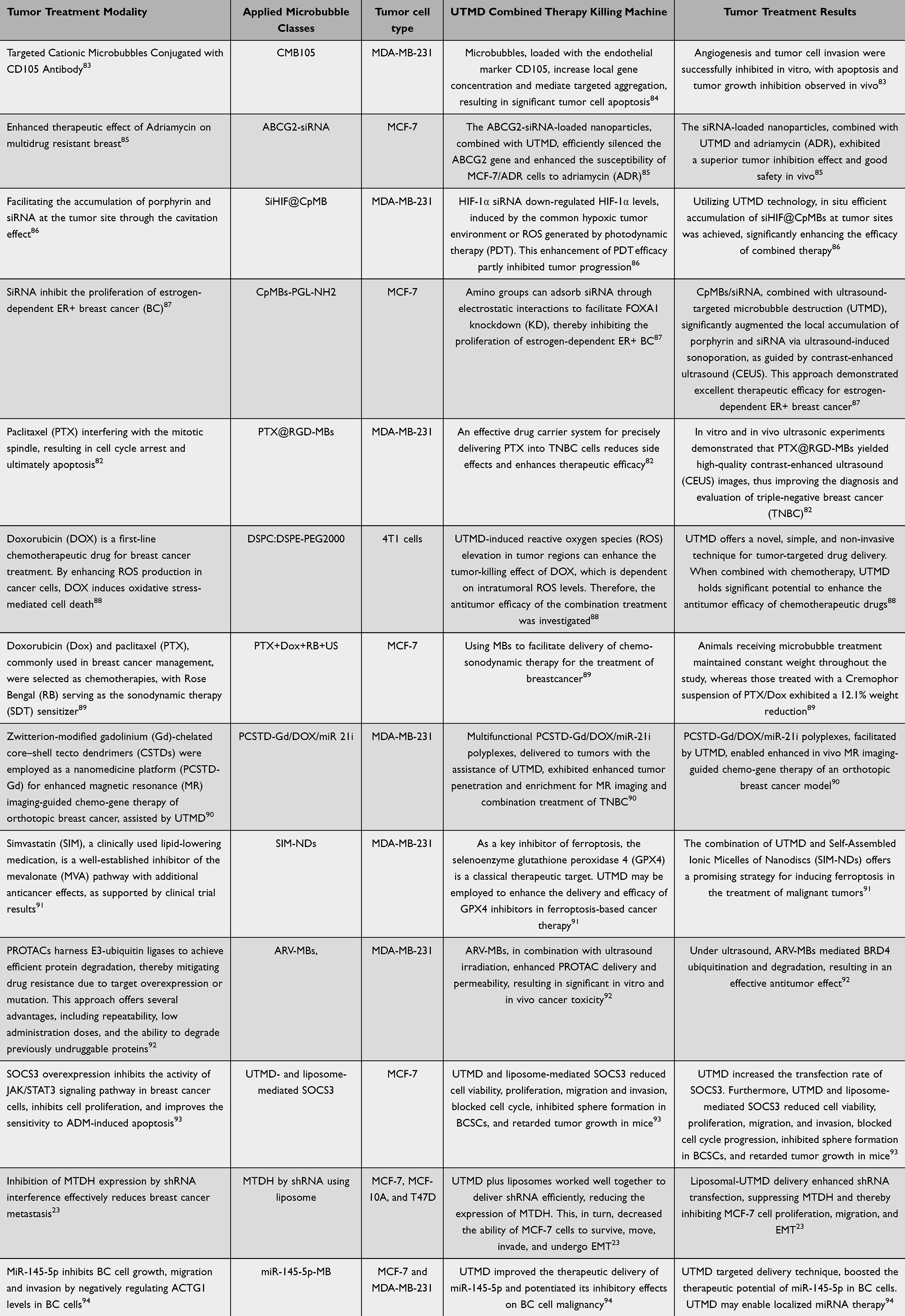 |
Table 1 | Studies on the Use of UTMB in the Treatment of Breast Cancer |
UMTD in the Radiotherapy of Breast Cancer
Radiation therapy (RT) plays a pivotal role in breast cancer management, reducing local recurrence following mastectomy or breast-conserving surgery.95,96 The seminal study at Guy’s Hospital in 1960 established the safety and efficacy of post-lumpectomy RT,97 with subsequent trials confirming its benefit in reducing local recurrence.98,99 However, the double-edged sword of RT’s tumoricidal power and associated toxicities, such as cardiotoxicity, has driven advances in our understanding of breast cancer biology and RT techniques.100,101 Subsequent advances in breast cancer biology and radiation oncology have enabled the personalization of RT, optimizing patient selection, treatment techniques, and fractionation schedules.102,103 However, despite these improvements, challenges remain, including radiation-induced skin reactions, cardiac toxicity, and myelosuppression.104 Ultrasound molecular tomography (UTMD) offers a potential role in enhancing treatment efficacy by targeting tumor microvasculature and blood flow dynamics.105,106 Several studies have investigated the synergistic effects of radiation therapy and ultrasound-guided focused ultrasound (FUS) in preclinical breast cancer models. These studies employed ultrasound stimulation of microbubbles to induce vascular damage, thereby enhancing tumor radiosensitivity. This approach has demonstrated compromised tumor vasculature and improved response to radiation therapy.107,108 Furthermore, the application of ultrasound-mediated drug delivery (UTMD) technology offers a promising avenue for targeted therapeutic interventions. For instance, UTMD has successfully facilitated the delivery of microRNAs, such as miR-21-5p inhibitors, into lung cancer cells, and this approach has demonstrated efficacy in preclinical breast cancer models, potentially improving treatment efficacy and mitigating adverse effects.109 Furthermore, research has demonstrated that ultrasound facilitates the infiltration of nanomaterials into tumor stroma, thereby enhancing the efficacy of in vivo radiotherapy and chemotherapy, and reducing tumor microvascular density and cell proliferation markers.110 Emerging evidence suggests that the combination of UTMD and radiotherapy holds significant promise for improving treatment outcomes and prognosis in breast cancer.
UTMD in Targeted Immunotherapy for Breast Cancer
Immune checkpoint inhibitors (ICIs) represent the foremost class of therapeutics currently employed in breast cancer immunotherapy. These agents potentiate the immune system’s anti-tumor response by inhibiting the PD-1/PD-L1 pathway, thereby relieving the suppression of T cells.111 A notable example is pembrolizumab (Keytruda), a PD-1 inhibitor that has demonstrated significant efficacy, particularly in the treatment of triple-negative breast cancer (TNBC).112 Notably, ICIs shift the therapeutic paradigm by explicitly targeting the tumor microenvironment (TME) rather than the tumor cells themselves. This strategy offers a novel approach to targeted therapy, even in cases where the tumor cells lack conventional therapeutic targets.113,114 However, thanks to immune checkpoint blockade (ICB), explicitly targeting the tumor microenvironment (TME) rather than the tumor cells themselves provides a new approach to targeted therapy, even if the tumor cells lack actionable targets. Components of the TME can contribute to disease progression by secreting and expressing factors that stimulate tumor cell proliferation and suppress anti-tumor immunity, or conversely, contribute to tumor control through adaptive immune mechanisms.115 The pivotal role of immunity in cancer underscores the necessity for immunotherapeutic agents to effectively penetrate the TME to reach tumor cells, engage with PD-1, and subsequently modulate the response of immune effector cells, including macrophages, natural killer (NK) cells, and dendritic cells, ultimately enabling effective tumor cell elimination.116–118 Recent research has demonstrated that ultrasound-targeted microbubble destruction (UTMD) can effectively modulate the tumor immune microenvironment, presenting a promising strategy for tumor immunotherapy.119,120 The synergistic action of ultrasound and microbubbles enhances the tumor immunosuppressive microenvironment by inducing antigen release from tumor cells, both mechanically and thermally. This process facilitates antigen presentation and promotes T-cell recognition and subsequent killing of tumor cells. Consequently, UTMD may address some of the challenges inherent in traditional therapeutic approaches such as immune checkpoint blockade (ICB) and chimeric antigen receptor (CAR)-T cell therapy.24,121 Several studies have investigated the efficacy of ultrasound-mediated drug delivery in targeting HER2-positive breast cancer. Elamir et al demonstrated that liposome-encapsulated anti-HER2 monoclonal antibodies, released via microbubble-triggered sonoporation at low frequencies, exhibited superior tumor growth inhibition compared to other treatment modalities. This enhanced efficacy was attributed to increased drug efficiency, reduced cytotoxicity, promotion of apoptosis, and stimulation of an antitumor immune response.122 These findings corroborate the work of Callmann et al, who also observed that ultrasound stimulation facilitated targeted drug release in HER2-positive breast cancer cells, resulting in improved tumor suppression and decreased systemic toxicity. Further research is needed to elucidate the precise mechanisms underlying these observed benefits and to optimize the parameters of ultrasound-mediated drug delivery for clinical translation.123 In a recent study, a novel ultrasound-responsive spherical nucleic acid (SNA) system was developed to target c-Myc and PD-L1 in triple-negative breast cancer (TNBC). This self-assembled, vector-free small interfering RNA (siRNA) system selectively inhibits c-Myc and PD-L1 in cancer cells when activated by ultrasound, thereby enhancing therapeutic efficacy against TNBC.124 Additionally, the combination of ultrasound-stimulated microbubbles and hyperthermia (USMB and HT) has shown promise in targeting breast tumor vasculature, with potential therapeutic benefits demonstrated in preclinical studies.125 Furthermore, ultrasound-stimulated microbubble cavitation (USMC) technology has been employed to enhance drug concentration and therapeutic efficacy in breast cancer. For instance, a study demonstrated that USMC technology significantly increased the drug concentration and therapeutic efficacy of orally administered gefitinib in mice with ovarian cancer.40 Additionally, the anti-tumor effects of PD-1 immunotherapy can be synergistically enhanced by modifying the local tumor microenvironment. This can be achieved by adjusting vascular permeability and inducing T-cell infiltration, thereby improving overall therapeutic outcomes.126,127 This study introduces a novel therapeutic modality that combines low-intensity focused ultrasound-targeted microbubble disruption (LIFU-TMD) with programmed death-ligand 1 (PD-L1) blockade immunotherapy. LIFU-TMD has been demonstrated to induce the disruption of aberrant tumor vasculature, thereby reducing tumor blood perfusion and facilitating the transformation of the tumor microenvironment (TME). This transformation sensitizes the TME to anti-PD-L1 immunotherapy, resulting in significant inhibition of 4T1 mammary carcinoma growth in murine models.121 The mechanistic underpinnings of this synergistic effect include the enhancement of CD8+ T cell infiltration within the tumor, alleviation of the immunosuppressive TME, and the induction of systemic anti-tumor immune responses. These effects collectively augment the local and distal therapeutic efficacy of anti-PD-L1 antibodies. Additionally, ultrasound-targeted microbubble destruction (UTMD) has been found to play a pivotal role in the optimization of chimeric antigen receptor T-cell (CAR-T) therapies. UTMD can precisely guide CAR-T cells to specific tumor sites, either through ultrasound-mediated delivery or by enhancing the infiltration and activation of CAR-T cells within the tumor. These mechanisms collectively improve the efficacy of CAR-T therapies.128
Ultrasound technology serves dual purposes in clinical settings, with significant applications in both therapeutic and diagnostic modalities. In the context of breast cancer diagnostics, ultrasound is an indispensable tool for the early detection of malignancies, particularly in regions such as China, where its usage is more prevalent than conventional mammography. This preference is attributable to the fact that most Chinese women possess small and dense breast tissue, which can obscure the sensitivity of traditional mammographic imaging. In the diagnostic workflow for breast cancer, ultrasound offers several advantages over radiography, especially in dense breast tissue. Ultrasound imaging provides more detailed information about the internal structure and composition of the breast, enhancing the detectability of lesions that may be masked by dense tissue in mammograms. This capability is crucial for early intervention and improved patient outcomes.129,130
While the above presentation clearly showcases the significant clinical potential of UTMD in breast cancer immunotherapy, several challenges remain. These include the unpredictability of UTMD’s interaction with adjuvant radiotherapy, chemotherapy, and other therapies; the potential for immune-related adverse reactions; and the intricacy of the tumor’s anti-immune mechanisms. Consequently, further research and clinical trials are essential to refine these therapeutic strategies and to establish the optimal use of UTMD in breast cancer treatment.
UTMD in Hormonal Therapy of Breast Cancer
HER2+ breast cancer is a highly aggressive subtype, and precise assessment of HER2 expression status is crucial for determining patient eligibility for targeted anti-HER2 therapy.131,132 While this treatment effectively reduces recurrence and mortality risks, it also presents various side effects.133 Hormone therapy for breast cancer targets hormone receptor-positive (ER+/PR+) subtypes.134 Moreover, microbubble-enhanced and ultrasound-guided (MB+US) drug delivery has demonstrated promise across various therapies, including HER2+ breast cancer, due to its tunable, noninvasive, and spatially targeted nature.86,135,136 Researchers have developed ultrasound imaging combined with poly (lactic acid-hydroxyacetic acid) nanocarriers to dynamically monitor tamoxifen resistance.137 This technology, which integrates hormone therapy with ultrasound to create targeted nanobubbles for ultrasound molecular imaging, offers a promising tool for real-time monitoring of drug resistance in the clinic. While these therapies have proven effective in clinical settings, challenges such as drug resistance and side effects persist.138,139 UTMD has shown potential in managing side effects and providing new insights into drug resistance. Consequently, there is a need to continually explore and develop more effective treatment options for breast cancer, particularly in the application of ultrasound nanobubbles.
UTMD in Radiofrequency Ablative Therapy for Breast Cancer
Imaging-guided Radiofrequency ablation (RFA) of breast cancer has become an important tool in modern breast therapy140,141(Figure 3). Among these techniques, ultrasound-guided RFA is a new minimally invasive breast surgical modality widely used in clinical practice. RFA generates heat locally by causing ionic oscillations through the high-frequency alternating current flowing around the electrodes, leading to protein denaturation and coagulative necrosis of tissues, as well as apoptosis and inactivation of tumor cells.142–144 As a localized radical treatment, it offers the advantages of easy mastery, small incisions, and good cosmetic effects.145 Currently, RFA has shown good results in the treatment of breast cancer. Early studies have confirmed that radiofrequency ablation can cause complete necrosis of ablated tumor cells in various types of breast diseases. The rate of complete tumor necrosis after ablation, as observed through pathological examination, ranges from 76% to 100%.146,147 Subsequent studies have shown that the 5-year progression-free survival rate of patients who underwent radiofrequency ablation was significantly higher than that of patients who underwent post-conservative radiotherapy, with 5-year survival rates ranging from 87% to 97%.148–150 Zhang et al combined RFA with lumpectomy, applying RFA to inactivate the peritumoral invasive cavity after the surgical procedure. This approach resulted in improved breast appearance, reduced local recurrence rates, and decreased secondary surgery rates.151 The application of RFA in early breast cancer demonstrated good local control rates, safety, and high patient quality of life and satisfaction,152 underscoring the clinical advantages of radiofrequency ablation in breast cancer treatment and the potential for local treatment of breast cancer tumors. Regarding the potential for widespread use of radiofrequency ablation in the treatment of malignant breast tumors, the key challenges currently faced are primarily twofold: efficiently and thoroughly ablating the tumor to achieve complete inactivation, and non-invasively detecting recurrence post-procedure.153,154 In addressing these challenges, researchers have discovered that ultrasound, on one hand, induces rapid expansion and contraction of microbubbles within tissues, thereby generating localized high temperatures that augment the thermal effects of radiofrequency ablation and enhance its efficacy. On the other hand, the combination of ultrasound with targeted microbubbles composed of specific nanomaterials enables the visualization of specific tumor sites.155,156 Consequently, recent studies have demonstrated that an approach combining ultrasound-targeted microbubbles with radiofrequency ablation techniques can enhance the therapeutic efficacy of ablation.157–159 It has been shown that the combined use of low-frequency ultrasound and microbubbles amplifies the inhibitory effect of radiofrequency ablation on pancreatic cancer cells, reducing cell migration and proliferation.160 Furthermore, in a study on breast cancer ablation therapy, ultrasound-guided cryoablation combined with endocrine therapy in ER-positive, HER2-negative locally advanced breast cancer was found to be effective in improving ablation efficiency and promoting tumor cell apoptosis using this combined treatment method.161 This further confirms that the combined effect of ultrasound microbubbles plays a positive role in enhancing ablation efficiency, thereby promoting tumor cell apoptosis and inactivation in ablation therapy. The underlying mechanism may be that microbubbles, under ultrasound irradiation, act as bioaugmentation agents, enhancing the efficiency of thermal ablation therapy by altering the acoustic impedance difference and thus increasing ultrasound energy deposition in the tissue environment.162 Moreover, in another study, researchers discovered that perfluorocarbon (PFC) nanoparticles, when subjected to focused ultrasound (FUS), transform into microbubbles during tumor thermal ablation. This transformation alters the acoustic environment of the tissues and enhances ultrasound energy deposition, thereby achieving synergistic FUS-assisted ablation therapy for tumors.84,163,164
 |
Figure 3 Influence of complete radiofrequency ablation on prognosis of breast cancer. |
In a comparative trial, the study demonstrated that the strategy of combining ultrasound with microbubbles significantly improved the accuracy and efficacy of ablation, particularly in the treatment of breast cancer tumors, and showed great potential for use in breast cancer treatment. However, several issues require further investigation and optimization: Microbubble stability and safety: The materials and types of microbubbles used in the study need to be optimized to ensure in vivo stability and safety, and to minimize potential side effects. Equipment and parameter optimization: The equipment and parameter settings used for the ultrasound-microbubble combination need to be further optimized to achieve the best treatment outcomes. This includes optimizing the ultrasound frequency and power, as well as the microbubble concentration and size. Clinical validation: Although numerous studies are currently in the animal experimentation and theoretical research stages, large-scale clinical trial data are scarce. Consequently, further clinical validation is required to evaluate the actual efficacy and safety of this approach in treating various tumor types and stages. The aforementioned review indicates that the application of ultrasound combined with microbubbles in radiofrequency ablation holds promise; however, it still necessitates continued exploration and optimization to play a more significant role in clinical practice.
Conclusion and Prospect
Ultrasound-targeted microbubble destruction (UTMD) has emerged as a potent, non-invasive adjuvant therapy for breast cancer treatment, demonstrating significant efficacy in enhancing the immune microenvironment and augmenting the effectiveness of radiotherapy, immunotherapy, and ablative therapies. This novel approach challenges the traditional paradigm, which has primarily focused on the direct cytotoxic effects of therapeutic agents on tumor cells. Despite the development of diverse therapeutic modalities, the lack of optimal delivery vehicles and strategies to improve the tumor microenvironment has hindered the full potential of these treatments. UTMD addresses this gap by enhancing therapeutic outcomes. To fully exploit the therapeutic potential of UTMD in breast cancer adjuvant treatment, several key areas of challenges and future research directions are identified. Optimizing ultrasound parameters, such as frequency and intensity, to maximize drug efficacy, and enhancing the biocompatibility and in vivo stability of microbubbles or nanocarriers to minimize side effects on normal tissues are crucial. Moreover, while preclinical studies and theoretical research have shown promise, there is a dearth of large-scale clinical trial data. More clinical validation is necessary to assess the efficacy and safety of UTMD across different types and stages of breast cancer. As the application of UTMD in breast cancer treatment continues to evolve, it is expected to play a pivotal role in the future of oncology. The academic community must rigorously investigate and refine the parameters and safety profiles of UTMD to ensure its maximum clinical benefit. By addressing these challenges, UTMD can become a cornerstone in the multidisciplinary approach to breast cancer treatment.
Data Sharing Statement
The analyzed data sets generated during the study are available from the corresponding author on reasonable request.
Funding
The authors report there is no funding associated with the work featured in this article.
Disclosure
No potential conflict of interest was reported by the authors.
References
1. Sung H, Ferlay J, Siegel RL, et al. Global cancer statistics 2020: GLOBOCAN estimates of incidence and mortality worldwide for 36 cancers in 185 countries. CA Cancer J Clin. 2021;71(3):209–249. doi:10.3322/caac.21660
2. Arnold M, Morgan E, Rumgay H, et al. Current and future burden of breast cancer: global statistics for 2020 and 2040. Breast. 2022;66:15–23. doi:10.1016/j.breast.2022.08.010
3. Winters S, Martin C, Murphy D, Shokar NK. Breast cancer epidemiology, prevention, and screening. Prog mol Biol Transl Sci. 2017;151:1–32.
4. Li X, Yang J, Peng L, et al. Triple-negative breast cancer has worse overall survival and cause-specific survival than non-triple-negative breast cancer. Breast Cancer Res Treat. 2017;161(2):279–287. doi:10.1007/s10549-016-4059-6
5. Qi J, Li M, Wang L, et al. National and subnational trends in cancer burden in China, 2005-20: an analysis of national mortality surveillance data. Lancet Public Health. 2023;8(12):e943–e955. doi:10.1016/S2468-2667(23)00211-6
6. Katsura C, Ogunmwonyi I, Kankam HK, Saha S. Breast cancer: presentation, investigation and management. Br J Hosp Med. 2022;83(2):1–7. doi:10.12968/hmed.2021.0459
7. Roy M, Fowler AM, Ulaner GA, Mahajan A. Molecular classification of breast cancer. PET Clin. 2023;18(4):441–458. doi:10.1016/j.cpet.2023.04.002
8. Sarhangi N, Hajjari S, Heydari SF, Ganjizadeh M, Rouhollah F, Hasanzad M. Breast cancer in the era of precision medicine. Mol Biol Rep. 2022;49(10):10023–10037. doi:10.1007/s11033-022-07571-2
9. Gao JJ, Swain SM. Luminal A breast cancer and molecular assays: a review. Oncologist. 2018;23(5):556–565. doi:10.1634/theoncologist.2017-0535
10. Waks AG, Winer EP. Breast cancer treatment: a review. JAMA. 2019;321(3):288–300.
11. Asleh K, Riaz N, Nielsen TO. Heterogeneity of triple negative breast cancer: current advances in subtyping and treatment implications. J Exp Clin Cancer Res. 2022;41(1):265.
12. Yin L, Duan JJ, Bian XW, Yu SC. Triple-negative breast cancer molecular subtyping and treatment progress. Breast Cancer Res. 2020;22(1):61. doi:10.1186/s13058-020-01296-5
13. Harbeck N, Gnant M. Breast cancer. Lancet. 2017;389(10074):1134–1150. doi:10.1016/S0140-6736(16)31891-8
14. Yang F, Xiao Y, Ding JH, et al. Ferroptosis heterogeneity in triple-negative breast cancer reveals an innovative immunotherapy combination strategy. Cell Metab. 2023;35(1):84–100.e8. doi:10.1016/j.cmet.2022.09.021
15. Li Y, Zhang H, Merkher Y, et al. Recent advances in therapeutic strategies for triple-negative breast cancer. J Hematol Oncol. 2022;15(1):121. doi:10.1186/s13045-022-01341-0
16. Zhu X, Chen L, Huang B, et al. Efficacy and mechanism of the combination of PARP and CDK4/6 inhibitors in the treatment of triple-negative breast cancer. J Exp Clin Cancer Res. 2021;40(1):122. doi:10.1186/s13046-021-01930-w
17. O’Sullivan CC, Clarke R, Goetz MP, Robertson J. Cyclin-dependent kinase 4/6 inhibitors for treatment of hormone receptor-positive, ERBB2-negative breast cancer: a review. JAMA Oncol. 2023;9(9):1273–1282. doi:10.1001/jamaoncol.2023.2000
18. Jilishitz I, Quiñones JL, Patel P, et al. NP-ALT, a liposomal:peptide drug, blocks p27Kip1 phosphorylation to induce oxidative stress, necroptosis, and regression in therapy-resistant breast cancer cells. Mol Cancer Res. 2021;19(11):1929–1945. doi:10.1158/1541-7786.MCR-21-0081
19. Nardin S, Ruelle T, Giannubilo I, Del Mastro L. Adjuvant treatment in hormone receptor-positive early breast cancer: new approaches of endocrine therapy. Tumori. 2024;110(3):162–167. doi:10.1177/03008916231216830
20. Mehraj U, Dar AH, Wani NA, Mir MA. Tumor microenvironment promotes breast cancer chemoresistance. Cancer Chemother Pharmacol. 2021;87(2):147–158. doi:10.1007/s00280-020-04222-w
21. Tharp KM, Kersten K, Maller O, et al. Tumor-associated macrophages restrict CD8(+) T cell function through collagen deposition and metabolic reprogramming of the breast cancer microenvironment. Nat Cancer. 2024;5(7):1045–1062. doi:10.1038/s43018-024-00775-4
22. Park M, Kim D, Ko S, Kim A, Mo K, Yoon H. Breast cancer metastasis: mechanisms and therapeutic implications. Int J Mol Sci. 2022;23(12):6806. doi:10.3390/ijms23126806
23. Xu J, Wang Y, Li Z, Wang Q, Zhou X, Wu W. Ultrasound-targeted microbubble destruction (UTMD) combined with liposome increases the effectiveness of suppressing proliferation, migration, invasion, and epithelial- mesenchymal transition (EMT) via targeting metadherin (MTDH) by ShRNA. Med Sci Monit. 2019;25:2640–2648. doi:10.12659/MSM.912955
24. Liu S, Zhang Y, Liu Y, et al. Ultrasound-targeted microbubble destruction remodels tumour microenvironment to improve immunotherapeutic effect. Br J Cancer. 2023;128(5):715–725. doi:10.1038/s41416-022-02076-y
25. Lin L, Du Y, Hao J, Wu R, Du L. UTMD inhibits pancreatic cancer growth and metastasis by inducing macrophage polarization and vessel normalization. Biomed Pharmacother. 2023;160:114322. doi:10.1016/j.biopha.2023.114322
26. Kopechek JA, Carson AR, McTiernan CF, et al. Ultrasound targeted microbubble destruction-mediated delivery of a transcription factor decoy inhibits STAT3 signaling and tumor growth. Theranostics. 2015;5(12):1378–1387. doi:10.7150/thno.12822
27. Zhang Y, Lv W, Li H, et al.Exploring the relationship between autophagy and Gefitinib resistance in NSCLC by silencing PDLIM5 using ultrasound-targeted microbubble destruction technology. Cancer Cell Int. 2022;22(1):293 doi:10.1186/s12935-022-02718-4.
28. Shi D, Guo L, Sun X, et al. UTMD inhibit EMT of breast cancer through the ROS/miR-200c/ZEB1 axis. Sci Rep. 2020;10(1):6657. doi:10.1038/s41598-020-63653-w
29. Zhang W, Nan SL, Bai WK, Hu B. Low-frequency ultrasound combined with microbubbles improves gene transfection in prostate cancer cells in vitro and in vivo. Asia Pac J Clin Oncol. 2022;18(1):93–98. doi:10.1111/ajco.13521
30. Sitta J, Howard CM. Applications of ultrasound-mediated drug delivery and gene therapy. Int J Mol Sci. 2021;22(21):11491. doi:10.3390/ijms222111491
31. Awad NS, Paul V, AlSawaftah NM, et al. Ultrasound-responsive nanocarriers in cancer treatment: a review. ACS Pharmacol Transl Sci. 2021;4(2):589–612. doi:10.1021/acsptsci.0c00212
32. Gunderman A, Montayre R, Ranjan A, Chen Y. Review of robot-assisted HIFU therapy. Sensors. 2023;23(7):3707. doi:10.3390/s23073707
33. Ter Haar G. HIFU tissue ablation: concept and devices. Adv Exp Med Biol. 2016;880:3–20.
34. Wang C, Li Z, Bai J. Bubble-assisted HIFU ablation enabled by calcium peroxide. J Mater Chem B. 2022;10(23):4442–4451. doi:10.1039/D2TB00587E
35. Chen J, Nan Z, Zhao Y, et al. Enhanced HIFU theranostics with dual-frequency-ring focused ultrasound and activatable perfluoropentane-loaded polymer nanoparticles. Micromachines. 2021;12(11):1324. doi:10.3390/mi12111324
36. Zhou K, Strunk H, Dimitrov D, et al. US-guided high-intensity focused ultrasound in pancreatic cancer treatment: a consensus initiative between Chinese and European HIFU centers. Int J Hyperthermia. 2024;41(1):2295812. doi:10.1080/02656736.2023.2295812
37. Mei L, Zhang Z, Song X, Zhao X. Microbubbles activated by low-frequency ultrasound enhance the anti-tumor effects of curcumin in glioma cells by suppressing the TGF-β1/Smad/VEGF/NCAM signaling pathway. Gen Physiol Biophys. 2024;43(1):73–84. doi:10.4149/gpb_2023024
38. Wang LY, Zheng SS. Advances in low-frequency ultrasound combined with microbubbles in targeted tumor therapy. J Zhejiang Univ Sci B. 2019;20(4):291–299. doi:10.1631/jzus.B1800508
39. He J, Liu Z, Zhu X, Xia H, Gao H, Lu J. Ultrasonic microbubble cavitation enhanced tissue permeability and drug diffusion in solid tumor therapy. Pharmaceutics. 2022;14(8):1642. doi:10.3390/pharmaceutics14081642
40. Amate M, Goldgewicht J, Sellamuthu B, Stagg J, Yu F. The effect of ultrasound pulse length on microbubble cavitation induced antibody accumulation and distribution in a mouse model of breast cancer. Nanotheranostics. 2020;4(4):256–269. doi:10.7150/ntno.46892
41. Chen J, Wang J, Yan X, et al. Enhancing the therapeutic efficacy of gefitinib on subcutaneously transplanted SKOV3 ovarian cancer tumors in nude mice via ultrasound‑stimulated microbubble cavitation. Exp Ther Med. 2024;28(3):336. doi:10.3892/etm.2024.12625
42. Chowdhury SM, Abou-Elkacem L, Lee T, Dahl J, Lutz AM. Ultrasound and microbubble mediated therapeutic delivery: underlying mechanisms and future outlook. J Control Release. 2020;326:75–90. doi:10.1016/j.jconrel.2020.06.008
43. Chapla R, Huynh KT, Schutt CE. Microbubble-nanoparticle complexes for ultrasound-enhanced cargo delivery. Pharmaceutics. 2022;14(11):2396. doi:10.3390/pharmaceutics14112396
44. Edwards IA, De Carlo F, Sitta J, Varner W, Howard CM, Claudio PP. Enhancing targeted therapy in breast cancer by ultrasound-responsive nanocarriers. Int J Mol Sci. 2023;24(6):5474. doi:10.3390/ijms24065474
45. Fan CH, Ho YJ, Lin CW, Wu N, Chiang PH, Yeh CK. State-of-the-art of ultrasound-triggered drug delivery from ultrasound-responsive drug carriers. Expert Opin Drug Deliv. 2022;19(8):997–1009. doi:10.1080/17425247.2022.2110585
46. Li Y, Du M, Fang J, Zhou J, Chen Z. UTMD promoted local delivery of miR-34a-mimic for ovarian cancer therapy. Drug Deliv. 2021;28(1):1616–1625. doi:10.1080/10717544.2021.1955041
47. Zhang H, Li Y, Rao F, Liufu C, Wang Y, Chen Z. A novel UTMD system facilitating nucleic acid delivery into MDA-MB-231 cells. Biosci Rep. 2020;40(2):BSR20192573. doi:10.1042/BSR20192573
48. Liao Y, Luo H, He Z, et al. A combination of UTMD-mediated HIF-1α shRNA transfection and TAE in the treatment of hepatic cancer. Biomed Res Int. 2019;2019:1937460. doi:10.1155/2019/1937460
49. Qu F, Wang P, Zhang K, et al. Manipulation of mitophagy by ”all-in-one” nanosensitizer augments sonodynamic glioma therapy. Autophagy. 2020;16(8):1413–1435. doi:10.1080/15548627.2019.1687210
50. Zhou C, Xie X, Yang H, et al. Novel class of ultrasound-triggerable drug delivery systems for the improved treatment of tumors. Mol Pharm. 2019;16(7):2956–2965. doi:10.1021/acs.molpharmaceut.9b00194
51. Hu J, Li X, Yang L, Li H. Hypoxia, a key factor in the immune microenvironment. Biomed Pharmacother. 2022;151:113068. doi:10.1016/j.biopha.2022.113068
52. Jing X, Yang F, Shao C, et al. Role of hypoxia in cancer therapy by regulating the tumor microenvironment. Mol Cancer. 2019;18(1):157. doi:10.1186/s12943-019-1089-9
53. Tan H, Tian Y, Yang H, et al. Oxygen-sufficient lipid nanobubbles combined with UTMD for enhanced sonodynamic therapy of Hep-G2 cells. J Biomed Mater Res B Appl Biomater. 2021;109(11):1796–1806. doi:10.1002/jbm.b.34839
54. Logan KA, Nesbitt H, Callan B, et al. Synthesis of a gemcitabine-modified phospholipid and its subsequent incorporation into a single microbubble formulation loaded with paclitaxel for the treatment of pancreatic cancer using ultrasound-targeted microbubble destruction. Eur J Pharm Biopharm. 2021;165:374–382. doi:10.1016/j.ejpb.2021.05.018
55. Entzian K, Aigner A. Drug delivery by ultrasound-responsive nanocarriers for cancer treatment. Pharmaceutics. 2021;13(8):1135. doi:10.3390/pharmaceutics13081135
56. Zavala VA, Serrano-Gomez SJ, Dutil J, Fejerman L. Genetic epidemiology of breast cancer in Latin America. Genes. 2019;10(2):153. doi:10.3390/genes10020153
57. Britt KL, Cuzick J, Phillips KA. Key steps for effective breast cancer prevention. Nat Rev Cancer. 2020;20(8):417–436. doi:10.1038/s41568-020-0266-x
58. Xu X, Zhang M, Xu F, Jiang S. Wnt signaling in breast cancer: biological mechanisms, challenges and opportunities. Mol Cancer. 2020;19(1):165. doi:10.1186/s12943-020-01276-5
59. Prat A, Pineda E, Adamo B, et al. Clinical implications of the intrinsic molecular subtypes of breast cancer. Breast. 2015;24 Suppl 2:S26–35. doi:10.1016/j.breast.2015.07.008
60. Corti C, Giugliano F, Nicolò E, Tarantino P, Criscitiello C, Curigliano G. HER2-low breast cancer: a new subtype. Curr Treat Options Oncol. 2023;24(5):468–478. doi:10.1007/s11864-023-01068-1
61. Tarantino P, Viale G, Press MF, et al. ESMO expert consensus statements (ECS) on the definition, diagnosis, and management of HER2-low breast cancer. Ann Oncol. 2023;34(8):645–659. doi:10.1016/j.annonc.2023.05.008
62. Nicolò E, Tarantino P, Curigliano G. Biology and treatment of HER2-low breast cancer. Hematol Oncol Clin North Am. 2023;37(1):117–132. doi:10.1016/j.hoc.2022.08.013
63. Stettler S, Aebi S. Endocrine treatments in breast cancer. Praxis. 2022;111(10):550–556. doi:10.1024/1661-8157/a003878 Spanish
64. Bartlett J, Sgroi DC, Treuner K, et al. Breast cancer index and prediction of benefit from extended endocrine therapy in breast cancer patients treated in the adjuvant tamoxifen-to offer more? (aTTom) trial. Ann Oncol. 2019;30(11):1776–1783. doi:10.1093/annonc/mdz289
65. Obidiro O, Battogtokh G, Akala EO. Triple negative breast cancer treatment options and limitations: future outlook. Pharmaceutics. 2023;15(7):1796. doi:10.3390/pharmaceutics15071796
66. Vagia E, Mahalingam D, Cristofanilli M. The landscape of targeted therapies in TNBC. Cancers. 2020;12(4):916. doi:10.3390/cancers12040916
67. Singh DD, Yadav DK. TNBC: potential targeting of multiple receptors for a therapeutic breakthrough, nanomedicine, and immunotherapy. Biomedicines. 2021;9(8):876. doi:10.3390/biomedicines9080876
68. Chaudhuri A, Kumar DN, Dehari D, et al. Endorsement of TNBC biomarkers in precision therapy by nanotechnology. Cancers. 2023;15(9):2661. doi:10.3390/cancers15092661
69. Mouabbi JA, Hassan A, Lim B, Hortobagyi GN, Tripathy D, Layman RM. Invasive lobular carcinoma: an understudied emergent subtype of breast cancer. Breast Cancer Res Treat. 2022;193(2):253–264. doi:10.1007/s10549-022-06572-w
70. Reed AE M, Kalinowski L, Simpson PT, Lakhani SR. Invasive lobular carcinoma of the breast: the increasing importance of this special subtype. Breast Cancer Res. 2021;23(1):6. doi:10.1186/s13058-020-01384-6
71. Karim AM, Eun Kwon J, Ali T, et al. Triple-negative breast cancer: epidemiology, molecular mechanisms, and modern vaccine-based treatment strategies. Biochem Pharmacol. 2023;212:115545. doi:10.1016/j.bcp.2023.115545
72. Budny A, Starosławska E, Budny B, et al. Epidemiology and diagnosis of breast cancer. Pol Merkur Lekarski. 2019;46(275):195–204.
73. Zraik IM, Heß-Busch Y. Management of chemotherapy side effects and their long-term sequelae. Urologe A. 2021;60(7):862–871. doi:10.1007/s00120-021-01569-7
74. Pan K, Farrukh H, Chittepu V, Xu H, Pan CX, Zhu Z. CAR race to cancer immunotherapy: from CAR T, CAR NK to CAR macrophage therapy. J Exp Clin Cancer Res. 2022;41(1):119. doi:10.1186/s13046-022-02327-z
75. Xian C, Chen H, Xiong F, Fang Y, Huang H, Wu J. Platinum-based chemotherapy via nanocarriers and co-delivery of multiple drugs. Biomater Sci. 2021;9(18):6023–6036. doi:10.1039/D1BM00879J
76. Li B, Shao H, Gao L, Li H, Sheng H, Zhu L. Nano-drug co-delivery system of natural active ingredients and chemotherapy drugs for cancer treatment: a review. Drug Deliv. 2022;29(1):2130–2161. doi:10.1080/10717544.2022.2094498
77. Ren WW, Xu SH, Sun LP, Zhang K. Ultrasound-based drug delivery system. Curr Med Chem. 2022;29(8):1342–1351. doi:10.2174/0929867328666210617103905
78. Yan F, Li X, Jin Q, et al. Therapeutic ultrasonic microbubbles carrying paclitaxel and LyP-1 peptide: preparation, characterization and application to ultrasound-assisted chemotherapy in breast cancer cells. Ultrasound Med Biol. 2011;37(5):768–779. doi:10.1016/j.ultrasmedbio.2011.02.006
79. Amiri-Kordestani L, Basseville A, Kurdziel K, Fojo AT, Bates SE. Targeting MDR in breast and lung cancer: discriminating its potential importance from the failure of drug resistance reversal studies. Drug Resist Updat. 2012;15(1–2):50–61. doi:10.1016/j.drup.2012.02.002
80. Subhan MA. Advances with metal oxide-based nanoparticles as MDR metastatic breast cancer therapeutics and diagnostics. RSC Adv. 2022;12(51):32956–32978. doi:10.1039/D2RA02005J
81. Li Y, Gao X, Yu Z, et al. Reversing multidrug resistance by multiplexed gene silencing for enhanced breast cancer chemotherapy. ACS Appl Mater Interfaces. 2018;10(18):15461–15466. doi:10.1021/acsami.8b02800
82. Bai M, Dong Y, Huang H, et al. Tumour targeted contrast enhanced ultrasound imaging dual-modal microbubbles for diagnosis and treatment of triple negative breast cancer. RSC Adv. 2019;9(10):5682–5691. doi:10.1039/C8RA09737B
83. Zhou Y, Gu H, Xu Y, et al. Targeted antiangiogenesis gene therapy using targeted cationic microbubbles conjugated with CD105 antibody compared with untargeted cationic and neutral microbubbles. Theranostics. 2015;4:399–417. doi:10.7150/thno.10351
84. Alphandéry E. Ultrasound and nanomaterial: an efficient pair to fight cancer. J Nanobiotechnol. 2022;20(1):139. doi:10.1186/s12951-022-01243-w
85. Bai M, Shen M, Teng Y, et al. Enhanced therapeutic effect of Adriamycin on multidrug resistant breast cancer by the ABCG2-siRNA loaded polymeric nanoparticles assisted with ultrasound. Oncotarget. 2015;41:43779–43790. doi:10.18632/oncotarget.6085
86. Sun S, Xu Y, Fu P, et al. Ultrasound-targeted photodynamic and gene dual therapy for effectively inhibiting triple negative breast cancer by cationic porphyrin lipid microbubbles loaded with HIF1α-siRNA. Nanoscale. 2018;10(42):19945–19956. doi:10.1039/C8NR03074J
87. Zhao R, Liang X, Zhao B, et al. Ultrasound assisted gene and photodynamic synergistic therapy with multifunctional FOXA1-siRNA loaded porphyrin microbubbles for enhancing therapeutic efficacy for breast cancer. Biomaterials. 2018;173:58–70. doi:10.1016/j.biomaterials.2018.04.054
88. Wu M, Song Z, Zhang S, et al. Local tumor ischemia-reperfusion mediated by ultrasound-targeted microbubble destruction enhances the anti-tumor efficacy of doxorubicin chemotherapy. Cancer Manag Res. 2019;Volume 11:9387–9395. doi:10.2147/CMAR.S225607
89. Logan K, Foglietta F, Nesbitt H, et al. Targeted chemo-sonodynamic therapy treatment of breast tumours using ultrasound responsive microbubbles loaded with paclitaxel, doxorubicin and Rose Bengal.Eur. J Pharm Biopharm. 2019;139:224–231. doi:10.1016/j.ejpb.2019.04.003
90. Gong J, Song C, Li G, et al. Ultrasound-enhanced theranostics of orthotopic breast cancer through a multifunctional core-shell tecto dendrimer-based nanomedicine platform. Biomater Sci. 2023;11:4385–4396. doi:10.1039/D3BM00375B
91. Liu R, Shi D, Guo L, et al. Ultrasound-targeted microbubble disruption with key nanodroplets for effective ferroptosis in triple-negative breast cancer using animal model. Int J Nanomed. 2024;2037–2052. doi: 10.2147/IJN.S400495
92. He H, Li F, Tang R, et al. Ultrasound controllable release of proteolysis targeting chimeras for triple-negative breast cancer treatment. Biomater Res. 2024;2024:0064. doi:10.34133/bmr.0064
93. Tang X, Hao N, Zhou Y, et al. Ultrasound targeted microbubble destruction-mediated SOCS3 attenuates biological characteristics and epithelial-mesenchymal transition (EMT) of breast cancer stem cells. Bioengineered. 2022;2:3896–3910. doi:10.1080/21655979.2022.2031384
94. Ren L, Wang L, Yi X, et al. Ultrasound microbubble-stimulated miR-145-5p inhibits malignant behaviors of breast cancer cells by targeting ACTG1.[J]. Ultrasound Q. 1970;2:136–143.
95. Gage I, Harris JR. Radiation therapy and breast cancer. Curr Opin Oncol. 1998;10(6):513–516. doi:10.1097/00001622-199811000-00006
96. Haque W, Butler EB, Teh BS. Personalized radiation therapy for breast cancer. Curr Oncol. 2024;31(3):1588–1599. doi:10.3390/curroncol31030121
97. Atkins H, Hayward JL, Klugman DJ, Wayte AB. Treatment of early breast cancer: a report after ten years of a clinical trial. Br Med J. 1972;2(5811):423–429. doi:10.1136/bmj.2.5811.423
98. Louis-Sylvestre C, Clough K, Asselain B, et al. Axillary treatment in conservative management of operable breast cancer: dissection or radiotherapy? Results of a randomized study with 15 years of follow-up. J Clin Oncol. 2004;22(1):97–101. doi:10.1200/JCO.2004.12.108
99. Kaidar-Person O, Gentilini O, Poortmans P. Not only volumes matter for breast radiation therapy. Radiother Oncol. 2022;177:236–237. doi:10.1016/j.radonc.2022.10.021
100. Loap P, Vu-Bezin J, Monceau V, Jacob S, Fourquet A, Kirova Y. Dosimetric evaluation of the benefit of deep inspiration breath hold (DIBH) for locoregional irradiation of right breast cancer with volumetric modulated arctherapy (VMAT). Acta Oncol. 2023;62(2):150–158. doi:10.1080/0284186X.2023.2177976
101. Douek M, De Silva-Minor S, Davies L, Jones B. Breast cancer radiation therapy. Lancet. 2020;396(10262):1558–1559. doi:10.1016/S0140-6736(20)32323-0
102. Mutter RW, Choi JI, Jimenez RB, et al. Proton therapy for breast cancer: a consensus statement from the particle therapy cooperative group breast cancer subcommittee. Int J Radiat Oncol Biol Phys. 2021;111(2):337–359. doi:10.1016/j.ijrobp.2021.05.110
103. Moran MS. Advancements and personalization of breast cancer treatment strategies in radiation therapy. Cancer Treat Res. 2018;173:89–119.
104. Moran MS, Ho AY. Radiation therapy for low-risk breast cancer: whole, partial, or none. J Clin Oncol. 2022;40(36):4166–4172. doi:10.1200/JCO.22.01751
105. Jayasankar G, Koilpillai J, Narayanasamy D. A systematic study on long-acting nanobubbles: current advancement and prospects on theranostic properties. Adv Pharm Bull. 2024;14(2):278–301. doi:10.34172/apb.2024.042
106. Owen J, Negussie AH, Burks SR, et al. Microbubbles bound to drug-eluting beads enable ultrasound imaging and enhanced delivery of therapeutics. Sci Rep. 2024;14(1):20929. doi:10.1038/s41598-024-71831-3
107. Sharma D, Hussein F, Law N, et al. Focused ultrasound stimulation of microbubbles in combination with radiotherapy for acute damage of breast cancer xenograft model. Technol Cancer Res Treat. 2022;21:15330338221132925. doi:10.1177/15330338221132925
108. Lai P, Tarapacki C, Tran WT, et al. Breast tumor response to ultrasound mediated excitation of microbubbles and radiation therapy in vivo. Oncoscience. 2016;3(3–4):98–108. doi:10.18632/oncoscience.299
109. Zhou X, Liu H, Pang Y, Wang M, Liu S. UTMD-mediated delivery of miR-21-5p inhibitor suppresses the development of lung cancer. Tissue Cell. 2022;74:101719. doi:10.1016/j.tice.2021.101719
110. Bhardwaj P, Goda JS, Pai V, et al.Ultrasound augments on-demand breast tumor radiosensitization and apoptosis through a tri-responsive combinatorial delivery theranostic platform. Nanoscale. 2021;13(40):17077–17092. doi:10.1039/D1NR04211D
111. Yi M, Zheng X, Niu M, Zhu S, Ge H, Wu K. Combination strategies with PD-1/PD-L1 blockade: current advances and future directions. Mol Cancer. 2022;21(1):28. doi:10.1186/s12943-021-01489-2
112. Keenan TE, Tolaney SM. Role of immunotherapy in triple-negative breast cancer. J Natl Compr Canc Netw. 2020;18(4):479–489. doi:10.6004/jnccn.2020.7554
113. Li Q, Han J, Yang Y, Chen Y. PD-1/PD-L1 checkpoint inhibitors in advanced hepatocellular carcinoma immunotherapy. Front Immunol. 2022;13:1070961. doi:10.3389/fimmu.2022.1070961
114. Wu Q, Jiang L, Li SC, He QJ, Yang B, Cao J. Small molecule inhibitors targeting the PD-1/PD-L1 signaling pathway. Acta Pharmacol Sin. 2021;42(1):1–9. doi:10.1038/s41401-020-0366-x
115. Wu M, Huang Q, Xie Y, et al. Improvement of the anticancer efficacy of PD-1/PD-L1 blockade via combination therapy and PD-L1 regulation. J Hematol Oncol. 2022;15(1):24. doi:10.1186/s13045-022-01242-2
116. Huang Q, Wu X, Wang Z, et al. The primordial differentiation of tumor-specific memory CD8(+) T cells as bona fide responders to PD-1/PD-L1 blockade in draining lymph nodes. Cell. 2022;185(22):4049–4066.e25. doi:10.1016/j.cell.2022.09.020
117. Wu X, Gu Z, Chen Y, et al. Application of PD-1 blockade in cancer immunotherapy. Comput Struct Biotechnol J. 2019;17:661–674. doi:10.1016/j.csbj.2019.03.006
118. Pei L, Liu Y, Liu L, et al. Roles of cancer-associated fibroblasts (CAFs) in anti- PD-1/PD-L1 immunotherapy for solid cancers. Mol Cancer. 2023;22(1):29. doi:10.1186/s12943-023-01731-z
119. Zhang H, Liu L, Liu J, et al. Roles of tumor-associated macrophages in anti-PD-1/PD-L1 immunotherapy for solid cancers. Mol Cancer. 2023;22(1):58. doi:10.1186/s12943-023-01725-x
120. Xingang W, Ming S, Yingying S, et al. Ultrasound-triggered cascade reaction via MnO2-CpG nanoparticles for boosting pyroptosis-mediated cancer immunotherapy. Nano Today. 2024;102394. doi:10.1016/j.nantod.2024.102394
121. Wu N, Cao Y, Liu Y, et al. Low-intensity focused ultrasound targeted microbubble destruction reduces tumor blood supply and sensitizes anti-PD-L1 immunotherapy. Front Bioeng Biotechnol. 2023;11:1173381. doi:10.3389/fbioe.2023.1173381
122. Elamir A, Ajith S, Sawaftah NA, et al. Ultrasound-triggered herceptin liposomes for breast cancer therapy. Sci Rep. 2021;11(1):7545. doi:10.1038/s41598-021-86860-5
123. Callmann CE, Kusmierz CD, Dittmar JW, Broger L, Mirkin CA. Impact of liposomal spherical nucleic acid structure on immunotherapeutic function. ACS Cent Sci. 2021;7(5):892–899. doi:10.1021/acscentsci.1c00181
124. Wang R, Li G, Gao F, et al. Ultrasound-responsive spherical nucleic acid against c-Myc/PD-L1 to enhance anti-tumoral macrophages in triple-negative breast cancer progression. Sci China Life Sci. 2024;67(4):698–710. doi:10.1007/s11427-023-2433-y
125. Sharma D, Cartar H, Quiaoit K, Law N, Giles A, Czarnota GJ. Effect of ultrasound-stimulated microbubbles and hyperthermia on tumor vasculature of breast cancer xenograft. J Ultrasound Med. 2022;41(11):2659–2671. doi:10.1002/jum.15950
126. Zhu Z, Shibata R, Hoffman KL, et al. Integrated nasopharyngeal airway metagenome and asthma genetic risk endotyping of severe bronchiolitis in infancy and risk of childhood asthma. Eur Respir J. 2024;64:2401130. doi:10.1183/13993003.01130-2024
127. Si Y, Yue J, Liu Z, et al. Phase-transformation nanoparticle-mediated sonodynamic therapy: an effective modality to enhance anti-tumor immune response by inducing immunogenic cell death in breast cancer. Int J Nanomed. 2021;16:1913–1926. doi:10.2147/IJN.S297933
128. Meng X, Wu X, Zheng Y, et al. Exploiting Ca(2+) signaling in T cells to advance cancer immunotherapy. Semin Immunol. 2020;49:101434. doi:10.1016/j.smim.2020.101434
129. Wang Y, Li Y, Song Y, et al. Comparison of ultrasound and mammography for early diagnosis of breast cancer among Chinese women with suspected breast lesions: a prospective trial. Thorac Cancer. 2022;13(22):3145–3151. doi:10.1111/1759-7714.14666
130. Sasikala S, Ezhilarasi M, Arunkumar S. Feature selection algorithm based on binary BAT algorithm and optimum path forest classifier for breast cancer detection using both echographic and elastographic mode ultrasound images. J Cancer Res Ther. 2023;19(2):191–197. doi:10.4103/jcrt.JCRT_324_19
131. Hamilton E, Shastry M, Shiller SM, Ren R. Targeting HER2 heterogeneity in breast cancer. Cancer Treat Rev. 2021;100:102286. doi:10.1016/j.ctrv.2021.102286
132. Iqbal N, Iqbal N. Human epidermal growth factor receptor 2 (HER2) in cancers: overexpression and therapeutic implications. Mol Biol Int. 2014;2014:852748. doi:10.1155/2014/852748
133. Shirman Y, Lubovsky S, Shai A. HER2-low breast cancer: current landscape and future prospects. Breast Cancer. 2023;15:605–616. doi:10.2147/BCTT.S366122
134. Schettini F, Prat A. Dissecting the biological heterogeneity of HER2-positive breast cancer. Breast. 2021;59:339–350. doi:10.1016/j.breast.2021.07.019
135. Ali-Thompson S, Daly GR, Dowling GP, et al. A bibliometric analysis of HER2-positive breast cancer: 1987-2024. Front Oncol. 2024;14:1355353. doi:10.3389/fonc.2024.1355353
136. Song KH, Trudeau T, Kar A, Borden MA, Gutierrez-Hartmann A. Ultrasound-mediated delivery of siESE complexed with microbubbles attenuates HER2± cell line proliferation and tumor growth in rodent models of breast cancer. Nanotheranostics. 2019;3(2):212–222. doi:10.7150/ntno.31827
137. Li Y, Chen X, Zhou Z, et al. Dynamic surveillance of tamoxifen-resistance in ER-positive breast cancer by CAIX-targeted ultrasound imaging. Cancer Med. 2020;9(7):2414–2426. doi:10.1002/cam4.2878
138. Ferraro E, Drago JZ, Modi S. Implementing antibody-drug conjugates (ADCs) in HER2-positive breast cancer: state of the art and future directions. Breast Cancer Res. 2021;23(1):84. doi:10.1186/s13058-021-01459-y
139. Ogitani Y, Hagihara K, Oitate M, Naito H, Agatsuma T. Bystander killing effect of DS-8201a, a novel anti-human epidermal growth factor receptor 2 antibody-drug conjugate, in tumors with human epidermal growth factor receptor 2 heterogeneity. Cancer Sci. 2016;107(7):1039–1046. doi:10.1111/cas.12966
140. Dhamija E, Singh R, Mishra S, Hari S. Image-guided breast interventions: biopsy and beyond. Indian J Radiol Imaging. 2021;31(2):391–399. doi:10.1055/s-0041-1734223
141. Kinoshita T. RFA experiences, indications and clinical outcomes. Int J Clin Oncol. 2019;24(6):603–607. doi:10.1007/s10147-019-01423-z
142. Xia LY, Hu QL, Xu WY. Efficacy and safety of radiofrequency ablation for breast cancer smaller than 2 cm: a systematic review and meta-analysis. Front Oncol. 2021;11:651646. doi:10.3389/fonc.2021.651646
143. Peek M, Douek M. Ablative techniques for the treatment of benign and malignant breast tumours. J Ther Ultrasound. 2017;5:18. doi:10.1186/s40349-017-0097-8
144. Chan WK, Sun JH, Liou MJ, et al. Novel and advanced ultrasound techniques for thyroid thermal ablation. Endocrinol Metab. 2024;39(1):40–46. doi:10.3803/EnM.2024.1917
145. Kawamoto H, Tsugawa K, Furuya Y, et al. Percutaneous ultrasound-guided cryoablation for early-stage primary breast cancer: a follow-up study in Japan. Breast Cancer. 2024;31(4):695–704. doi:10.1007/s12282-024-01584-4
146. Manenti G, Bolacchi F, Perretta T, et al. Small breast cancers: in vivo percutaneous US-guided radiofrequency ablation with dedicated cool-tip radiofrequency system. Radiology. 2009;251(2):339–346. doi:10.1148/radiol.2512080905
147. Chen J, Zhang C, Li F, et al. A meta-analysis of clinical trials assessing the effect of radiofrequency ablation for breast cancer. Onco Targets Ther. 2016;9:1759–1766. doi:10.2147/OTT.S97828
148. Zhao Q, Wang J, Fu YL, Hu B. Radiofrequency ablation for stage <IIB non-small cell lung cancer: opportunities, challenges, and the road ahead. Thorac Cancer. 2023;14(32):3181–3190. doi:10.1111/1759-7714.15114
149. Han ZY, Dou JP, Zheng L, et al. Safety and efficacy of microwave ablation for the treatment of low-risk papillary thyroid microcarcinoma: a prospective multicenter study. Eur Radiol. 2023;33(11):7942–7951. doi:10.1007/s00330-023-09802-x
150. Wang Q. Comparison of clinical efficacy and quality of life between breast-conserving surgery and modified radical surgery for early breast cancer. Electronic J Pract Gynecol Endocrinol. 2019;6(25):100, 108.
151. Zhang HM, Wu HR, Wang ZH, et al. Clinical study of lumpectomy with radiofrequency ablation for early breast cancer. Int J Surg. 2017;44(6):392–396,cover3.
152. Peta KT, Durandt C, van Heerden MB, Joubert AM, Pepper MS, Ambele MA. Effect of 2-methoxyestradiol treatment on early- and late-stage breast cancer progression in a mouse model. Cell Biochem Funct. 2023;41(7):898–911. doi:10.1002/cbf.3842
153. Bai XM, Yang W, Zhang ZY, et al. Long-term outcomes and prognostic analysis of percutaneous radiofrequency ablation in liver metastasis from breast cancer. Int J Hyperthermia. 2019;35(1):183–193. doi:10.1080/02656736.2018.1488279
154. Junhua A, Yun J, Zhenzhou W, Ling Y, Ding L. Treatment of malignant liver tumors by radiofrequency ablation combined with low-frequency ultrasound radiation with microbubbles. PLoS One. 2013;8(1):e53351. doi:10.1371/journal.pone.0053351
155. Jiang F, Wang L, Tang Y, et al. US/MR bimodal imaging-guided bio-targeting synergistic agent for tumor therapy. Int J Nanomed. 2022;17:2943–2960. doi:10.2147/IJN.S363645
156. Xie J, Yan C, Yan Y, et al. Multi-modal Mn-Zn ferrite nanocrystals for magnetically-induced cancer targeted hyperthermia: a comparison of passive and active targeting effects. Nanoscale. 2016;8(38):16902–16915. doi:10.1039/C6NR03916B
157. Shi Y, Liu X, Luo Z, et al. The impact of microbubble-enhanced therapeutic ultrasound combined with prothrombin on microwave ablation in the rabbit liver. Med Ultrason. 2016;18(4):438–445. doi:10.11152/mu-869
158. Li SY, Huang PT, Fang Y, et al. Ultrasonic cavitation ameliorates antitumor efficacy of residual cancer after incomplete radiofrequency ablation in rabbit VX2 liver tumor model. Transl Oncol. 2019;12(8):1113–1121. doi:10.1016/j.tranon.2019.05.007
159. Wang H, Ding W, Shi H, Bao H, Lu Y, Jiang TA. Combination therapy with low-frequency ultrasound irradiation and radiofrequency ablation as a synergistic treatment for pancreatic cancer. Bioengineered. 2021;12(2):9832–9846. doi:10.1080/21655979.2021.1995581
160. Chen Z, Zhao H, Qiao X, et al. Effect of microbubble-enhanced ultrasound on radiofrequency ablation of rabbit liver. Ultrasound Med Biol. 2018;44(7):1451–1459. doi:10.1016/j.ultrasmedbio.2018.03.008
161. Tingting L, Kunlun C, Lin L, et al. A preliminary study on the therapeutic effect of radiofrequency ablation for anterior sentinel lymph node-negative early breast cancer. Chin J Ultrasonography. 2021;30(3):236–242.
162. Wang Y, Tang Y, Du Y, et al. Genetically engineered bacteria-mediated multi-functional nanoparticles for synergistic tumor-targeting therapy. Acta Biomater. 2022;150:337–352. doi:10.1016/j.actbio.2022.07.056
163. Liu S, Hou X, Zhu W, et al. Lipid perfluorohexane nanoemulsion hybrid for MRI-guided high-intensity focused ultrasound therapy of tumors. Front Bioeng Biotechnol. 2022;10:846446. doi:10.3389/fbioe.2022.846446
164. Namen AV, Jandhyala S, Jordan T, Luke GP. Repeated acoustic vaporization of perfluorohexane nanodroplets for contrast-enhanced ultrasound imaging. IEEE Trans Ultrason Ferroelectr Freq Control. 2021;68(12):3497–3506. doi:10.1109/TUFFC.2021.3093828
 © 2025 The Author(s). This work is published and licensed by Dove Medical Press Limited. The
full terms of this license are available at https://www.dovepress.com/terms.php
and incorporate the Creative Commons Attribution
- Non Commercial (unported, 3.0) License.
By accessing the work you hereby accept the Terms. Non-commercial uses of the work are permitted
without any further permission from Dove Medical Press Limited, provided the work is properly
attributed. For permission for commercial use of this work, please see paragraphs 4.2 and 5 of our Terms.
© 2025 The Author(s). This work is published and licensed by Dove Medical Press Limited. The
full terms of this license are available at https://www.dovepress.com/terms.php
and incorporate the Creative Commons Attribution
- Non Commercial (unported, 3.0) License.
By accessing the work you hereby accept the Terms. Non-commercial uses of the work are permitted
without any further permission from Dove Medical Press Limited, provided the work is properly
attributed. For permission for commercial use of this work, please see paragraphs 4.2 and 5 of our Terms.
Recommended articles
Cost-Effectiveness Analysis of the Oncotype DX Breast Recurrence Score® Test in Node-Negative Early Breast Cancer
Berdunov V, Millen S, Paramore A, Griffin J, Reynia S, Fryer N, Brown R, Longworth L
ClinicoEconomics and Outcomes Research 2022, 14:619-633
Published Date: 19 September 2022
Evaluating the Fragility of Long-Term Outcomes for Neoadjuvant versus Adjuvant Chemotherapy Prescription in Early Breast Cancer: Pooled Data from 10 Randomised Clinical Trials
Davey MG, Kerin MJ
Breast Cancer: Targets and Therapy 2022, 14:343-350
Published Date: 17 October 2022
Advice from One Patient to Another: Qualitative Analysis of Patients’ Perspectives About Chemotherapy Initiation
Malinowski C, Paredes E, Housten AJ, Chavez-MacGregor M
Patient Preference and Adherence 2022, 16:3283-3289
Published Date: 14 December 2022
Dietary Behavior and Its Association with Nutrition Literacy and Dietary Attitude Among Breast Cancer Patients Treated with Chemotherapy: A Multicenter Survey of Hospitals in China
Tang H, Wang R, Yan P, Zhang W, Yang F, Guo S, Li T, Yi L, Bai X, Lin S, Zhang Y, Shang L
Patient Preference and Adherence 2023, 17:1407-1419
Published Date: 9 June 2023
Breast Cancer: An Overview of Current Therapeutic Strategies, Challenge, and Perspectives
Wang J, Wu SG
Breast Cancer: Targets and Therapy 2023, 15:721-730
Published Date: 20 October 2023