")
Back to Journals » International Journal of Chronic Obstructive Pulmonary Disease » Volume 20
Clinical Characteristics and Outcomes of Eosinophilic Exacerbations of COPD
Authors Donnan M , Liu TL, Gvalda M, Chen X, Foo CT, MacDonald MI, Thien F
Received 2 July 2024
Accepted for publication 8 April 2025
Published 13 April 2025 Volume 2025:20 Pages 1061—1070
DOI https://doi.org/10.2147/COPD.S485246
Checked for plagiarism Yes
Review by Single anonymous peer review
Peer reviewer comments 2
Editor who approved publication: Dr Jill Ohar
Matthew Donnan,1 Tong Lei Liu,1 Matthew Gvalda,1 Xinye Chen,1 Chuan T Foo,1,2 Martin Ian MacDonald,1,2 Francis Thien1,2
1Department of Respiratory Medicine, Eastern Health, Melbourne, Victoria, Australia; 2Faculty of Medicine, Nursing and Health Sciences, Monash University, Melbourne, Victoria, Australia
Correspondence: Francis Thien, Department of Respiratory Medicine, Eastern Health, 5 Arnold St, Box Hill, Melbourne, Victoria, 3128, Australia, Email [email protected]
Introduction: The role of eosinophilic inflammation in exacerbations of chronic obstructive pulmonary disease (COPD) is increasingly recognised. Eosinophilic exacerbations have previously been associated with shorter hospital length of stay and lower inpatient mortality. The objective of this study was to examine clinical characteristics of hospitalised COPD exacerbations stratified by admission eosinophil count.
Methods: We performed a retrospective cohort study of exacerbations of COPD at an Australian tertiary health service between 1st October 2019 and 30th September 2020 that were identified using ICD-10 discharge codes. Patients were excluded if they received any systemic corticosteroids prior to hospitalisation. Admissions were stratified according to blood eosinophil count as high eosinophil (HE, ≥ 2% total white blood cell count), or low eosinophil (LE, < 2%).
Results: Four hundred and six patients were analysed. HE patients were younger (74.7 vs 77.7 years, p=0.001) and had fewer co-morbidities (1 [1– 2] vs 2 [1– 3], p=0.044). Patients with HE were less likely to be taking inhaled corticosteroids (59% vs 71%, p=0.017). HE exacerbations had a higher blood eosinophil count (0.31 vs 0.06 × 10⁹/L, p< 0.0001), lower total white cell count (8.45 vs 10.6, p< 0.001), lower CRP (10.4 vs 26.7, p< 0.001), fewer infections (29.5% vs 52.1%, p< 0.001) and less oxygen requirement (35.2% vs 46.8%, p=0.036). HE exacerbations had a shorter length of stay (3.56 vs 4.40 days, p=0.047) but similar inpatient mortality.
Discussion: Eosinophilic exacerbations of COPD were phenotypically different, affect a younger, less co-morbid population and were associated with shorter length of stay. This may be useful to help prognosticate clinical outcomes and guide clinical management.
Plain Language Summary: Exacerbations of COPD are common and carry high morbidity and mortality. Some exacerbations are associated with eosinophilic inflammation, but greater detail surrounding the relationship between eosinophils and exacerbations of COPD is needed. In our study, eosinophilic exacerbations of COPD were found to affect a younger, less co-morbid population, were less likely to be associated with infection, require less oxygen and were associated with shorter length of stay, but had similar rates of inpatient mortality.
Keywords: COPD, exacerbation, eosinophils, mortality, length of stay
Introduction
Chronic obstructive pulmonary disease (COPD) is a chronic lung disease resulting in irreversible airflow obstruction.1 COPD can be complicated by acute exacerbations (AECOPDs) that manifest as worsening respiratory symptoms including cough, dyspnoea and wheeze.1,2 Exacerbations of COPD carry significant morbidity and mortality.3 While COPD is recognised to be a heterogeneous condition, exacerbations themselves are also varied in cause and associated clinical outcomes.1 The inflammatory changes seen in chronic COPD are largely driven by alveolar macrophages, T lymphocytes and neutrophils, although some patients with COPD exhibit type 2 inflammation characterised by elevated airway and serum eosinophils.4–7 Most exacerbations of COPD are driven by bacterial or viral infection; however, a significant proportion are associated with eosinophilic inflammation.8
Eosinophilia in stable COPD is associated with an increased risk of exacerbation and can predict the likelihood of exacerbation reduction by inhaled corticosteroid therapy.1,9,10 However, the utility of eosinophils as a biomarker during an exacerbation of COPD is less well established. When compared to non-eosinophilic exacerbations, eosinophilic exacerbations of COPD have been associated with lower mortality and shorter hospital length of stay.11–24
There is a limited armamentarium in the management of exacerbations of COPD, and treatment is generally “one size fits all”; however, there is increasing evidence that peripheral eosinophil count measured at the time of an exacerbation predicts those AECOPDs most likely to benefit from oral corticosteroids.25–27 In order to better evaluate the prevalence and outcomes of eosinophilic exacerbations of COPD, we aimed to describe characteristics and associations of COPD exacerbations across a tertiary health service.
Methods
The study was approved by the Eastern Health Human Research Ethics Committee (Reference: QA21-056) and informed consent waived due to the low-risk nature of the retrospective review. Patient data remained anonymous and was held securely, and the study was conducted in accordance with the Declaration of Helsinki. All hospital presentations across the three acute sites of our health service between 1st October 2019 and 30th September 2020 were screened for the discharge diagnosis of AECOPD using ICD-10 Codes J.440 (Chronic obstructive pulmonary disease with acute lower respiratory infection), J.441 (Chronic obstructive pulmonary disease with [acute] exacerbation) and J.449 (Chronic obstructive pulmonary disease, unspecified).
Electronic medical records (EMR) were manually screened. Only patients whose primary discharge diagnosis was an AECOPD were included for analysis. All patients with a clinical diagnosis of AECOPD were assessed with exclusion if their primary underlying respiratory disease was not COPD or where AECOPD was not the primary reason for admission. Only the first clinical encounter for each patient was included, with subsequent admissions within the study period for the same patient excluded. Patients were excluded if they had received systemic corticosteroids prior to hospitalisation. This included maintenance treatment for COPD or other conditions or acute pre-hospital treatment for an AECOPD. Admissions to subacute care, rehabilitation wards or the Hospital in the Home service were excluded, as were those with incomplete or incorrectly coded admission data.
Data Collection
Baseline demographics including age, gender, co-morbidities, and smoking status were recorded. AECOPD were stratified by peripheral blood eosinophil count into high eosinophil (HE, ≥2% total white blood cell count) and low eosinophil (LE, <2% total white blood cell count) using the first available blood sample during index admission, prior to any corticosteroid administration. An infective exacerbation was defined by any of; positive respiratory viral polymerase chain reaction (PCR), consolidation on admission chest x-ray, or C-reactive protein (CRP) ≥20 mg/L.14 Blood gas parameters were recorded based on the first arterial or venous sample available during index admission, if occurring within 24 hours of presentation. Baseline COPD treatment was defined as the recorded home medications at the time of admission. Pre-hospital treatment was determined by file review. Respiratory function test parameters including post-bronchodilator spirometry values were recorded if they had occurred within 12 months prior to admission. In-hospital spirometry is not a part of routine clinical practice in Australia, as such, most patients do not undergo respiratory function tests during an admission for an exacerbation of COPD. The most recent lung function tests available prior to admission were used.
Data were recorded on the administration of antibiotics, corticosteroids (oral or intravenous, and initial dosing regimen), and requirement for supplemental oxygen, non-invasive ventilation (NIV), mechanical ventilation (MV) and intensive care (ICU) admission. Patients using domiciliary continuous positive airway pressure (CPAP) or NIV were only recorded as requiring this therapy if a change in usual care was instigated. Inpatient mortality was defined as death during incident admission or in an extension of that admission to an inpatient rehabilitation or palliative care facility. 12-month mortality was defined as documented death within our hospital’s EMR.
Statistical Analysis
SPSS Version 28 (IBM Corp, 2021, Armonk, New York) was used for all statistical analyses. Continuous variables were assessed for normality using the Shapiro–Wilk test and summarized using mean, median, standard deviation, or interquartile range according to their distribution. Categorial variables are presented as number and percentage. Group comparisons were performed using χ2 or Fisher’s exact test for categorial variables and Student’s t-test or Mann–Whitney U-test for continuous variables. A p value of <0.05 was considered statistically significant. Simple linear regression was performed to examine the relationships between the variables of age, total comorbidities, need for ICU admission, need for inpatient oxygen therapy, provision of antibiotics, infective/non-infective exacerbations, FEV1, a range of eosinophil thresholds and clinical outcomes including length of stay. To determine the independent contribution of the above variables to the length of stay, stepwise multivariable linear regression models were generated.
Results
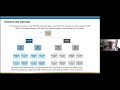
A total of 895 admissions were identified, with 406 included for the final analysis (Figure 1).
 |
Figure 1 Consort Diagram. |
Baseline characteristics are presented in Table 1. The median age of the overall cohort was 76.6 years (IQR 68.0–84.6), and 56.7% were male. HE exacerbations comprised 30.0% of admissions (n=122). Patients with HE were younger (74.7 vs 77.7 years, p=0.001) with fewer comorbidities (median [IQR] co-morbidities 1 [1–2] vs 2 [1–3], p=0.044). The proportion of current smokers was similar (30.3% vs 29.9%, p=0.873). Respiratory function tests within the 12 months prior to admission were available for 166/406 (40.9%). Patients with an HE exacerbation were more likely to have severe (GOLD 3, 14.6% vs 0.9%) and very severe (GOLD 4, 2.1% vs 0.0%) airflow obstruction (both p<0.001), but there was no difference in mean FEV1% predicted (53 [38.3–68.8] vs 55 [37–72], p=0.536). Patients with HE were less likely to be taking inhaled corticosteroids (ICS) pre-admission (58.1% vs 71.0%, p=0.017), but had similar domiciliary oxygen prescription rates.
 |
Table 1 Baseline Characteristics Stratified by Peripheral Eosinophils Less Than or Greater Than 2% |
Patients with HE exacerbations had a higher blood eosinophil count (0.31 vs 0.06 × 10⁹/L, p<0.001), lower total white cell count (8.45 vs 10.6 × 10⁹/L, p<0.001), lower neutrophil count (5.24 vs 7.77 × 10⁹/L, p<0.001), and lower CRP (10.4 vs 26.7 mg/L, p<0.001) (see Table 2). HE exacerbations were associated with a lower urea (5.8 vs 6.7 mmol/L, p=0.007) and higher eGFR (75 v 69 mL/min/1.73m2, p=0.007), but there were no differences in arterial or venous blood gas parameters on presentation. HE exacerbations were less likely to be associated with infection (29.5% vs 52.1%, p<0.001), and less likely to be treated with antibiotics (68.9% vs 82.4%, p=0.02) (see Table 2 and Table 3). Most patients received systemic corticosteroids (79.5% vs 76.1%, p=0.448), and there was no difference in the rate of oral or intravenous prescription between the groups.
 |
Table 2 Exacerbation Parameters Stratified by Peripheral Eosinophils Less Than or Greater Than 2% |
 |
Table 3 Exacerbation Management and Outcomes Stratified by Peripheral Eosinophils Less Than or Greater Than 2% |
HE exacerbations were less likely to require oxygen (35.2% vs 46.8%, p=0.036), but there was no difference in rates of ICU admission or need for ventilatory support. Patients with HE had a shorter hospital stay (3.56 vs 4.40 days, p=0.047).
Simple linear regression showed a significant relationship between length of stay and eosinophils ≥2% (unstandardised B value −0.844, p=0.047). However, only age and need for inpatient oxygen therapy remained significant when adjusted for the other variables in the final multivariable regression model.
Discussion
Patients with eosinophilic exacerbations of COPD reflect a different clinical phenotype to those without eosinophilia. Patients with an eosinophilic exacerbation are younger and less co-morbid than their non-eosinophilic counterparts. Eosinophilic exacerbations were less likely to be associated with infection, less likely to require supplementary oxygen, and have a shorter length of hospital stay.
Previous studies have demonstrated lower mortality in eosinophilic versus non-eosinophilic exacerbations of COPD.11,14,15,20,22–24 Eosinophilia in stable COPD has also been associated with improved long-term mortality.24 We did not find a difference in inpatient or 12-month mortality between our two cohorts. Mortality was four times higher in the low eosinophil group (1/122 [0.8%] in HE group and 9/284 [3.2%] in the LE group) but this fell short of statistical significance. This may reflect an inadequate sample size given the low overall mortality rate of only 2.46%, similar to some previous estimates28 but lower than other large studies.29 We also demonstrated an overall 12-month mortality of 6.4% post-hospitalizations for AECOPD, far less than previously published rates in excess of 20%.30,31 This is likely explained by lack of data linkage, as only patient’s whose deaths were recorded in our health service’s EMR were included.
Eosinophilic AECOPD required a shorter hospital admission compared to non-eosinophilic exacerbations, findings in keeping with previous reports.11–17,19–21 This may reflect corticosteroid responsiveness in eosinophilic AECOPD but could also reflect a younger and less co-morbid population in our study. Among patients with COPD, serum eosinophilia has been associated with both lower and higher FEV1.9,32 Eosinophilic inflammation is highly responsive to corticosteroid treatment; patients with elevated eosinophils in stable COPD demonstrate significant improvement in FEV1 and symptom burden when treated with systemic corticosteroids.33 Eosinophil count among stable COPD patients can predict exacerbations, with higher eosinophil count leading to an increased exacerbation rate, the risk of which is attenuated by ICS.10,34 Conversely, eosinopenia may predict harm from ICS treatment, with an increased risk of pneumonia seen in this population.35 ICS use appears to only have a small impact on the number of circulating peripheral eosinophils and largely does not affect eosinophilic/non-eosinophilic stratification.36 In our cohort, patients with eosinophilic exacerbations of COPD were less likely to be taking ICS prior to admission, suggesting a potential benefit of the addition of ICS in this population.
Part of the significant variation in reported clinical outcomes associated with eosinophilic exacerbations of COPD may be due in part to a lack of consensus on the definition of eosinophilia in this population. While the primary mechanistic biomarker of interest is airway eosinophilia, there is a reasonable correlation between sputum eosinophils and peripheral blood eosinophils.37,38 There is a strong concordance between two most commonly used definitions of eosinophilia; >2% of total white blood cells and absolute eosinophil count >150μL,32 although it has been demonstrated that the best predictor of sputum eosinophils >3%, is a serum eosinophil count >2%.8,25
Systemic corticosteroids are a mainstay of COPD exacerbation management and have been shown to reduce treatment failure, improve time to recovery and reduce severity of exacerbations.39 However, corticosteroid treatment is associated with a range of both short- and long-term complications including psychological side effects, myopathy, hyperglycaemia, weight gain, cardiovascular disease and osteoporosis. Previous studies have explored the utility of eosinophil count to guide systemic corticosteroid treatment in AECOPD, but this has yet to be translated into mainstream clinical practice.25,27,40 A double blinded-RCT among community managed AECOPDs demonstrated benefit from oral corticosteroids (OCS) for eosinophilic AECOPDs (treatment failure rate of 11% vs 66%, p<0.001). Among non-eosinophilic AECOPDs, no benefit from OCS was observed with similar treatment failure rate between the two cohorts (26% vs 20%, p=0.35).26 Sivapalan et al demonstrated that OCS use for inpatient AECOPDs only on days when peripheral eosinophils >300 cells/μL was non-inferior to standard care and resulted in less corticosteroid exposure, less resultant hyperglycaemia, and no difference in short-term hospital-free survival. There was, however, a trend towards greater re-admission with an exacerbation of COPD within 30 days in the eosinophil guided cohort.40 More recently, Ramakrishnan et al demonstrated in a community-based cohort that eosinophil guided corticosteroid therapy was non-inferior to standard care with regard to treatment failure (19% vs 32%, RR 0.60, 95% CI 0.33 to 1.04, p=0.070), and multiple quality of life measures. Notably, in a small subgroup analysis, they identified possible harm from OCS in non-eosinophilic AECOPDs. Fewer treatment failures were observed when low eosinophil exacerbations treated with placebo compared with prednisolone (16.7% vs 42%, relative risk 0.47, 95% CI 0.18 to 0.99, p=0.048).27
In keeping with our findings, eosinophilic exacerbations have been shown to be less often associated with infection than non-eosinophilic exacerbations,16 with an inverse relationship seen between eosinophil count and markers of inflammation including CRP.14,15,17,18,41 Kolsum et al also demonstrated an inverse relationship between the isolation of common respiratory tract infection organisms and serum eosinophils.42 While much of the research focus into the relationship between eosinophils and COPD has been on those with eosinophilia, eosinopenia during an exacerbation of COPD is associated with increased length of stay and higher inpatient mortality.11,14,43 This may reflect a lack of treatable eosinophilic inflammation, or indicate a concurrent infection, and perhaps should be used preferentially to guide treatment. The majority of our cohort (70%) had an eosinophil count <2% at presentation, suggesting a large proportion of patients who may benefit from further understanding in this area, with targeted prescription of corticosteroids and reduction in potential harm.
COPD is a heterogeneous disease, but current exacerbation management is largely generic. There have been limited recent advances in exacerbation management, and the typical approach still consists of systemic corticosteroids, inhaled bronchodilators, and in many cases antibiotics.44 While various exacerbation phenotypes have been defined, this has yet to result in significant changes in targeted clinical management or outcomes.8,21 An increasing appreciation of the underlying COPD phenotype, as well as the acute exacerbation phenotype, is needed in order to better guide management and minimise potential adverse effects of treatment. While peripheral eosinophilia is an imperfect biomarker – with no standardised definition of eosinophilia, concerns regarding individual variation and the impact of corticosteroid treatment on measurement of eosinophils – blood eosinophils remain easily accessible and readily available. Thus, eosinophils remain the most promising biomarker to guide the management of COPD exacerbations. Despite this, eosinophils do not routinely form part of the evaluation or definition of exacerbations of COPD.2 Given the lack of progress over many years in management of exacerbation of COPD, a more nuanced, personalised and targeted approach is needed. Targeted therapy against eosinophilic inflammation with monoclonal antibodies mepolizumab, benralizumab and dupilumab has demonstrated varying results in the reduction of exacerbation frequency, with further research required in this area.45–49
Strengths
We present the largest cohort of eosinophilic exacerbations of COPD reported in an Australian population, with clinically relevant, real-world data collected across three metropolitan hospitals.
Limitations
This study had several limitations, largely driven by its single health service and retrospective nature. Exacerbations were defined as eosinophilic or non-eosinophilic based on an individual blood test taken at the time of admission, with an acknowledgement that eosinophils may fluctuate in some patients. The diagnosis of COPD was made based on EMR review and coding diagnoses. Patients were excluded if their underlying disease was not COPD, however not all patients had spirometry confirmed diagnosis. However, this is reflective of the real-world clinical experience in which many patients have not had spirometry prior to index admission, and thus we are confident our findings can be applied broadly. While all efforts were made to ensure as complete a dataset as possible, missing data-points were present in some categories.
Conclusion
Eosinophilic exacerbations of COPD are common and are associated with phenotypical differences that may be used to prognosticate and help guide management. Eosinophil count at presentation can be used to stratify risk of important clinical outcomes, including length of stay, and may soon be utilised to guide treatment.
Author Contributions
All authors made a significant contribution to the work reported, whether that is in the conception, study design, execution, acquisition of data, analysis and interpretation, or in all these areas; took part in drafting, revising or critically reviewing the article; gave final approval of the version to be published; have agreed on the journal to which the article has been submitted; and agree to be accountable for all aspects of the work.
Funding
There is no funding to report.
Disclosure
The authors report no conflicts of interest in this work.
References
1. GOLD. Global Strategy for the Diagnosis, Management, and Prevention of Chronic Obstructive Pulmonary Disease; 2023.
2. Celli BR, Fabbri LM, Aaron SD, et al. An updated definition and severity classification of chronic obstructive pulmonary disease exacerbations: the Rome proposal. Am J Resp Crit Care. 2021;204(11):1251–1258.
3. NACAP. Outcomes of patients included in the 2017/2018 COPD clinical audit. 2020.
4. Barnes PJ. Inflammatory mechanisms in patients with chronic obstructive pulmonary disease. J Allergy Clin Immun. 2016;138(1):16–27. doi:10.1016/j.jaci.2016.05.011
5. Barnes PJ. Cellular and molecular mechanisms of chronic obstructive pulmonary disease. Clin Chest Med. 2014;35(1):71–86. doi:10.1016/j.ccm.2013.10.004
6. Papi A, Luppi F, Franco F, Fabbri LM. Pathophysiology of exacerbations of chronic obstructive pulmonary disease. Proc Am Thorac Soc. 2006;3(3):245–251. doi:10.1513/pats.200512-125SF
7. Tashkin DP, Wechsler ME. Role of eosinophils in airway inflammation of chronic obstructive pulmonary disease. Int J Chronic Obstr. 2018;13:335–349.
8. Bafadhel M, McKenna S, Terry S, et al. Acute exacerbations of chronic obstructive pulmonary disease. Am J Resp Crit Care. 2012;184(6):662–671. doi:10.1164/rccm.201104-0597OC
9. Vedel-Krogh S, Nielsen SF, Lange P, Vestbo J, Nordestgaard BG. Blood eosinophils and exacerbations in chronic obstructive pulmonary disease. The Copenhagen general population study. Am J Resp Crit Care. 2016;193(9):965–974. doi:10.1164/rccm.201509-1869OC
10. Pascoe S, Locantore N, Dransfield MT, Barnes NC, Pavord ID. Blood eosinophil counts, exacerbations, and response to the addition of inhaled fluticasone furoate to vilanterol in patients with chronic obstructive pulmonary disease: a secondary analysis of data from two parallel randomised controlled trials. Lancet Respir Med. 2015;3(6):435–442. doi:10.1016/S2213-2600(15)00106-X
11. Holland M, Alkhalil M, Chandromouli S, Janjua A, Babores M. Eosinopenia as a marker of mortality and length of stay in patients admitted with exacerbations of chronic obstructive pulmonary disease. Respirology. 2010;15(1):165–167. doi:10.1111/j.1440-1843.2009.01651.x
12. Duman D, Aksoy E, Karakurt Z, et al. The utility of inflammatory markers to predict readmissions and mortality in COPD cases with or without eosinophilia. Int J Chronic Obstr. 2015;10(1):2469–2478.
13. Bafadhel M, Greening NJ, Harvey-Dunstan TC, et al. blood eosinophils and outcomes in severe hospitalized exacerbations of COPD. Chest. 2016;150(2):320–328. doi:10.1016/j.chest.2016.01.026
14. MacDonald MI, Osadnik CR, Bulfin L, et al. Low and high blood eosinophil counts as biomarkers in hospitalized acute exacerbations of COPD. Chest. 2019;156(1):92–100. doi:10.1016/j.chest.2019.02.406
15. Jabarkhil A, Moberg M, Janner J, et al. Elevated blood eosinophils in acute COPD exacerbations: better short- and long-term prognosis. Eur Clin Respir J. 2020;7(1):1757274.
16. Wu CW, Lan CC, Hsieh PC, Tzeng IS, Wu YK. Role of peripheral eosinophilia in acute exacerbation of chronic obstructive pulmonary disease. World J Clin Cases. 2020;8(13):2727–2737. doi:10.12998/wjcc.v8.i13.2727
17. Cui Y, Zhan Z, Zeng Z, et al. Blood eosinophils and clinical outcomes in patients with acute exacerbation of chronic obstructive pulmonary disease: a propensity score matching analysis of real-world data in China. Front Med. 2021;8:653777. doi:10.3389/fmed.2021.653777
18. Greulich T, Tüffers J, Mager S, et al. High eosinophil blood counts are associated with a shorter length of hospital stay in exacerbated COPD patients – a retrospective analysis. Respir Res. 2020;21(1):106. doi:10.1186/s12931-020-01365-5
19. Ho J, He W, Chan MTV, et al. Eosinophilia and clinical outcome of chronic obstructive pulmonary disease: a meta-analysis. Sci Rep-Uk. 2017;7(1):13451. doi:10.1038/s41598-017-13745-x
20. You Y, Chao SG. Blood eosinophils and clinical outcome of acute exacerbations of chronic obstructive pulmonary disease: a systematic review and meta-analysis. Respiration. 2021;100(3):228–237. doi:10.1159/000510516
21. MacDonald MI, Osadnik CR, Bulfin L, et al. MULTI-PHACET: multidimensional clinical phenotyping of hospitalised acute COPD exacerbations. Erj Open Res. 2021;7(3):00198–2021. doi:10.1183/23120541.00198-2021
22. Casanova C, Bartolome RC, de T Juan P, et al. Prevalence of persistent blood eosinophilia: relation to outcomes in patients with COPD. Eur Respir J. 2017;50(5):1701162. doi:10.1183/13993003.01162-2017
23. Zhang Y, Liang LR, Zhang S, et al. Blood eosinophilia and its stability in hospitalized COPD exacerbations are associated with lower risk of all-cause mortality. Int J Chronic Obstr. 2020;15:1123–1134.
24. Prudente R, Ferrari R, Mesquita CB, et al. Peripheral blood eosinophils and nine years mortality in COPD patients. Int J Chronic Obstr. 2021;16:979–985.
25. Bafadhel M, McKenna S, Terry S, et al. Blood eosinophils to direct corticosteroid treatment of exacerbations of chronic obstructive pulmonary disease. Am J Resp Crit Care. 2012;186(1):48–55. doi:10.1164/rccm.201108-1553OC
26. Bafadhel M, Davies L, Calverley PMA, Aaron SD, Brightling CE, Pavord ID. Blood eosinophil guided prednisolone therapy for exacerbations of COPD: a further analysis. Eur Respir J. 2014;44(3):789–791. doi:10.1183/09031936.00062614
27. Ramakrishnan S, Jeffers H, Langford-Wiley B, et al. Blood eosinophil-guided oral prednisolone for COPD exacerbations in primary care in the UK (STARR2): a non-inferiority, multicentre, double-blind, placebo-controlled, randomised controlled trial. Lancet Respir Med. 2024;12(1):67–77. doi:10.1016/S2213-2600(23)00298-9
28. Patil SP, Krishnan JA, Lechtzin N, Diette GB. In-hospital mortality following acute exacerbations of chronic obstructive pulmonary disease. Arch Intern Med. 2003;163(10):1180–1186. doi:10.1001/archinte.163.10.1180
29. Hartl S, Lopez-Campos JL, Pozo-Rodriguez F, et al. Risk of death and readmission of hospital-admitted COPD exacerbations: European COPD Audit. Eur Respir J. 2016;47(1):113–121. doi:10.1183/13993003.01391-2014
30. Soltani A, Reid D, Wills K, Walters EH. Outcomes of acute exacerbations of COPD. Intern Med J. 2015;45(9):925–933. doi:10.1111/imj.12816
31. Lindenauer PK, Dharmarajan K, Qin L, Lin Z, Gershon AS, Krumholz HM. Risk trajectories of readmission and death in the first year after hospitalization for chronic obstructive pulmonary disease. Am J Respir Crit Care Med. 2018;197(8):1009–1017. doi:10.1164/rccm.201709-1852OC
32. Singh D, Kolsum U, Brightling CE, et al. Eosinophilic inflammation in COPD: prevalence and clinical characteristics. Eur Respir J. 2014;44(6):1697–1700. doi:10.1183/09031936.00162414
33. Brightling CE, Monteiro W, Ward R, et al. Sputum eosinophilia and short-term response to prednisolone in chronic obstructive pulmonary disease: a randomised controlled trial. Lancet. 2000;356(9240):1480–1485. doi:10.1016/S0140-6736(00)02872-5
34. Bafadhel M, Peterson S, Blas MAD, et al. Predictors of exacerbation risk and response to budesonide in patients with chronic obstructive pulmonary disease: a post-hoc analysis of three randomised trials. Lancet Respir Med. 2018;6(2):117–126. doi:10.1016/S2213-2600(18)30006-7
35. Pavord ID, Lettis S, Anzueto A, Barnes N. Blood eosinophil count and pneumonia risk in patients with chronic obstructive pulmonary disease: a patient-level meta-analysis. Lancet Respir Med. 2016;4(9):731–741. doi:10.1016/S2213-2600(16)30148-5
36. Kreindler JL, Watkins ML, Lettis S, Tal-Singer R, Locantore N. Effect of inhaled corticosteroids on blood eosinophil count in steroid-naïve patients with COPD. BMJ Open Respir Res. 2016;3(1):e000151. doi:10.1136/bmjresp-2016-000151
37. Negewo NA, McDonald VM, Baines KJ, et al. Peripheral blood eosinophils: a surrogate marker for airway eosinophilia in stable COPD. Int J Chronic Obstr. 2016;11:1495–1504.
38. Hastie AT, Martinez FJ, Curtis JL, et al. Association of sputum and blood eosinophil concentrations with clinical measures of COPD severity: an analysis of the SPIROMICS cohort. Lancet Respir Med. 2017;5(12):956–967. doi:10.1016/S2213-2600(17)30432-0
39. Walters JA, Tan DJ, White CJ, Gibson PG, Wood‐Baker R, Walters EH. Systemic corticosteroids for acute exacerbations of chronic obstructive pulmonary disease. Cochrane db Syst Rev. 2014;2014(9):CD001288.
40. Sivapalan P, Lapperre TS, Janner J, et al. Eosinophil-guided corticosteroid therapy in patients admitted to hospital with COPD exacerbation (CORTICO-COP): a multicentre, randomised, controlled, open-label, non-inferiority trial. Lancet Respir Med. 2019;7(8):699–709.
41. Lv MY, Qiang LX, Li ZH, Jin SD. The lower the eosinophils, the stronger the inflammatory response? The relationship of different levels of eosinophils with the degree of inflammation in acute exacerbation COPD (AECOPD). J Thorac Dis. 2021;13(1):232–243. doi:10.21037/jtd-20-2178
42. Kolsum U, Donaldson GC, Singh R, et al. Blood and sputum eosinophils in COPD; relationship with bacterial load. Respir Res. 2017;18(1):88. doi:10.1186/s12931-017-0570-5
43. Steer J, Gibson J, Bourke SC. The DECAF Score: predicting hospital mortality in exacerbations of chronic obstructive pulmonary disease. Thorax. 2012;67(11):970. doi:10.1136/thoraxjnl-2012-202103
44. Jeyachandran V, Hurst JR. Advances in chronic obstructive pulmonary disease: management of exacerbations. Br J Hosp Med. 2022;83(7):1–7.
45. Pavord ID, Chanez P, Criner GJ, et al. Mepolizumab for eosinophilic chronic obstructive pulmonary disease. New Engl J Med. 2017;377(17):1613–1629.
46. Pavord I, Chapman K, Bafadhel M, et al. Mepolizumab for eosinophil-associated COPD: analysis of METREX and METREO. Int J Chronic Obstr. 2021;16:1755–1770.
47. Criner GJ, Celli BR, Brightling CE, et al. Benralizumab for the prevention of COPD exacerbations. N Engl J Med. 2019;381(11):1023–1034. doi:10.1056/NEJMoa1905248
48. Bhatt SP, Rabe KF, Hanania NA, et al. Dupilumab for COPD with type 2 inflammation indicated by eosinophil counts. N Engl J Med. 2023;389(3):205–214. doi:10.1056/NEJMoa2303951
49. Agusti A. Biologics for COPD — finally here. N Engl J Med. 2023;389(3):274–275. doi:10.1056/NEJMe2305752
 © 2025 The Author(s). This work is published and licensed by Dove Medical Press Limited. The
full terms of this license are available at https://www.dovepress.com/terms.php
and incorporate the Creative Commons Attribution
- Non Commercial (unported, 4.0) License.
By accessing the work you hereby accept the Terms. Non-commercial uses of the work are permitted
without any further permission from Dove Medical Press Limited, provided the work is properly
attributed. For permission for commercial use of this work, please see paragraphs 4.2 and 5 of our Terms.
© 2025 The Author(s). This work is published and licensed by Dove Medical Press Limited. The
full terms of this license are available at https://www.dovepress.com/terms.php
and incorporate the Creative Commons Attribution
- Non Commercial (unported, 4.0) License.
By accessing the work you hereby accept the Terms. Non-commercial uses of the work are permitted
without any further permission from Dove Medical Press Limited, provided the work is properly
attributed. For permission for commercial use of this work, please see paragraphs 4.2 and 5 of our Terms.
Recommended articles
Pulmonologists’ Opinion on the Use of Inhaled Corticosteroids in Chronic Obstructive Pulmonary Disease Patients in Spain: A Cross-Sectional Survey
Miravitlles M, González-Torralba F, Represas-Represas C, Pomares X, Márquez-Martín E, González C, Amado C, Forné C, Alonso S, Alcázar B, Barrecheguren M, Jurado Mirete JM, Naval E
International Journal of Chronic Obstructive Pulmonary Disease 2022, 17:1577-1587
Published Date: 12 July 2022
The Effect of Maintenance Treatment with Erdosteine on Exacerbation Treatment and Health Status in Patients with COPD: A Post-Hoc Analysis of the RESTORE Dataset
Calverley PMA, Papi A, Page C, Rogliani P, Dal Negro RW, Cazzola M, Cicero AF, Wedzicha JA
International Journal of Chronic Obstructive Pulmonary Disease 2022, 17:1909-1920
Published Date: 22 August 2022
In-Hospital Outcomes of Heart Failure Patients with Valvular Heart Disease: Insights from Real-World Claims Data

Izumi C, Matsuyama R, Yamabe K, Iwasaki K, Takeshima T, Murphy SME, Teng L, Igarashi A
ClinicoEconomics and Outcomes Research 2023, 15:349-360
Published Date: 18 May 2023
Characteristics and Outcomes of People With COPD Who Experience Exacerbations While on Inhaled Triple Therapy: Results of the SIRIUS I Cohort Study in the US (2015–2019)
Nordon C, Carstens D, Fagerås M, Müllerová H, Veeranki PS, Alves JA, Germack HD, Barnes TL, McCormack MC
International Journal of Chronic Obstructive Pulmonary Disease 2025, 20:1851-1864
Published Date: 11 June 2025
Withholding of Life-Sustaining Treatment and Mortality in ICU Patients with Severe Acute COPD Exacerbations: A Retrospective French Cohort
Puechoultres P, Jamme M, Abi-Abdallah G, Diop S, Legriel S, Ferré A
International Journal of Chronic Obstructive Pulmonary Disease 2025, 20:1995-2009
Published Date: 18 June 2025