")
Back to Journals » International Journal of Nanomedicine » Volume 20
How Advanced are Conductive Nanocomposite Hydrogels for Repairing and Monitoring Myocardial Infarction?
Authors Liu Y, Liu D, Xue Y, Sun H, Zhan X, Sun L, Kang K
Received 29 October 2024
Accepted for publication 17 May 2025
Published 28 May 2025 Volume 2025:20 Pages 6777—6812
DOI https://doi.org/10.2147/IJN.S503445
Checked for plagiarism Yes
Review by Single anonymous peer review
Peer reviewer comments 5
Editor who approved publication: Professor Farooq A. Shiekh
Yang Liu,1,* Donghui Liu,1,* Yanghong Xue,1,* Haobo Sun,1 Xu Zhan,1 Lihua Sun,2 Kai Kang1,3,4
1Department of Cardiovascular Surgery, The First Hospital of Harbin Medical University, Harbin, 150000, People’s Republic of China; 2Department of Cardiovascular Surgery, The First Affiliated Hospital of Harbin Medical University, and Department of Pharmacology, College of Pharmacy, Harbin Medical University, Harbin, 150081, People’s Republic of China; 3Key Laboratory of NHC Cell Transplantation, The First Hospital of Harbin Medical University, Harbin, 150000, People’s Republic of China; 4Key Laboratory of Liver and Spleen Surgery, Ministry of Education, The First Affiliated Hospital of Harbin Medical University, Harbin, 150000, People’s Republic of China
*These authors contributed equally to this work
Correspondence: Kai Kang, Department of Cardiovascular Surgery, The First Affiliated Hospital of Harbin Medical University, Harbin, 150000, People’s Republic of China, Email [email protected] Lihua Sun, Department of Cardiovascular Surgery, The First Affiliated Hospital of Harbin Medical University, and Department of Pharmacology, School of Pharmacy, Harbin Medical University, Harbin, 150081, People’s Republic of China, Email [email protected]
Abstract: Myocardial infarction (MI) remains the leading cause of death worldwide. Cardiomyocytes, being terminally differentiated cells, have limited regenerative capacity. Following an MI, myocyte necrosis and ventricular dilation can lead to heart failure. While current treatments for heart disease—such as pharmaceuticals, coronary interventions, coronary artery bypass grafting, cellular therapy, and heart transplantation—offer some relief, their effectiveness is limited, particularly in patients with severe myocardial damage. Recent advancements in cardiac tissue engineering have introduced a range of materials aimed at repairing the heart, with conductive hydrogels emerging as a promising approach. These materials, which include metallic nanomaterials, conductive polymers, carbon-based conductive materials, and other specialized types of conductive substances, exhibit excellent electrical conductivity, tunable mechanical properties, and biomimetic features. As a result, they are increasingly being considered for myocardial repair. This review explores the application of conductive hydrogels in treating myocardial infarction, highlighting recent research in various types of conductive hydrogels. These are categorized by their nanomaterial composition, including hydrogels designed for cell culture scaffolds, patch-type hydrogels, and injectable conductive hydrogels. Additionally, electrophysiological monitoring during MI is gaining importance in understanding disease progression and prognosis. In recent years, conductive hydrogels have rapidly evolved to serve as tools for real-time monitoring of signal changes, while their electroresponsive properties open new possibilities for targeted drug delivery in infarct therapy.
Keywords: myocardial infarction, conductivity, nanomaterials, hydrogel, cardiac tissue engineering
Introduction
Cardiovascular disease (CVD) represents a significant health issue that impacts both the physical and mental well-being of individuals, with myocardial infarction (MI) being one of the leading causes of death worldwide. MI is characterized by severe damage to cardiomyocytes due to coronary artery obstruction, which results in ischemic necrosis of cardiac tissue and triggers a complex inflammatory response.1 The pathophysiology of myocardial infarction involves structural changes in the heart tissue, adverse myocardial remodeling, and subsequent cardiac dysfunction, which can eventually progress to refractory heart failure. Current treatments, including pharmacological therapies, cellular therapies, and percutaneous coronary interventions, do not fully restore the mechanical and electrical properties of the damaged cardiac tissue.2 In contrast, while circulatory assist devices and transplantation offer more effective solutions, they are constrained by factors such as a limited supply of donors, immune rejection, and concerns about durability.3 Therefore, there is a pressing need for new therapies that can significantly restore cardiac function and potentially regenerate damaged cardiac tissue.4 A critical aspect of this process is restoring the mechanical and electrical conductivity of myocardial tissue. In experimental stem cell therapies for myocardial infarction, the aim is to enhance therapeutic outcomes by differentiating stem cells into cardiomyocytes at the injury site. However, the low retention of stem cells remains a significant challenge. Although combining stem cells with injectable hydrogels has shown promising synergistic effects, the issue of poor cell retention has yet to be fully resolved.5 As a result, some researchers have shifted their focus to tissue-engineered materials, hoping to directly restore the mechanical and electrical properties of damaged cardiac regions, effectively “filling” the diseased area and supporting cardiac function.
Synchronized ventricular contraction is crucial for normal cardiac function. After myocardial infarction, scar tissue interrupts the electrical conductivity between surviving cardiomyocytes, leading to electro-mechanical separation that disrupts synchronized ventricular contraction and, consequently, overall cardiac function. Tissue-engineered materials have the potential to functionally replace the damaged myocardium, acting as an “electrical bridge” while providing mechanical support to the affected areas. Some of these materials can also deliver drugs, improving the local tissue environment, promoting cardiomyocyte survival, reducing further cell loss, and significantly enhancing recovery outcomes.6 Among various materials, hydrogels are widely used in tissue engineering due to their unique properties. These materials can transition from a sol-gel state into a polymer with a three-dimensional network structure. With a high water content similar to that of natural tissues, hydrogels effectively encapsulate cells or drugs, offering structural and functional support. Additionally, hydrogels are biodegradable, breaking down gradually without causing significant damage to surrounding healthy tissue, making them particularly well-suited for biomedical applications.7 Conductive materials come in several forms, including carbon-based nanomaterials, gold nanomaterials, conductive polymers, ionic liquids, and silicon nanowires (SiNWs). These materials possess both conductive and mechanical properties, making them ideal for applications that require electrical connectivity and structural support restoration in myocardial tissues.8
Nanomaterials combined with biomimetic structures could provide an excellent three-dimensional framework for cell and drug delivery. Adding conductive nanoparticles to hydrogels can further enhance their electrical conductivity, offering great potential for repairing infarcted heart muscle. This combination could improve electrical signal transmission and promote repair and regeneration of damaged heart tissue.9 In this review, we begin with a brief overview of the fundamental principles of cardiac electrophysiology and then summarize recent advances in cardiac tissue engineering. Our focus is on hydrogel patches and injectable hydrogels used in cardiac tissue regeneration and repair. Additionally, we explore research on conductive hydrogel scaffolds in cell culture applications and conclude by discussing the future potential of conductive hydrogels in the development of tissue engineering.
How Cardiac Electrophysiological Microenvironment and Conductivity Works?
The major cell types in the heart include cardiomyocytes, endothelial cells, fibroblasts, and immune cells. Together with the extracellular matrix, these cells provide mechanical support and are essential for maintaining cardiac function. Cardiomyocytes constitute approximately 75% of the total cell population in the heart and are connected to the endocardium.10 Cardiomyocytes, fibroblasts, endothelial cells, and vascular smooth muscle cells work together as structural support cells, playing a crucial role in cardiac pathological remodeling. The cardiac extracellular matrix (ECM), though devoid of cellular components, provides vital structural support to these cells. It contains key factors that protect cardiac cells and is integral to intercellular signaling, helping to maintain the overall structure and function of the heart.11 Coordinated contraction is crucial for proper heart function, and pacemaker and conduction cells form the heart’s conduction system. A key aspect of cardiomyocyte interaction is the gap junction, located at the intercalated disc, which enables electrical impulses to propagate throughout the heart. The most important component of this structure is connexin-43 (CX-43), a protein that facilitates ion exchange and ensures the synchronized transmission of action potentials. Aberrant expression of CX-43 disrupts electrical conduction, leading to cardiac arrhythmias, which pose a significant risk of death.12 The heart functions in a synchronized, rhythmic manner, making it crucial to understand its conductivity. Let us explore how the heart’s electrical system maintains this coordinated activity.
The sinus node (SN) is the origin of the heart’s electrical activity. Electrical impulses travel through the internodal pathway to the atrioventricular (AV) node, where a slight delay occurs, allowing the atria to contract before the ventricles. From the AV node, the signal moves through the His bundle to the Purkinje network, spreading throughout the ventricular myocytes and generating action potentials. Throughout this process, cardiomyocytes are continuously exposed to periodic electrical activity, and their metabolism is adapted to this rhythm, ensuring proper function and normal contraction of the heart.13 This contraction ultimately forces blood into the aorta and pulmonary arteries, maintaining circulation throughout the body and lungs.14 Natural myocardial tissue exhibits an electrical conductivity of approximately 10⁻4S/cm. In cases of ischaemic heart disease, particularly myocardial infarction (MI), fibroblasts and the extracellular matrix (ECM) undergo compensatory proliferation and hyperplasia. This results in excessive fibrin deposition and fibrotic scarring, which disrupts normal electrical conduction. Such alterations compromise both systolic and diastolic functions, potentially causing severe cardiac dysfunction and life-threatening arrhythmias.15
In summary, electrical conduction in the heart is crucial not only for maintaining its rhythmic contractions but also for ensuring the proper metabolic activity of cardiomyocytes.
Prospects for the Use of Conductive Hydrogels in Ischaemic Cardiomyopathy
Given the pathophysiological changes associated with myocardial ischaemia, conductive hydrogels offer a highly promising therapeutic strategy to overcome the limitations of traditional treatments and facilitate in vivo repair of ischaemic cardiac tissue. Different hydrogel designs possess unique functions, typically categorized into patch and injectable forms, combining electrical conductivity with physical support. These properties help slow ventricular remodelling and enable microenvironmental responsiveness for controlled drug release. The use of hydrogels in ischaemic cardiac tissue enables localized, targeted delivery of cells and growth factors, promoting tissue repair and enhancing therapeutic outcomes.16 The hydrogel provides a customizable three-dimensional microenvironment for the encapsulated cells, shielding them from mechanical stress and abrasion caused by myocardial contractions. This protective environment enhances cell retention at the target site, improves their effectiveness, and ultimately promotes more efficient cardiac tissue repair.17 He et al conducted a pioneering randomised double-blind clinical trial on cell-injected ischaemic cardiac repair. They developed an injectable hydrogel scaffold containing bovine collagen components and integrated allogeneic human umbilical cord mesenchymal stem cells (hUC-MSCs) into the scaffold. After injecting this hydrogel into patients with chronic ischaemic heart disease following coronary artery bypass grafting, both the function and structure of the damaged myocardium improved, and the treatment was found to be biologically safe. While the trial was limited by a small sample size, it lays the foundation for larger clinical studies in the future.18 Another approach in hydrogel scaffold development focuses on creating fibre networks with exceptional elastic properties. These scaffolds are designed not only to provide strong mechanical support but also to act as carriers for cells and biological factors. The aim is to promote vascular regeneration, support cardiac tissue generation, and reverse ventricular remodelling, ultimately enhancing cardiac function and facilitating repair following injury.19 Cardiac hydrogel patches are currently undergoing preclinical trials. Chachques et al developed three-dimensional bioabsorbable polycaprolactone scaffolds filled with peptide hydrogels and combined them with adipose stem cells to create cardiac patches. These patches were implanted into a sheep model of heart disease, showing promising therapeutic results. While the surgically induced heart attack model used in this study does not fully replicate a clinical disease episode, it represents a significant step toward the potential clinical application of wrapped-supported biological precursors.20
Arslan et al developed a fully vascularizable and perfusable human cardiac microtubule (MT) system in vitro using human induced pluripotent stem cells (hiPSCs). In this system, vascular cells were co-cultured with MTs in fibrin hydrogels, creating a hybrid vascular network. This innovative system holds significant potential for drug screening and disease modeling applications, offering a promising platform for studying cardiovascular diseases.21
As advances in detection materials progress, flexible electronic components are increasingly integrated into tissue engineering. Studies have demonstrated improvements in myocardial infarct size following treatment. However, a key challenge remains: the difficulty of continuously and directly monitoring tissue function after the implantation of tissue-engineered scaffolds. To tackle this issue, hybrid integrated systems have been developed to sense pH, mechanical motion, and relevant biomarkers. These systems facilitate remote monitoring of tissue function and enable precise control over drug release, ultimately improving outcomes after implantation.22
Overall, tissue engineering technologies are advancing rapidly, with notable progress in areas such as stem cell development, material patches, injectable hydrogels, flexible electronic components, and 3D and 4D bioprinting. The aim is to address the limitations of biomaterials, including immunoreactivity, histocompatibility, and degradation, while ensuring the stability of stem cells. These advancements offer significant promise for enhancing the success rate of clinical applications and improving patient outcomes. (Figure 1)
 |
Figure 1 Schematic diagram of conductive composite hydrogel for the treatment of myocardial infarction. (Created in BioRender. Liu, (Y) (2025) https://BioRender.com/r42j314). Notes: Nanoconductive composite hydrogels hold significant potential for repairing and monitoring myocardial infarction. A variety of nanoparticles can be doped into composite hydrogels, which can be classified into three types: cell scaffolds, patches, and injectable hydrogels. These hydrogels offer a range of functions, including improving electrical conduction, reducing inflammation, promoting angiogenesis, enhancing regeneration, preventing fibrosis, and providing physical support. These multifunctional properties make nanoconductive composite hydrogels a promising approach for heart repair and the restoration of cardiac function after myocardial infarction. |
Conductive Hydrogel for Repairing Heart-Damaged Tissue
Conductive hydrogels are regarded as one of the most promising options for cardiac regeneration and functional recovery. The spatial structure, electrical conductivity, cross-linking, degradation, and drug release properties of these hydrogels are influenced by their material composition, allowing their functionality to be customized to the specific microenvironment of the affected area. Whether used independently or in combination with therapeutic drugs, hydrogels hold significant potential for advancing cardiac repair.23 Effective cardiac tissue reconstruction requires the establishment of suitable cellular pathways to support the extracellular matrix of the myocardium, electrically stimulate the cells, form contractile bands, and reconstruct the vascular network. One of the key features of cardiomyocytes is their electrical conductivity, making the restoration of the conductive network between these cells crucial for successful myocardial repair. Conductive hydrogels play a critical role in this process by enhancing electrical coupling, promoting the formation of myocardial isoelectric syncytia, and supporting the maintenance of spontaneous cardiac contractility.24,25 Electrical stimulation activates multiple cellular signaling pathways, enhances the intracellular microenvironment, and regulates cell migration, proliferation, and differentiation. When integrated with tissue-engineered scaffolds, this strategy merges biocompatibility with electrical conductivity, representing a significant advancement in the field of regenerative medicine.26 Conductive hydrogels possess excellent deformation and strain capacity, making them ideal for the creation of flexible, stretchable electrodes and sensors. This capability offers promising prospects for expanding the use of hydrogels in real-time health monitoring and flexible electronic circuits.27 Next, we will explore various types of conductive nanoparticle hydrogels for infarct repair, as well as hydrogels with flexible electronic capabilities for real-time monitoring during myocardial infarction.(Figure 2)
 |
Figure 2 Primarily presents the chemical structures of several nanomaterials and their associated application scenarios. (Created in BioRender. Liu, (Y) (2025) https://BioRender.com/weowssh). Notes: The chemical structure of a conductive material governs its functional properties. Through ongoing exploration of its structure-function relationship, its potential applications in various fields can be enhanced. |
Metal Nanoparticles
Gold Nanoparticle
Gold nanoparticles (AuNPs) are regarded as the most stable nanoparticles in nature. Their optical properties are influenced by surface plasmon resonance (SPR), which can be tuned by altering the shape and structure of the nanoparticles, such as by forming nanorods, nanocages, and other configurations. These structural modifications impact the SPR, leading to variations in the optoelectronic properties of gold nanoparticles. Furthermore, gold nanoparticles are biocompatible, highly conductive, and photoresponsive, making them ideal candidates for biomedical and electronic applications.28 The customizable size and shape of AuNPs are crucial for tissue engineering applications. Scaffold materials synthesized with AuNPs can be easily adjusted for compliance, conductivity, and surface activity. Increasing the concentration of gold nanoparticles improves conductivity and reduces impedance across the test range (~1 hz to 1 MHz). Additionally, AuNP-doped scaffolds enhance the ability of stem cells to differentiate into various cell types, making them highly effective for regenerative medicine and tissue engineering applications.29 Studies have shown that scaffolds enriched with AuNPs enhance calcium ion propagation and increase the expression of the gap junction protein Cx43 in cardiac tissue. This improvement in intercellular coordination and contractile synchronization highlights the potential of AuNP-enriched scaffolds to promote more efficient cardiac tissue repair and function.30
Navaei et al developed hybrid hydrogels by doping GelMA with gold nanorods (GNR) to enhance mechanical stiffness and electrical conductivity. Cardiomyocytes cultured on these GelMA-GNR hydrogels demonstrated excellent cell retention, activity, functional integrity, and rhythmic pacing. Additionally, the levels of cellular matrix integrin β-1 were increased, while cardiac-specific markers such as troponin I, α-actinin, and connexin-43 (Cx43) showed enhanced tissue connectivity, refinement of conduction structures, and improved electrical conductivity. These findings suggest that this hybrid hydrogel can serve as an effective matrix for cardiac tissue engineering.31
There is increasing interest in cell therapies to repair damaged heart tissue, particularly through the transplantation of human induced pluripotent stem cells (hiPSCs). However, this approach faces challenges such as low survival rates and the risk of induced arrhythmias. To address these concerns, Roshanbinfar et al developed a biohybrid hydrogel aimed at enhancing the contractility of engineered cardiac tissue while releasing pharmacological therapeutic factors. The hydrogel, composed of branched polyethyleneimine (bPEI), gold nanoparticles (AuNPs), and collagen, offers improved mechanical properties and electrical conductivity, with conductivity measurements ranging from 0.048 to 0.068 S/cm. The bPEI coatings on the AuNPs are positively charged, enabling the loading of various negatively charged drugs, peptides, and oligonucleotides. This novel hydrogel enhances electrical conductivity and drug delivery capabilities, potentially circumventing immune rejection and tumorigenic risks associated with cell transplantation. It presents a promising new approach to cell-free systemic therapies for heart disease treatment.32
In a study by Sesena-Rubfiaro et al, conducting polymers and nanomaterials were incorporated into the extracellular matrix to enhance both mechanical strength and electrical conductivity between cardiomyocytes. They embedded gold nanorods (GNRs) into fibronectin hydrogels—one of the primary components of the extracellular matrix—to create a GNR-fibronectin matrix, which was then used to construct three-dimensional human engineered cardiac tissue (hECT) structures. The GNR-hECTs demonstrated exceptional support for long-term cell survival. In a 9-month culture study, the hydrogel cavities within the hECTs still housed viable cells with preserved cellular structure, active contractile function, and sustained electrical conductivity. This scaffold significantly advances the clinical potential of 3D cell culture platforms for cardiac cells and lays a strong foundation for the next generation of hECT development.33
Pournemati et al incorporated gold nanoparticles (AuNPs) into injectable hydrogels composed of alginate (Alg) and gelatin (Gel), achieving nanoparticle doping without the use of stabilisers and thereby avoiding their associated side effects. Alginate hydrogels are notable as the first injectable decellularised biomaterials to enter clinical trials for myocardial infarction (MI) treatment, offering a non-immunogenic and non-toxic therapeutic option. The resulting nanocomposite hydrogel features a three-dimensional porous network with excellent electrical conductivity (2.04 × 10⁻4 S/cm) and a pore size of approximately 40 μm, providing an ideal environment for cell growth and tissue regeneration. This structure significantly enhances cell adhesion and differentiation, leading to improved therapeutic outcomes following MI, including increased ejection fraction, reduced infarct size, and decreased fibrotic tissue. In summary, this injectable conductive scaffold represents a promising, safe, and efficient strategy for targeted cell delivery in infarcted cardiac tissue.34
Providing sufficient oxygen to the ischaemic region of myocardial infarction is a critical therapeutic strategy. Xu et al developed an injectable oxygen-generating biomacromolecular hydrogel by combining catalase (CAT) with alginate (Alg), fibronectin (Fib), and exosomes (Exo) derived from mesenchymal stem cells (MSCs). This composite hydrogel also incorporated gold nanoparticles (AuNPs), with a conductivity of 8.51 × 10⁻4 S/cm, which closely matches that of myocardial tissue. The addition of AuNPs and CAT accelerated gelation and improved the mechanical properties of the hydrogel. Under hypoxic conditions, the oxygen-releasing hydrogels provided oxygen for nearly 5 days. After 7 days of in vitro cell culture, the hydrogels produced paracrine factors similar to those secreted by rat neonatal cardiomyocytes (RNCs), rat cardiac fibroblasts (RCFs), and human umbilical vein endothelial cells (HUVECs), which promoted angiogenesis. However, further studies and validation in large animal models are necessary before clinical application.35
Biopolymer injectable hydrogels hold significant promise for myocardial regeneration, but challenges remain in improving their electrical conductivity, mechanical properties, and ability to scavenge reactive oxygen species (ROS). To overcome these issues, Carvalho et al developed a novel injectable hydrogel composed of AuNPs@LNFs doped with gelatin and hyaluronic acid (HA). The incorporation of lysozyme nanofibres (LNFs), a class of protein nanostructures known for their excellent mechanical properties and ROS scavenging ability, enhanced both the mechanical and electrical properties of the hydrogel. The composite hydrogel exhibited an electrical conductivity of 0.003–0.004 S/cm. Furthermore, in vitro computed tomography (CT) was used to visualize the AuNPs within the hydrogel, ensuring proper dosage and enabling the monitoring of hydrogel degradation and AuNP metabolism. This innovative hydrogel formulation addresses critical challenges in electrical conductivity and ROS management, offering promising potential for advancing myocardial regenerative therapeutics.36
To enhance electromechanical coupling and facilitate monitoring of the hydrogel with the host myocardial tissue, Zhu et al developed a thermosensitive composite hydrogel (GNR@SN/Gel) consisting of gold nanorods (GNR), silicate nanosheets (SN), and polymer. The silicate nanosheets effectively protected the gold nanorods and created a homogeneous and stable dispersion of GNR@SN, representing the optimal filling state. Additionally, the inclusion of Ga cations enables non-invasive monitoring of the hydrogel’s in vivo implantation site using positron emission tomography (PET) and CT imaging, allowing for individualized and precise treatment. The combination of the conductive hydrogel with mesenchymal stem cells (MSCs) promotes neovascularization, enhances cardiac blood supply, reduces myocardial fibrosis, inhibits apoptosis, and protects myocardial viability during both the early and late stages of therapy.37
The high water content of conductive hydrogels often reduces their electrical conductivity, limiting their effectiveness as conductors. To address this challenge, a study developed a composite hydrogel using whiskered gold nanosheets. By incorporating a dry network of these nanosheets into an aqueous hydrogel matrix, the resulting gold-hydrogel nanocomposite exhibited an electrical conductivity of 520 S/cm and could be stretched three times without dehydration, regardless of the hydrogel matrix type. This composite hydrogel also demonstrated strong adhesion to wet rat epicardium and sciatic nerve, making it ideal for applications such as in vivo epicardial electrocardiographic recordings, epicardial pacing, and electrical stimulation of the sciatic nerve. Furthermore, after two weeks of subcutaneous implantation in rats, the hydrogel showed no significant inflammatory response, indicating its good biosafety for biomedical applications.38
Hydrogel scaffolds embedded with gold nanoparticles exhibit excellent physicochemical and electromechanical properties. These hydrogels serve not only as carriers for loading therapeutic drugs but also enhance the differentiation, proliferation, and maturation of transplanted cells. However, further studies are required to fully understand the mechanisms by which gold nanoparticles influence cardiac cell maturation. Additionally, more experimental research is needed to comprehensively evaluate the biocompatibility of these hybrid hydrogels.
Platinum Nanoparticles
Gold nanoparticles (AuNPs) are valued for their excellent electrical conductivity, low cytotoxicity, and good biocompatibility. However, their use in myocardial infarction treatment is hindered by the increased levels of reactive oxygen species (ROS) following infarction, which can damage transplanted cells. Platinum nanoparticles (PtNPs) offer both catalytic and antioxidant properties, but their high cost remains a challenge. One way to reduce this cost is by synthesizing bimetallic nanoparticles with a high platinum surface area. The dendritic platinum shell in Au@Pt nanoparticles minimizes platinum use while enhancing ROS scavenging capabilities. Liu et al developed Au@Pt nanoparticle/alginate (Au@Pt/Alg) hydrogels, which have a dual function: they regulate oxidative stress and improve conductivity. When the concentration of Au@Pt core-shell nanoparticles was 1 mg/mL, the conductivity of the Au@Pt/Alg hydrogel reached 2.66 x 10^-4 S/cm, closely matching the conductivity of natural heart muscle. In a novel approach, adipose-derived stem cells (ADSCs) were encapsulated in Au@Pt/Alg hydrogels for treating early heart disease. In their experiment, the researchers ligated the left anterior descending coronary artery of SD rats and injected the hydrogel into the infarct zone, followed by thoracic suture. The results indicated that the Au@Pt/Alg hydrogel loaded with ADSCs exhibited antioxidant, anti-inflammatory, and angiogenic effects in the infarct area, offering a promising new treatment for early myocardial infarction.39
Nanosilver
Silver is known for its exceptional electrical conductivity at room temperature and is available in various nanomaterial forms, such as powders, nanoparticles, inks, nanosheets, and nanowires. Common methods for synthesizing silver nanowires include photochemical, wet chemical, hydrothermal, templating, and solution-phase techniques. Due to its high electrical conductivity, transparency, flexibility, and large aspect ratio, silver enhances the stability and sensitivity of strain sensors.40
Smart hydrogel scaffolds incorporating silver nanowires (AgNW) are gaining significant attention. In one study, researchers combined gelatin methacryloyl (GelMA), collagen, and AgNW to create a composite hydrogel. They optimized the hydrogel ratio, determining that a 7:3:1 ratio of GelMA, collagen, and AgNW provided the best results. The hydrogel was then cross-linked using ultraviolet (UV) light and heat to enhance its mechanical properties and electrical conductivity. Human umbilical vein endothelial cells (HUVECs) encapsulated within this three-dimensional (3D) conductive hydrogel exhibited high viability. Additionally, the conductive hybrid hydrogel demonstrated the ability to release small molecules through physical changes triggered by electrical stimulation. This GelMA-collagen-AgNW hybrid hydrogel shows great potential as a smart actuator for drug delivery.41
Liquid Metal
Liquid metals, including eutectic gallium-indium (EGaIn) and gallium-indium-tin (Galinstan), can be synthesized using methods like ultrasound, immersion mixing, laser processing, and pressure sintering. These materials offer excellent electrical conductivity, self-healing capabilities, malleability, and biocompatibility. Due to these properties, they are well-suited for applications in flexible sensors, implantable medical devices, and drug delivery systems.42
Wang et al developed a composite hydrogel consisting of a crosslinked chitosan quaternary ammonium salt and liquid metal (CHACC-LM) for smart responsive applications. The liquid metal serves as a filler, enhancing the hydrogel matrix’s toughness and ductility, while enabling it to respond to various external stimuli, including temperature and aqueous solutions. The CHACC-LM hydrogel is electrically conductive, exhibits excellent antibacterial properties, electrical self-healing abilities, and strain sensitivity (GF = 1.6). When the optimal amount of liquid metal is added, the conductivity reaches 0.6 S/cm. Additionally, the hydrogel offers long-term wear resistance, prevents bacterial growth upon direct contact with human skin, and is responsive to environmental factors such as low and high temperatures and water contact, making it highly suitable for sensor applications.43
Lee et al incorporated liquid metal particles (LMPs) into PNIPAM hydrogels to enhance electrical properties through volume conversion. This improvement is attributed to the minimal nucleation effect of LMPs during polymerization and their liquid-like behavior. The PNIPAM/LMP hydrogels exhibit an exceptional 6.1 orders of magnitude increase in switching volume, and can maintain electrical switching of over 4.5 orders of magnitude even after multiple swelling/dissolution cycles. The hydrogel demonstrates excellent biomimetic mechanical properties, biocompatibility, and self-healing capabilities. After co-culturing 3T3 cells with the composite hydrogel for 3 days, the cell survival rate remained high, suggesting the hydrogel is suitable for long-term implantation. Additionally, its adaptability to dynamic human tissue further enhances its potential for in vivo applications.44
The properties of metal nanoparticles are influenced by factors such as their shape, size, and distribution, enabling a wide range of optical and conductive applications in tissue engineering. However, achieving uniform dispersion in a medium remains a challenge, requiring continuous monitoring to ensure long-term stability. Smart conductive hydrogels that respond to external stimuli like pH and temperature are anticipated to play a key role in the future treatment of heart disease.
Table 1 Examples of metal nanoparticle-conducting composite hydrogels containing metal nanoparticles in myocardial infarctions.
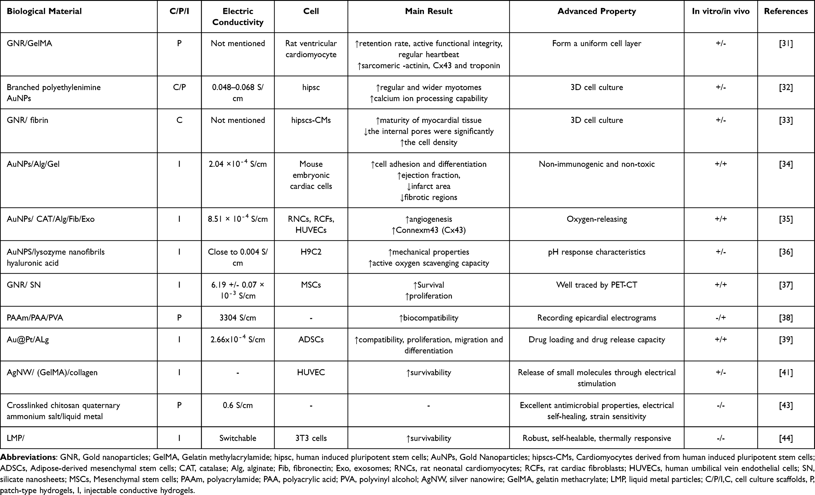 |
Table 1 Examples of Metal Nanoparticle-Conducting Composite Hydrogels Containing Metal Nanoparticles in Myocardial Infarctions |
Conductive Polymer Nanoparticle
Polypyrrole
Polypyrrole(PPy) is a positively charged aromatic polymer that is electroactive in water and electrolytes, although it is an insulator when undoped. Its conductivity ranges from 10⁻4S/cm to 104 S/cm, depending on the synthesis method and the type of oxidant used. PPy synthesized through chemical oxidation polymerization can achieve conductivity as high as 105 S/cm, while thin-film PPy materials can exhibit conductivity exceeding 380 S/cm.45 Oxidants and dopants can improve the mechanical properties and electrical conductivity of PPy-based conductive composites.46 PPy can be synthesized by chemical or electrochemical methods of oxidative polymerisation.47 The electrochemical synthesis method is more efficient for producing poly(pyrrole). This process involves oxidizing the pyrrole monomer on the electrode surface to generate pyrrole radical cations (C₄H₄NH+). These cations then couple to form long-chain polymers, with various microstructures developing depending on the electrochemical conditions. Typically, the microstructures are grown on a bubble template, and the resulting poly(pyrrole) film is often doped, exhibiting excellent electrical conductivity.48 Polypyrrole can be combined with other materials, such as hyaluronic acid hydrogels, to create conductive hydrogels that enhance cell proliferation and facilitate electrical signal transmission. This combination makes polypyrrole-based conductive hydrogels highly valuable and uniquely suited for tissue engineering applications.49
Recently, Yan et al developed an innovative injectable hydrogel by incorporating human endometrial mesenchymal stem cell-derived exosomes (hEMSC-Exo) into a polypyrrole-chitosan (PPY-CHI) matrix. This hydrogel continuously releases exosomes, and hEMSC-Exo has been shown to promote angiogenesis and reduce cardiomyocyte apoptosis. The hydrogel’s conductivity is measured at 4.82 ± 0.12 S/cm. In vitro experiments demonstrated that hEMSC-Exo promotes anti-apoptosis and pro-angiogenesis effects via the EGF/PI3K/AKT signaling pathway. In a rat myocardial infarction model, this composite hydrogel improved cardiac function post-infarction by promoting reperfusion and reducing ventricular remodeling. Furthermore, the hydrogel’s conductive properties helped synchronize cardiac conduction, reduce arrhythmias, and maintain enhanced cardiac conduction for 2–3 months.50
Several patches for treating heart attacks have been developed in recent years, but they often face challenges related to their fixation to the tissue, requiring specific responses or direct sutures. To overcome this, Liang et al designed a conductive, biocompatible hydrogel that can quickly adhere to the moist surface of the heart without the need for light or sutures, thus avoiding the tissue damage caused by traditional sutures. This hydrogel utilizes iron3+-polymerized pyrrole and dopamine to create a conductive crosslinked network of poly(pyrrole). With excellent mechanical properties and flexibility, the hydrogel remains stable during the heart’s beating and is resistant to breaking or detaching. Its electrical conductivity (6.51 ± 0.12 × 10⁻4 S/cm) closely matches that of natural heart muscle (approximately 10⁻4 S/cm). The hydrogel can adhere securely to a beating heart for up to 4 weeks, demonstrating good cytocompatibility. It enhances electrical conduction, helps re-open blood vessels, and improves blood flow and function in the infarct area. This research lays a solid foundation for developing sutureless patches in cardiac tissue engineering.51
In tissue engineering, both injectable hydrogels and cardiac biomaterial patches have unique advantages, but their effectiveness is often limited when used independently. Wu et al proposed a combined treatment approach involving an injectable hydrogel made from gel hyaluronic acid aldehyde (HA-CHO) and hydrazide hyaluronic acid (HHA), which is injected into the myocardial infarction area, while a conductive GelDA/DA-PPy hydrogel patch is applied to the outer layer of the heart muscle. The hydrogel patch offers excellent fatigue resistance, wet adhesion, and electrical conductivity, while the injectable hydrogel is known for its cell compatibility. The combination of these two hydrogels significantly enhances cardiac function, as demonstrated by echocardiography, histology, and angiography. Echocardiographic parameters, including EF, FS, LVIDd, LVIDs, EDV, and ESV, all showed significant improvement. Masson trichrome staining revealed a marked reduction in fibrosis in the infarct area, and the increased thickness of the left ventricular wall indicated protection and regeneration of the heart muscle. Furthermore, immunofluorescence staining for von Willebrand factor (vWF) and α-smooth muscle actin (α-SMA) demonstrated a significant increase in microvessel density and the number of small arteries, suggesting that the hydrogel not only promotes angiogenesis but also enhances blood supply to the infarcted tissue.52
Zhang et al developed a composite hydrogel (PAAM-SA-PPy NSs) made from double-crosslinked polyacrylamide (PAAM), sodium alginate (SA), and conductive polypyrrole nanospheres (PPy NSs). The hydrogel successfully formed a homogeneous SA-PPy conductive network, exhibiting high conductivity (6.44 S/cm), excellent mechanical strength, biocompatibility, self-healing properties, and strong adhesion. Strain sensors fabricated from this hydrogel are highly sensitive, have a broad sensing range, respond quickly to subtle muscle movements, and maintain high stability. This research offers novel insights into the development of advanced electronic components and sensors.53
Structural visualization is crucial for analyzing the internal components of electronic sensors. Tie et al successfully developed a transparent, mechanically enhanced, and highly conductive PPy@CNF-PAM hydrogel. The hydrogel achieves high conductivity through the in situ formation of PPy nanofibers. In this system, PPy nanofibers are crosslinked with cellulose nanofibers (CNF) to create a transparent network. Studies have demonstrated that the resistance of these composite hydrogels changes when deformed, allowing them to detect small body movements. This indicates that PPy@CNF-PAM hydrogels hold significant potential for the rapid development of wearable and implantable smart devices.54
In another study, the authors combined polypyrrole with gelatin to create a composite material, which was then incorporated into a gel system made from oxidized xanthan gum (OXG) and gelatin through a Schiff base reaction, resulting in an injectable conductive hydrogel. This hydrogel enhanced the conduction of electrical signals in rat heart muscle, reduced the incidence of ventricular arrhythmias, and promoted the expression of α-SMA and vWF, both of which are crucial for the reconstruction of blood flow after ischemia. Live/dead staining of H9C2 and RCF cells co-cultured with hydrogel extracts for 2 days revealed good cell viability, indicating its biocompatibility.55
Han et al synthesized silk-pyrrole hydrogels by in-situ polymerization of pyrrole into a silk protein network. The resulting composite hydrogels exhibited a conductivity of 26S/cm and demonstrated sensitivity to deformation. Among the two in-situ polymerization routes tested, the hydrogel prepared by route A showed a higher degree of cross-linking and an increased storage modulus. Fourier transform infrared spectroscopy (FTIR) analysis revealed that the main characteristic peaks of silk fibroin were retained, indicating that the introduction of PPy had minimal impact on the chemical environment of the silk fibroin matrix, preserving the stability of the overall structure. Resistance measurements using the four-probe method showed that the original silk hydrogel was insulating, but the addition of PPy significantly reduced resistance and enhanced conductivity. Strain sensors made from these composite hydrogels are capable of detecting subtle movements such as breathing, laughter, and pulse beats, making them a promising tool for future vital sign monitoring.56
Polypyrrole is known for its excellent electrical conductivity, biocompatibility, and mechanical properties, making it a promising material for tissue engineering. However, its lack of degradability and metabolism in the body raises concerns, presenting a challenge for its application in tissue engineering. Additionally, cardiac tissue functions in a dynamic environment of continuous contraction and relaxation, requiring composite hydrogels to be durable, elastic, mechanically strong, and coordinated. These properties need further exploration in future experiments to ensure their suitability for such applications.
Polyaniline
Polyaniline (PANI) is the second most widely used conductive polymer after polypyrrole (PPy). It exists in three oxidation states, with the semi-oxidized turquoise salt form being the most stable and exhibiting the strongest conductivity. PANI can be doped with benzene and quinone rings to varying degrees, and its properties are influenced by the synthesis method and the type of doping acid used. PANI polymers are synthesized from aniline monomers and their derivatives through chemical oxidation polymerization or electrochemical polymerization reactions.57 The chemical oxidation of aniline requires harsh conditions and is typically performed in a highly acidic solution using oxidizing agents such as ammonium persulfate or ferric chloride. The intrinsic conductivity of polyaniline (PANI) is 1.2 S/cm, but its conductivity can be altered through acid doping. The redox properties and the degree of protonation of the polymer are crucial in determining its overall electrical properties.58 An anisotropic composite fiber network containing polyaniline (PANI) exhibits conductive properties, allowing it to mimic the natural extracellular matrix. This network forms an open-pore scaffold that supports cell attachment and growth. Additionally, the structure has the potential to promote angiogenesis, making it a promising material for tissue engineering applications.59
Conductive polymer hydrogels (CPHs) combine the benefits of conductive polymers, the mechanical properties of hydrogels, and three-dimensional nanostructures, making them highly valuable in bioengineering. Chakraborty et al developed a self-healing CPH consisting of polyaniline (PANI) and N-Fmoc-diphenylalanine (Fmoc-FF) as a dipeptide gelling agent. The mechanical stiffness of this hydrogel can be adjusted by varying the peptide concentration, and its self-healing properties allow the non-metallic polymer hydrogel to regain its intrinsic conductivity and restore volume conductivity. The hydrogel has a conductivity of 5 × 10⁻4 S/cm, which remains stable even during sustained coordinated contractions of the heart. Cardiac cells inoculated onto the hydrogel adhere and proliferate well, indicating its non-toxicity. This composite hydrogel shows great promise as a carrier for cell therapy, with potential applications for repairing infarct areas and serving as a pressure-sensitive electronic component.60
Peptide-assembled nanostructured hydrogels have great potential for mimicking the extracellular matrix (ECM), but their fragile structural construction and difficulty in incorporating additional functional properties pose significant challenges. To address this, Chakraborty et al developed mechanically stable and self-healing hydrogels containing RGD cell adhesion fragments to enhance cell attachment. These hydrogels exhibit good electrical conductivity (4.25 × 10⁻4 S/cm). Polyaniline (PANI) nanoparticles were incorporated into the hydrogel’s three-dimensional network to further improve its mechanical properties and electrical conductivity. This composite hydrogel displays antibacterial activity, electrical conductivity, and DNA-binding ability, and can also induce relatively isolated cardiomyocytes to form a synchronized cell population with coordinated contractions. Therefore, this supramolecular composite hydrogel holds great promise as a cell scaffold for tissue engineering applications.61
Wu et al developed an injectable conductive hydrogel (Alg-P-AAV hydrogel) by incorporating lignosulfonate-doped polyaniline (PANI/LS) nanorods and an adenovirus encoding vascular endothelial growth factor (AAV9-VEGF) into a calcium-crosslinked alginate matrix. This hydrogel exhibits good electrical conductivity (1.2 S/cm), antioxidant properties, and the ability to promote angiogenesis. The conductive network formed by PANI/LS nanorods helps synchronize the contraction of cardiomyocytes in vitro, preventing arrhythmias caused by low-conductivity scar tissue that interferes with electrical signals. The large functionalized surface area of the nanorod network enhances antioxidant capacity, protects H9C2 cardiomyocytes from oxidative stress, and improves cell survival. In a rat myocardial infarction model, injection of the Alg-P-AAV hydrogel significantly improved cardiac function by increasing ejection fraction (EF) and shortening fraction (FS), reducing the left ventricular internal diameter, decreasing fibrosis in the infarct area, and thickening the left ventricular wall to prevent ventricular remodeling. The hydrogel also significantly increased the density of capillaries and small arteries, promoted neovascularization, and enhanced the local blood supply. Given the complexity of infarcted tissue, this multifunctional injectable hydrogel shows comprehensive therapeutic potential, making it a promising concept for future cardiac therapy.62
Yu et al developed an injectable electromechanically coupled hydrogel patch (MEHP) with a cross-linking mechanism based on reversible non-covalent interactions and borate ester bonds. This hydrogel can be implanted into the pericardial cavity in a minimally invasive manner while also loading cells. The b-PANI backbone in the hydrogel generates a nanoscale electric double layer (EDL), promoting rapid transmission of electrical signals along the π-π conjugated network and enhancing the uniform distribution of hydrophilic and conductive units. This peptide-based, nano-engineered conductive supramolecular hydrogel also exhibits excellent self-healing and fatigue resistance properties, with conductivity ranging between 0.4 and 0.6 S/cm. The MEHP performs well under physiological conditions and can adapt sensitively to the pulsatile nature of the heartbeat. The hydrogel demonstrates good cytocompatibility and supports the encapsulation and proliferation of adipose-derived stem cells (ADSCs). Its therapeutic effects significantly contribute to infarct repair. The combination of this dynamically conductive biomaterial and electromechanical coupling strategies shows great potential for other applications in electroactive tissue repair.63
Micro-patterning technology is an ideal method for promoting cell alignment, growth, and monitoring. Noh et al used a UV-induced method to incorporate polyaniline (PANi) into a polyethylene glycol (PEG) hydrogel matrix, rapidly synthesizing conductive hydrogel micro-patterns. The PANi/PEG hydrogel exhibited a conductivity of 30.5 ± 0.5 × 10⁻³ S/cm. Mouse C2C12 myoblasts were successfully deposited on the micro-patterned hydrogel, and the MTT assay confirmed good cell proliferation. Furthermore, this composite hydrogel induced myogenic differentiation in C2C12 cells, with neatly arranged myotubes and the expression of myosin heavy chain. These results suggest that this composite hydrogel has great potential as an extracellular matrix for tissue engineering applications.64
Long-term and accurate electrical monitoring is crucial for studying cell growth and development. Wu et al developed a hydrogel sensor for detecting electrical activity by synthesizing a polyaniline (PAni) and flexible carbon cloth (CC) composite material. The electrochemical activity of the hydrogel was enhanced by incorporating catalytic platinum nanoparticles (Pt NPs) onto the polyaniline network, which enabled the effective detection of metabolites such as hydrogen peroxide (H2O2). The hydrogel electrochemical sensor demonstrated high sensitivity and specificity in detecting H2O2. Additionally, MCF-7 and K562 cells exhibited good long-term viability on the hydrogel. The sensor can directly detect electrophysiological changes in cells and distinguish the physiological states of living cells. These characteristics make the hydrogel an ideal tool for fabricating biomimetic sensors and monitoring cell-related biomarkers.65
Polyaniline (PANI) offers advantages such as good electrical conductivity, biocompatibility, and relatively simple synthesis, making it a promising material for tissue engineering. However, it also faces challenges related to cytotoxicity and doping stability, which need further experimental verification to ensure its suitability for long-term applications in this field.
Aniline Oligomer
Low-polyphenyleneamine retains the chemical properties of polymers while exhibiting characteristics typical of molecular semiconductors, particularly in terms of monodispersity and self-assembly. Through specific morphological processes induced by dopants, supramolecular structures such as nanowires, nanoribbons, rectangular nanoplates, and nanocrystals can be synthesized from low-polyphenyleneamine.66 Due to its low molecular weight, aniline can self-assemble into highly ordered nanostructures. In the first stage of aniline oxidation, non-conductive and insoluble aniline oligomers or intermediates are formed, which are thought to facilitate the extension of conductive polyaniline (PANI) nanorods and nanotubes in the second stage. Aniline oligomers, typically ranging from dimers to tetramers, are formed by oxidation in a weakly acidic medium. Under appropriate conditions, higher oligomers can be synthesized, leading to the formation of a variety of flower-like structures, such as dandelions, orchids, or roses.67 Chiral induction in aniline oligomers with a π-conjugated backbone is achieved through a chiral organization facilitated by intramolecular hydrogen bonding. This chiral arrangement influences the structure of the oligomers, and the π-conjugated polymers derived from these oligomers exhibit corresponding electrical properties.68 Electron-deficient materials have been studied less frequently due to their poor solubility. However, recent advancements in the synthesis and electrochemical oxidation coupling of electron-deficient aniline analogues with high electron deficiency have enabled the formation of soluble electron-deficient oligomers. Additionally, the isomerization of these oxidized oligomers can produce highly stable oxidation states, thereby expanding their potential applications.69 Aniline oligomers can be combined with other polymers by interacting with the side chain groups of the main polymer or by being incorporated into the main polymer backbone as a comonomer. The conductivity of conductive biomaterials based on aniline oligomers can be adjusted to mimic the electrical and mechanical properties of specific tissues or organs. This tunability provides promising potential for applications in areas such as cardiac tissue engineering and hydrogel development.70 Aniline oligomers have a significant impact on nanostructured organic conductive materials. The conductivity of aniline oligomers ranges from 10⁻6 to 10⁻² S/cm, and even small amounts used as additives can significantly enhance the conductivity of the main material. This makes them a crucial component in the development of advanced conductive materials.71 This greatly enhances its potential for use in tissue engineering, as improving conductivity is essential for promoting cell growth, communication, and tissue regeneration.
Hydrogen sulfide (H2S) is known for its anti-inflammatory and angiogenic effects, offering protection to the cardiovascular system. However, its short half-life and limited controlled release have hindered its therapeutic application. To address this, Liang et al developed a controlled-release hydrogel by grafting 2-aminopyridine-5-thioamide onto partially oxidized alginate (ALG-CHO). This material was combined with gelatin via the Schiff base reaction to form a conductive H2S-releasing hydrogel loaded with stem cells. The controlled release of H2S helps promote angiogenesis, reduce inflammation, and improve the microenvironment in the area of myocardial infarction. The composite hydrogel has an electrical conductivity of 3.7 × 10⁻4 S/cm, which is further increased by the addition of tetraaniline. In vitro studies demonstrated that this hydrogel has good electrical conductivity and can effectively release H2S to promote the survival and proliferation of adipose-derived stem cells (ADSCs). The hydrogel adheres well to a moist, beating heart and shows enhanced stem cell retention and higher sulfide concentration in rat myocardial tissue. Echocardiography revealed a significant improvement in ventricular ejection fraction, and histological staining showed a marked reduction in infarct size. Overall, the heart function of rats treated with the composite hydrogel improved significantly. This conductive composite hydrogel containing ADSCs holds great potential for the treatment of myocardial infarction.72
Wei et al developed a conductive, injectable, and biodegradable hydrogel for cardiac repair following myocardial infarction. The hydrogel is crosslinked using a matrix metalloproteinase-sensitive peptide (MMP-SP) and consists of partially oxidized sodium alginate (ALG-CHO), tetrabenzylamine (TA), the nanomedicine 1,4-dihydroxynaphthalene-4-one-3-carboxylic acid (DPCA), and thiol hyaluronan (HA-SH). DPCA stably promotes HIF-1α, a key factor in the regeneration process, and in combination with the other components, they exert a synergistic effect, enhancing the hydrogel’s regenerative capacity. By incorporating tetraamine, the conductivity of the hydrogel reached 8.8 × 10⁻5 S/cm. MMP-SP makes the hydrogel sensitive to matrix metalloproteinases, allowing for targeted degradation specifically in the myocardial infarction area. The composite hydrogel demonstrated good cytocompatibility with H9C2 cardiomyocytes and L929 fibroblasts, and in vivo subcutaneous injection confirmed its favorable histocompatibility. In addition to its biocompatibility, the hydrogel significantly reduced the expression of inflammatory and apoptotic factors, while maintaining high levels of HIF-1α, promoting angiogenesis, and enhancing the expression of connexin 43 (Cx43). Histological staining and ultrasound imaging revealed that the multifunctional hydrogel significantly reduced infarct size and markedly improved cardiac function, as shown by increases in ejection fraction (EF) and fractional shortening (FS). This composite hydrogel, combining electrical conductivity, degradability, and tissue regeneration promotion, offers a promising approach for the treatment of myocardial infarction.73
Aniline oligomer nanomaterials exhibit excellent electrical conductivity, adaptive modulation, and biocompatibility, making them highly promising for tissue engineering applications. However, the interactions between aniline oligomers and biologically active molecules or cells are not yet fully understood. Further in vivo and in vitro experiments are necessary to explore these interactions and comprehensively assess the potential of aniline oligomers in biomedical applications.
Polymerisation
Polythiophene (PTH) has a heterocyclic structure with positively charged ionizable side groups. Unsubstituted polythiophenes or 2,5-coupled polythiophenes are non-conductive, with typical conductivity below 10⁻6 S/cm. However, external doping along the π-orbital domains of the polymer backbone can increase the conductivity to between 10 and 100 S/cm. Polythiophenes can be synthesized using both electropolymerization and chemical polymerization methods.74 Polymerization of polythiophene occurs through complex chain extension and sequential reactions, where the reactivity of the thiophene monomers decreases as the oligomer chain lengthens. The widely accepted mechanism suggests that oligomers are initially formed, followed by redox reactions that extend these oligomers into longer polymer chains.75 The use of polythiophene (PTH) copolymers can overcome the limitations associated with using PTH alone. These copolymers can be synthesized through various methods, including macromonomer technology and reactive polymerization technology.76
In cardiac tissue engineering, three-dimensional conductive scaffolds play a vital role in supporting stem cell adhesion, proliferation, and cardiomyocyte differentiation. Yang et al developed a straightforward method for fabricating poly(3,4-ethylenedioxythiophene)/alginate (PEDOT/Alg) scaffolds by using adipic dihydrazide as a cross-linking agent to form a chemically cross-linked alginate network, while PEDOT was synthesized in situ within the alginate matrix. Studies demonstrated that brown adipose-derived stem cells (BADSCs) could successfully adhere to, proliferate on, and differentiate into cardiomyocytes on these scaffolds. The composite hydrogels significantly upregulated the expression of key cardiac markers, including troponin T (cTnT), muscle junction protein α-actinin, and gap junction protein connexin 43 (Cx43). Moreover, electrical stimulation increased intracellular reactive oxygen species (ROS) levels, further activating the cardiomyocyte differentiation pathway and promoting myocardial regeneration—an essential factor in repairing damaged heart tissue through cardiac tissue engineering.77
With growing interest in the health benefits of monitoring electrophysiological signals, Zeng et al tackled the challenge of constructing a homogeneous nanoconductive network by employing dopamine (DA) modification. They developed a conductive hydrogel (HA-DA-PP) by uniformly blending hyaluronic acid (HA) with DA-modified PEDOT and PSS (collectively referred to as PP). This hydrogel demonstrated excellent stretchability, capable of elongating up to 4.7 times its original length. The introduction of DA-modified PP significantly enhanced the hydrogel’s conductivity, reaching up to 0.97 S/cm. The hydrogel exhibited high sensitivity and accuracy in detecting human electrocardiogram (ECG) signals, clearly identifying R, S, T, P, and Q wave peaks, and outperformed conventional commercial hydrogel electrodes. Biocompatibility was confirmed through co-culture experiments with 3T3 cells and 14-day subcutaneous implantation in SD rats. Moreover, when applied directly to the epicardium in vivo, no signs of arrhythmia were observed in the ECG, further confirming its histocompatibility. These results highlight the hydrogel’s strong potential as a platform for the development of implantable electrophysiological monitoring devices.78
In this study, Wahid et al developed a biocompatible conductive hydrogel (CH) composed of PEDOT: PSS and gelatin using the bioimprinting technique. This bioimprinted conductive hydrogel was designed to examine the response of biological cells to its morphological features and to explore the relationship between cell growth and the conductivity of the substrate. These high-resolution bioimprinted hydrogels can effectively simulate the natural cell environment while enabling precise monitoring. The average replication fidelity of the bioimprints is over 90%, providing an almost exact 3D replica of the cells. Additionally, the conductivity of the crosslinked CH membrane was significantly increased from 10⁻³ S/cm to 1 S/cm.79
The ability to deliver controlled electrical stimulation and directly analyze functional readouts is crucial for the electrophysiological assessment of cardiac chip platforms. Zhang et al developed a novel platform integrating two soft conductive hydrogel column electrodes. These electrodes were created from a gelatin-based solution modified with PEDOT: PSS, which facilitated the delivery of human induced pluripotent stem cell-derived cardiomyocytes (hiPSC-CM) and cardiac fibroblasts for constructing 3D human cardiac tissue. Electrical stimulation promoted cell maturation and improved cardiac function. The conductive hydrogel columns supported various research applications, including dynamic drug delivery and continuous monitoring. Additionally, it was observed that an increase in heart rate enhanced the tissue’s response to adrenergic stimulation.80
Damage to the heart muscle is challenging to repair naturally and can lead to complications such as arrhythmia or sudden death due to uncoordinated contractions. To address this issue, Luque et al designed a printable multifunctional hydrogel composed of polyvinyl alcohol (PVA) dynamically crosslinked with gallic acid (GA) and PEDOT. This hydrogel patch exhibits excellent electrical conductivity, tensile toughness, and mechanical strength, all crucial for enhancing weakened heart contractions and pulse propagation. Inkjet printing was performed immediately after melting and mixing the PVA-GA/PEDOT composition. In vitro studies using rat cardiomyocytes demonstrated that the transmission of pacing signals was not hindered during intracellular Ca2+ transients. Furthermore, magnetic resonance imaging (MRI) and electrocardiograms (ECG) showed that the functional parameters remained stable for two weeks after implantation in rats, suggesting the hydrogel’s potential as a long-term implant for cardiac repair.81
Li et al developed a hydrogel paper patch (HPP) with excellent electrical conductivity and hydrophilicity by self-assembling PEDOT: PSS hydrogels onto paper fibers. This material is highly sensitive to both electrical and chemical substances, enabling it to function as both a conductive electrode and a highly sensitive glucose sensor. The HPP can simultaneously monitor electrocardiogram (ECG) signals during exercise and biochemical signals in sweat in real time. It incorporates a paper-based microfluidic channel that facilitates the efficient transport of sweat to the detector surface, ensuring stable electrical conductivity.82
In this study, Peng et al incorporated PEDOT: PSS into a polyvinyl alcohol (PVA)/polyacrylic acid (PAA) double network (DN) hydrogel to develop a novel strain sensor. The mechanical properties of the hydrogel can be tailored by adjusting the polymer composition and the number of freeze-thaw cycles. Dynamic hydrogen bonding within the hydrogel contributes to its self-healing and adhesive properties, making it well-suited for wearable devices. Additionally, the hydrogel sensor demonstrates high sensitivity and a reliable strain response to physiological signals such as pulse and vocal cord vibrations.83
Sauvage et al developed a soft conductive hydrogel patch based on a composite of (3,4-ethylenedioxythiophene): poly(styrenesulfonate) (PEDOT: PSS) and poly(vinyl alcohol) (PVA). The average conductivity of this cardiac patch is high, reaching 40S/cm, and can be adjusted by modifying the PVA concentration. Their research indicates that the hydrogel remains flexible under 10% cyclic stretching at a frequency of 1 hz, with its conductivity remaining stable over time, demonstrating the potential for stable, long-term use in a dynamic cardiac environment. The biofunctionalization of the hydrogel with an N-cadherin mimetic peptide promotes the adhesion and proliferation of cardiac fibroblasts (CFbs), ensuring the hydrogel’s biocompatibility. Additionally, the hydrogel exhibits enhanced antibacterial properties, which is valuable for infarction repair, and its improved antibacterial ability can lead to better treatment outcomes. However, the current antibacterial properties are limited to Staphylococcus aureus, and further testing against a broader range of bacteria would enhance its potential for clinical applications.84
Polythiophene (PTH) and its derivatives are highly valued in biomaterials due to their biocompatibility, conductivity, and tunability. However, challenges such as low solubility, aggregation of side chain groups, and processing difficulties must be addressed in order to further enhance their properties and broaden their range of applications.
Table 2 Examples of conductive polymer nanoparticle-containing conductive composite hydrogels in myocardial infarctions.
 |
Table 2 Examples of Conductive Polymer Nanoparticle-Containing Conductive Composite Hydrogels in Myocardial Infarctions |
Carbon-Based Nanomaterials
Carbon Nanotube
Carbon nanotubes (CNTs) are cylindrical structures formed by curling graphene sheets and are classified into single-walled carbon nanotubes (SWCNTs) and multi-walled carbon nanotubes (MWCNTs). SWCNTs have a diameter of approximately 1–2 nm, while MWCNTs consist of multiple concentric layers with a diameter ranging from 2 to 50 nm. The electrical conductivity of SWCNTs is exceptionally high, reaching up to 104 S/cm, while MWCNTs generally have lower conductivity, ranging between 10² and 10³ S/cm. To improve the dispersibility of carbon nanotubes in polar solvents, oxygen-containing functional groups such as carboxyl and hydroxyl groups are often introduced on their surfaces. This oxidation treatment enhances dispersion stability and prepares the material for further chemical modification.89 Various methods are used to synthesize CNTs. High-temperature processes like arc discharge and laser ablation can produce CNTs with fewer structural defects. Chemical functionalization of carbon nanotube sheets can enhance their adsorption properties, making them suitable for interactions with particles and filtration applications. The incorporation of multifunctional carbon nanotubes into composite materials can improve their electrical properties, resolution, and solubility, thereby significantly enhancing their potential for tissue engineering applications.90
Cardiac tissue regeneration engineering involves implanting specific cells into three-dimensional biomaterials, which are then reshaped into contractile cardiac tissue and transplanted for heart repair. This process requires the biomaterial to provide strong support for the cells to maintain their biological activity. To address the weak mechanical properties and poor electrical conductivity of collagen (Col) hydrogels, Sun et al developed electrically active nanocomposite hydrogels by combining carbon nanotubes (CNTs) with collagen. The CNT/Col hydrogels exhibited electrical conductivity of 16S/cm (longitudinal) and 0.005 S/cm (transverse), enhancing the mechanical and electrical properties of neural tissue, while also demonstrating good cell compatibility in vitro. Immunostaining of α-SA and TnI on cardiomyocytes cultured on CNT/Col and Col hydrogels showed that neonatal rat ventricular myocytes (NRVMs) exhibited a well-arranged, elongated morphology with prominent actin filaments. Notably, the sarcomere structure became significantly longer and more regularly aligned, resembling the structure of native cardiac muscle. Additionally, there was a significant increase in the expression of connexin 43 (CX43), a key protein in cell communication, with no cytotoxic effects observed on the cardiomyocytes. This study highlights the potential of CNT/Col hydrogels to enhance cardiac regenerative therapies and suggests the need for further investigation.91
In their research, Wang et al developed a highly elastic conductive hydrogel composed of carbon nanotubes (CNTs) and polyvinyl alcohol (PVA). The CNT-PVA network exhibits excellent stretchability, self-healing, and stability due to the complementary action of covalent acetal bonds and hydrogen bonds. Remarkably, this hydrogel remains stable even at high loading rates. Experimental results demonstrate that the CNT-PVA hydrogel is sensitive enough to detect tiny strains caused by the pulsation of the radial artery and the electromyogram signal from forearm muscles. Additionally, the conductive hydrogel can self-recover to its original state at room temperature, allowing it to be reused.92
Recently, Feng et al developed a dual-crosslinked network composite hydrogel (MWCNTs/CNWs/PAM/SA) with high conductive sensitivity, low hysteresis, good tissue adaptability, and shape memory. By optimizing the composition, the addition of multi-walled carbon nanotubes (MWCNTs) significantly enhanced the hydrogel’s conductivity. Furthermore, the composite hydrogel was integrated into a triboelectric nanogenerator (TENG), which utilizes friction and electrostatic induction to generate a continuous and stable alternating current voltage, capable of powering small, self-sufficient electronic devices. This development broadens the potential applications of composite hydrogels in the field of bioelectronics.93
Carbon nanotubes (CNTs) are increasingly used in medicine due to their excellent electrical and mechanical properties. However, their small size and high reactivity may pose potential risks when they enter the human body. More research is needed to fully assess their cytotoxicity and biocompatibility. To address this limitation, efforts are underway to improve the chemical production process to reduce the cytotoxicity of carbon nanotubes, making them safer for medical applications.
Graphene and Its Derivatives
Graphene is a two-dimensional monolayer of carbon atoms arranged in an aromatic structure, characterized by sp² atomic orbital hybridization. Out-of-plane π bonds form a delocalized electronic network, which is responsible for graphene’s exceptional electronic conductivity.94 Graphene has a highly ordered crystal structure and excellent electronic mobility, which is the reason for its outstanding electronic properties.95 Graphene has superlative mechanical strength, with a Young’s modulus of 1.0TPa.96 The oxidation process of graphene produces graphene oxide (GO), a material characterized by the introduction of oxygen-containing functional groups, such as carboxyl and epoxy groups, into its structure. These functional groups enhance the material’s dispersibility in water and its ability to undergo further chemical modifications.97 Defects in the sp² orbitals of graphene oxide (GO) lead to its poor electrical conductivity. However, this can be improved through chemical reduction. Reduced graphene oxide (rGO), obtained by chemical graphitization, exhibits a significantly enhanced conductivity of up to 9.44 S/cm. This improvement makes rGO a promising material for various electronic and energy storage applications.98 Graphene-based nanomaterials (GBNs) have a wide range of applications, including as carriers for therapeutic factors, antibacterial agents, biosensors, and bioimaging materials. Their unique properties, such as high surface area, excellent conductivity, and biocompatibility, make them highly versatile for use in various biomedical and technological fields.99
In a study, Zhang et al developed a crosslinked basic factor gelatin hydrogel with a patterned microstructure, in which graphene oxide (GO) was incorporated to enhance its properties. This hydrogel can be produced in a simple and reproducible manner. The composite hydrogel facilitated the alignment and maturation of neonatal rat ventricular myocytes. The presence of GO significantly improved contractile amplitude and coordination, while also increasing the expression of key cardiac genes, including α-actinin (Actn), cardiac troponin T (Tnnt), sarcoplasmic reticulum Ca2+-ATPase 2a (Serca2a), and connexin-43 (Cx43). The cardiomyocytes achieved optimal synchronized contraction within 2 days of seeding and continued to beat for up to 3 months, demonstrating the scaffold’s ability to support long-term cell survival. These findings highlight the potential of the scaffold for use in cardiac tissue engineering.100
Mousavi et al developed an innovative in situ hybridization-forming double network hydrogel composed of oxidized alginate (OA) and cardiomyocyte extracellular matrix (ECM). The incorporation of 3-(2-aminoethylamino)propyltrimethoxysilane (APTMS)-functionalized reduced graphene oxide (Amine-rGO) significantly enhanced the electrical and mechanical properties of the system. The composite hydrogel includes decellularized myocardial ECM extracted from bovine hearts, providing a rich source of bioactive molecules that promote cell attachment, proliferation, differentiation, and angiogenesis, which are critical for myocardial regeneration. The introduction of amine-rGO greatly improves the conductivity of the hydrogel, supporting electrical signal transmission and synchronized contraction of cardiomyocytes. Cell compatibility analysis using the MTT method on human umbilical vein endothelial cells (HUVECs) showed optimal cell activity at a concentration of 25 μg/mL of amine-rGO. Additionally, the composite hydrogel demonstrates stability under large deformations, making it suitable for dynamic tissue environments.101
Umbilical cord stem cell (UCMSC) transplantation therapy shows promise for repairing heart disease, but it faces challenges due to the low cell retention rate. To address this, Zhu et al developed an injectable hydrogel composed of methacrylic acid gelatin (GelMA) and oxidized dextran (ODEX), with the addition of graphene oxide (GO), which was reduced to rGO by dopamine. In vitro experiments demonstrated that this hydrogel has good biocompatibility and cell delivery capacity, promoting the growth, proliferation, and cardiac differentiation of UCMSCs. It also enhanced the expression of cardiac-specific markers, such as cardiac troponin I (cTnI) and connexin 43 (Cx43). Further in vivo studies in a rat myocardial infarction (MI) model revealed that the GelMA-O5/rGO hydrogel significantly improved cardiac function, reduced infarct size, supported the survival of UCMSCs, increased cTnI and Cx43 expression, and decreased the expression of the apoptotic marker caspase-3. In conclusion, the development of injectable GelMA-O5/rGO/UCMSCs hydrogels marks a significant advancement in the treatment of myocardial infarction.102
Recently, Jaramillo introduced a novel method for electrophysiological measurements using a hybrid hydrogel composed of methacryloyl-modified small intestinal submucosa (SISMA) and graphene oxide-polyethylene glycol (GO-PEG). This composite hydrogel exhibits shear-thinning properties, making it ideal for applications such as bioprinting or extrusion. Umbilical cord blood cells (UCBCs) were cultured in a 3D hypoxic environment with the addition of weak electrical stimulation and growth factors, resulting in significant cell proliferation and high survival rates.103
Controlled and adjustable drug release is crucial for effective disease treatment in tissue engineering. Aycan et al developed a conductive hydrogel carrier for the release of anti-inflammatory drugs based on natural hyaluronic acid (HA). The electromechanical properties of the hydrogel were enhanced using both in-situ and post-polymerization mechanisms. Ibuprofen (IBU) was chosen as a model drug, as it helps prevent inflammatory responses between neural implants and tissues, inhibiting cell proliferation and differentiation. The composite hydrogel demonstrated an electro-responsive release of ibuprofen (IBU), with the release rate progressively increasing in response to the applied voltage. This development opens the door to future therapeutic applications involving controlled drug release in response to electrical stimuli.104
Recently, Wang et al developed a cardiac patch made of methoxylated triethylene glycol-functionalized graphene and dopamine-modified gelatin (TEG-GR/GelDA). This patch enhances myocardial electrical coupling, protects cardiac function, promotes angiogenesis, and inhibits cardiomyocyte apoptosis. Compared to traditional graphene, the incorporation of TEG-GR into the hydrogel accelerates its degradation rate, aligning it more closely with the tissue repair process following myocardial infarction. GelDA hydrogels containing 2.5 mg/mL armchair-type TEG-GR or graphene demonstrated conductivity similar to that of heart muscle (0.2–0.6 S/m) and effectively prevented chronic inflammation. The self-adhesive properties of GelDA simplify the surgical procedure, eliminating the need for sutures, reducing myocardial damage, and improving surgical safety. Additionally, the GelDA solution exhibits excellent flowability and no viscosity before gelation, which is initiated by Fe3+, making it easier to transport through a microcatheter. This characteristic enables minimally invasive implantation of hydrogel patches, reducing catheter-related trauma.105
In recent years, graphene and its derivatives have garnered increasing attention due to their exceptional physical and chemical properties. They play a crucial role in promoting the maturation of cardiomyocytes and the proliferation and differentiation of stem cells. However, further in vivo experiments are needed to fully assess their biosafety, degradability, and long-term effects to ensure their suitability for biomedical applications.
Table 3 Examples of the use of conductive composite hydrogel containing carbon-based nanoparticles in myocardial infarction.
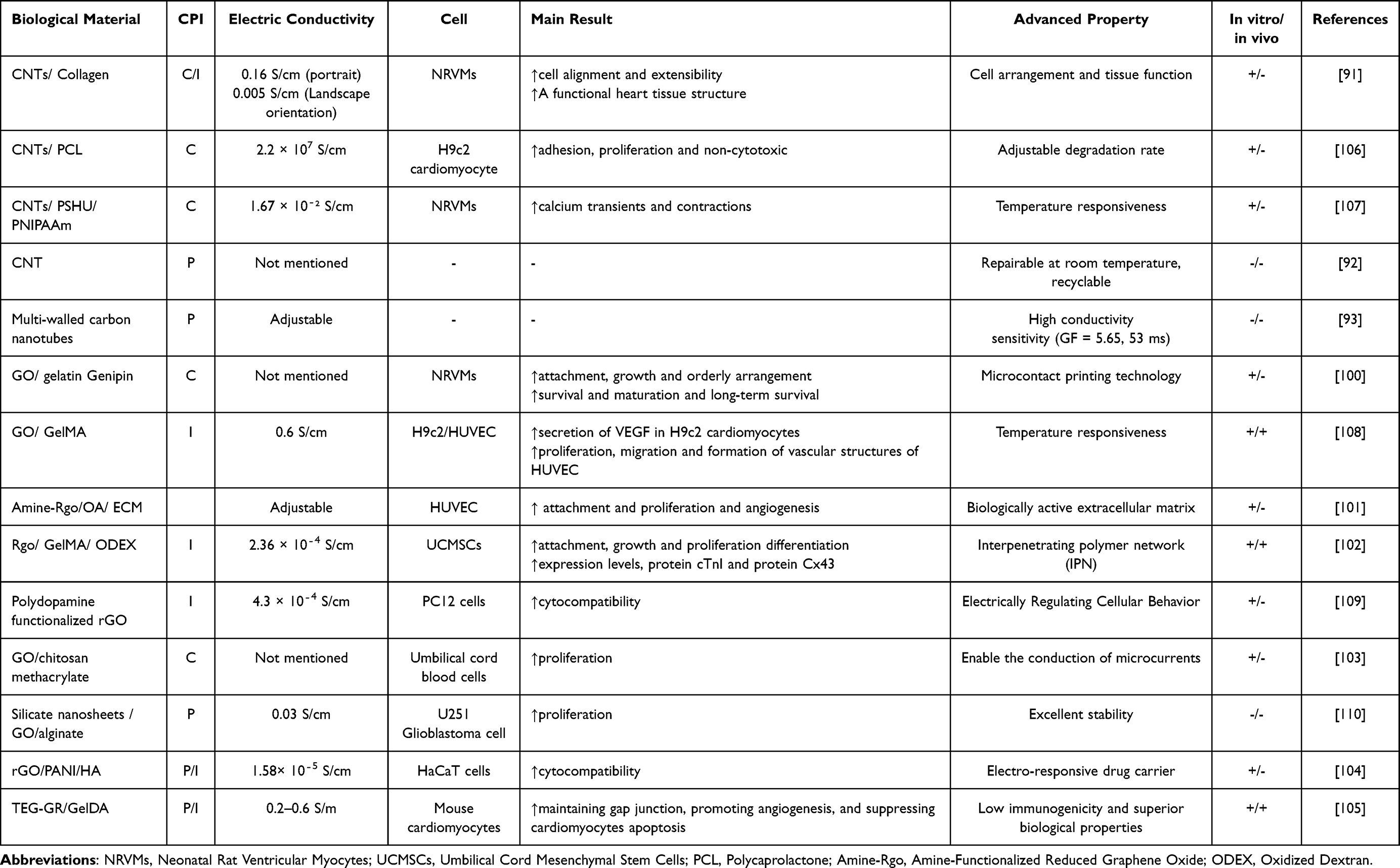 |
Table 3 Examples of the Use of Conductive Composite Hydrogel Containing Carbon-Based Nanoparticles in Myocardial Infarction |
Other Special Types of Conductive Hydrogels
MXene
MXenes are a large class of two-dimensional transition metal carbides, carbonitrides, and nitrides with the general formula Mn+1XnTx. In this formula, M represents an early transition metal, X represents carbon (C) or nitrogen (N), and Tx represents surface terminal atoms such as OH, O, or F. MXenes are derived from the MAX phase by selectively etching the “A” element (usually from group IIIA to VIA elements) of the MAX phase. Due to the free electrons on the transition metal carbide or nitride skeleton and the hydrophilicity of the surface terminations, MXenes exhibit extremely high electrical conductivity. For instance, Ti₃C2Tx MXene films can have a conductivity of up to 10,000 S/cm.111 Unlike the surfaces of other two-dimensional materials, such as graphene and transition metal disulfides, the surface functional groups of dioctylene can be chemically modified to impart various functional properties. This unique characteristic significantly enhances its versatility, making it suitable for a wide range of applications across different fields.112 Dielsene has ultra-high metal conductivity, high mechanical properties and multifunctional surfaces.113
Lee et al developed a new hydrogel heart patch by combining two-dimensional titanium carbide (Ti₃C2Tx) MXene with a natural biocompatible polymer. This patch can be rapidly synthesized and directly applied to heart tissue. MXene offers several key advantages, including even distribution, high conductivity (0.0183 S/cm), elasticity similar to heart tissue (30.4 kPa), strong tissue adhesion (6.8 kPa), and excellent mechanical properties. In vitro studies have shown that CAH hydrogels co-cultured with rat cardiomyocytes promote cardiomyocyte maturation, while in vivo experiments revealed that CAH hydrogels adhere stably to the beating epicardium. Animal experiments demonstrated that CAH promotes vascular reperfusion, reduces the inflammatory response, and increases the expression of connexin 43 (Cx43) in the infarcted heart, highlighting its potential for cardiac repair.114
In another study, Ye et al developed a new type of hydrophilic, biocompatible, and conductive material by incorporating MXene Ti2C and dopamine (inspired by mussel adhesion) into a PEGDA-GelMA cryogel. This biofunctionalized engineered cardiac patch (ECP) mimics natural cardiac tissue with excellent fatigue resistance, toughness, and an electrical conductivity of 0.087 S/cm, making it suitable for repairing highly infarcted myocardium. In vitro experiments demonstrated that new blood vessels formed in Ti2C-8 cryogels seeded with rat aortic endothelial cells. Additionally, Ti2C cryogel showed cell generation and tissue maturation after 7 days of co-culture with cardiomyocytes (CMs). Ti2C-8 cryogel ECP also exhibited contraction and beating behavior similar to that of cardiac tissue. After transplantation into the infarct area of myocardial infarction (MI) rats, Ti2C-8 cryogel ECP improved cardiac function, repaired damaged myocardium, and significantly promoted angiogenesis.115
Three-dimensional printing is increasingly gaining prominence in tissue engineering. Basara et al developed a novel conductive composite material with a conductivity of 0.1 S/cm for cardiac tissue engineering. They used aerosol jet printing to pattern polyethylene glycol (PEG) hydrogels and then 3D printed conductive titanium carbide (Ti₃C2Tx) MXene onto the hydrogels. Human induced pluripotent stem cell-derived cardiomyocytes (iCMs) were then seeded onto the 3D-printed scaffolds and cultured for 7 days. The results showed a significant increase in the expression of key cardiac markers, such as MYH7, SERCA2, and TNNT2, as well as improvements in Ca2+ handling and propagation during the spontaneous beating of iCMs. These findings suggest that 3D-printed Ti₃C2Tx MXene can be an effective carrier for therapeutic materials in the treatment of myocardial infarction (MI).116
Ge et al found in a study that Ti₃C₃Tx MXene can serve as a multifunctional cross-linking agent that gels rapidly. This is attributed to the multidimensional molecular interactions between MXene and the polymer, which promote radical degradation. Using MXene as a dynamic cross-linking agent enhances the mechanical properties, adhesion, and self-healing ability of the material. Furthermore, the combination of the photothermal properties of Ti₃C₃Tx and the phase change reaction of the polymer results in a unique thermosensitive response in the polymer-MXene hydrogel, which can gel quickly under near-infrared irradiation. These properties make polymer-MXene hydrogels promising base materials for 3D printing.117
Zhu et al proposed a “one-pot” method for synthesizing composite conductive hydrogels with varying MXene doping levels. The functional groups (-OH, -F, -O, etc.) on the surface of the MXene nanosheets improved the viscosity, elasticity, and mechanical properties of the composite hydrogels, resulting in a conductivity increase to 0.366±0.055 S/cm. Adding 2 w/v% MXene. CH enhanced the adhesion and migration growth of mouse fibroblasts (NH3T3s) and exhibited good cytocompatibility, making it a promising material for biomedical applications.118
A simple method for synthesizing conductive hydrogels is crucial for advancing research on biomaterials. Zhu et al proposed a “one-pot” method for synthesizing composite conductive hydrogels (CHs) with varying doping levels of MXene. Functional groups (-OH, -F, -O, etc.) on the surface of MXene nanosheets enhanced the viscosity, elasticity, and mechanical properties of the hydrogels. With the addition of 2 w/v% MXene, the conductivity of the composite hydrogel increased to 0.366 ± 0.055 S/cm. The composite hydrogel also demonstrated good cytocompatibility, promoting the adhesion and migration growth of mouse fibroblasts (NH3T3s).119
Advanced mixed-ion electronic conduction (MIEC) bioelectronic devices often face challenges such as uneven distribution of bioelectrical signals and low transmission efficiency. To address these issues, Luo et al developed a topological MXene network-enhanced MIEC hydrogel with improved adhesion energy and breaking strength, ensuring stability during repeated stretching and use. The topological MXene network effectively enhances the conductivity of the hydrogel by extending the Debye length, which improves its dielectric properties and electric field density. With excellent biocompatibility, this hydrogel is suitable for long-term ECG signal detection, enabling continuous and accurate monitoring of changes in the ECG signal over time.120
Iron ion (Fe3+) doping can enhance the conductivity and mechanical properties of hydrogels. Hua et al combined montmorillonite (MMT), polyacrylamide-acrylonitrile [P(AAm-co-AN)], xanthan gum (XG), and iron ions (Fe3+) to develop a xanthan gum hydrogel (XGH) with high extensibility, conductivity, and self-healing properties. The iron ions play a key role by forming ionic cross-links with the carboxyl groups (-COO-) on the xanthan gum chains and electrostatically interacting with MMT, which significantly improves the mechanical strength and self-healing ability of the hydrogel. Additionally, the migration of iron ions enhances the electrical conductivity, reaching 0.006788 ± 0.0005 S/cm. Due to these multi-species physical interactions, the hydrogel demonstrates excellent fatigue resistance, particularly in terms of compression recovery. In cell viability tests, fibroblasts (L929) cultured with the composite hydrogel at different concentrations showed cell viability above 90%, confirming its good biocompatibility. This research opens up new possibilities for flexible single-electron components and tissue engineering applications.121
With the rapid development of multifunctional hydrogels, conductive hydrogels are increasingly being utilized in wearable smart devices. In this study, conductive composite hydrogels were synthesized within 30 seconds at room temperature using a tannic acid–iron (TA@Fe3+) mediated dynamic catalytic system. TA@Fe3+ catalyzed the radical polymerization of acrylic acid (AA), leading to the rapid formation of gels. The dynamic and reversible coordination bond between Fe3+ and the carboxyl group (COO-) provides the hydrogel with self-healing abilities. These hydrogels exhibit remarkable extensibility, capable of stretching up to 35.6 times their original length, with a strain sensitivity coefficient of 2.11 and a conductivity of 33.58 S/cm. Additionally, the hydrogels possess highly effective electromagnetic interference (EMI) shielding properties, reducing the impact of EMI and radiation on electronic devices and health, thereby enhancing their use in electronic components. The addition of TA also imparts antibacterial properties to the hydrogel, making it suitable for direct contact with organs and tissues. These combined features significantly expand the potential applications of this composite hydrogel in sensors and wearable technology.122
Liu et al developed multifunctional AL-Fe3+/polyacrylic acid (PAA) hydrogels using a green approach by incorporating lignin. The radical generation from the AL-Fe3+ complex accelerated the gelation process. Thanks to the presence of abundant hydrogen bonds and metal coordination bonds, the AL-Fe3+/PAA hydrogels exhibited excellent mechanical strength, adhesion, and self-healing ability. Additionally, these hydrogel-based sensors are highly durable and feature a fast strain response, making them ideal for accurately monitoring physical activity or cardiac electrophysiological signals. Furthermore, the user’s movement and electrical signals can be remotely monitored via wireless devices, with ECG waves (P, Q, R, S, and T) clearly displayed. This development promotes the sustainable growth of flexible bioelectronics and enhances the potential for real-time health monitoring applications.123
In summary, MXene exhibits excellent electrical conductivity, mechanical strength, anti-inflammatory properties, and the ability to enhance cell viability. However, its multiple effects in the body require further investigation. Hydrogels doped with MXene show promising potential for repairing myocardial infarction, but more research is needed to fully understand their clinical applications and optimize their use in medical treatments.
TiO2
In recent years, the synthesis and application of titanium oxides have gained increasing attention. The performance of titanium-based biomaterials in biological systems can be enhanced through surface modification, as their in vivo response is largely influenced by their biocompatibility and surface properties. Titanium-based biomaterials with good porosity are particularly crucial for promoting cell survival, differentiation, and maturation, making them highly valuable in tissue engineering and related applications.124 Different types of TiO2 nanomaterials are produced using various preparation methods. TiO2 nanotube structures, with their large surface area and ion exchange capacity, are increasingly being utilized in the biomedical field. Studies have shown that electrochemical and topological changes on the micrometer or nanometer scale on the surface of implants can influence cell attachment. The electrical conductivity of TiO2 typically ranges from 10⁻10 S/cm to 10⁻6 S/cm. However, this conductivity can be significantly enhanced through doping or by exposing the material to high temperatures. Continued exploration of preparation methods is crucial to further expand the applications of TiO2 in bioengineering.125
The combination of high-performance TiO2 nanoparticles and a uniformly and stably dispersed matrix opens up new possibilities for composite hydrogels. Liu et al developed an injectable TiO2-PEG/CTS hydrogel that enhances the assembly of TiO2 nanoparticles and the matrix, with the goal of delivering cardiac repair cardiomyocytes (CMs). In this study, the novel spherical TiO2 particles were uniformly incorporated into the porous hydrogel structure, improving its elastic properties and physiological stability, while also enhancing cell adhesion and proliferation. Fluorescence imaging revealed that mesenchymal stem cells (MSCs) seeded into the hydrogel showed good interactions with the hydrogel matrix, enhanced intercellular connectivity, and maintained healthy cell morphology. Cardiomyocyte components in the injectable TiO2-PEG/CTS hydrogels demonstrated significant growth, and the hydrogels helped restore the mechanical properties of heart tissue.126
TiO2 nanomaterials hold significant potential in tissue engineering, but they still face several technical challenges. Firstly, the production of high-purity and optimally shaped TiO2 nanomaterials is costly, and the production process needs further optimization for large-scale applications. Additionally, combining TiO2 with other materials, such as carbon nanomaterials and metal nanoparticles, is essential for creating composite materials that can enhance the stability and performance of TiO2, thereby expanding its potential for broader applications in bioengineering and related fields.
Black Phosphorus
Black phosphorus (BP) is a thermodynamically stable allotropic form of phosphorus and the only element with a natural layered structure. Its structure consists of layers of hexagonal rings arranged in a chair conformation. In each layer, the phosphorus atoms are triple-connected, forming a unique two-dimensional hexagonal system. BP is often referred to as a “post-graphene” material due to its favorable properties compared to graphene, including tunable electronic properties, high mobility, and a direct bandgap, which make it highly promising for various applications in electronics, optoelectronics, and energy storage.127 In 2014, black phosphorus was exfoliated into single or few-layer structures, resulting in a new type of two-dimensional material called phosphorene. These two-dimensional black phosphorus materials possess adjustable direct band gaps, ultra-high charge mobilities, low resistances, high mechanical strengths, large specific surface areas, and anisotropic structures. These unique properties make phosphorene highly promising for various applications in electronics, optoelectronics, and nanotechnology, including transistors, photodetectors, energy storage devices, and sensors.128 The base and edge planes have different electron transport rates, and the edge plane has metallic properties.129 The electrical conductivity of black phosphorus (BP) at room temperature ranges from 0.1 S/cm to 1 S/cm. BP is one of the few materials that can change its electrical conductivity and even exhibit superconductivity under high pressure, making it a unique material for exploring pressure-induced electronic phase transitions. This ability to tune its electronic properties under pressure opens up exciting possibilities for BP in various high-performance applications, including sensors, transistors, and pressure-responsive devices.130 Black phosphorus (BP) plays a crucial role in hybrid hydrogels due to its unique properties, such as high charge mobility, tunable electronic properties, and strong mechanical strength. When synthesizing hydrogels, BP is typically prepared in the form of black phosphorus nanosheets (BPNS). Various methods are used to prepare BP composite hydrogels, including mixing, grafting, in-situ precipitation, and freeze-thaw techniques. These approaches help incorporate BPNS into the hydrogel matrix, enhancing its mechanical, electrical, and thermal properties, making BP-based hydrogels highly suitable for applications in electronics, sensors, and biomedical fields.131 Black phosphorus nanosheets (BPNS) have poor stability, but this limitation can be alleviated by covalent or non-covalent chemical functionalisation methods.132 Black phosphorus (BP) possesses several unique properties that distinguish it from other conductive materials. Firstly, it is biologically safe, as it can be broken down in the body into non-toxic by-products such as phosphates and phosphinates, making it suitable for biomedical applications. BP has also been shown to enhance cell adhesion to the extracellular matrix, stimulate cell proliferation, and promote angiogenesis, which are essential for myocardial infarction repair. Additionally, BP exhibits anti-inflammatory effects, further supporting its potential for use in cardiac repair treatments. These combined properties make BP a promising material for applications in tissue engineering and regenerative medicine.133
Conductive hydrogel scaffolds are essential for repairing electrically active tissues, but their non-degradability limits their application. To address this, Xu et al developed a BP@PDA composite hydrogel combined with GelMA (methacryloyl gelatin). The poly(dopamine) (PDA) modification not only improved the environmental stability of black phosphorus nanosheets (BPNS) but also enhanced their binding to the hydrogel matrix. The BP@PDA nanosheets significantly improved the electrical conductivity of the GelMA hydrogel, achieving a conductivity of 0.19 S/cm. Additionally, the hydrogel promoted the proliferation, migration, and gene expression of mesenchymal stem cells (MSCs) within a three-dimensional scaffold. The combination of conductive biocomposite hydrogels and electrical stimulation further enhanced the differentiation of mesenchymal stem cells. The degradation rate of GelMA-BP@PDA biocomposite hydrogels was faster in vivo than in vitro, likely due to the infiltration of enzymes and cell secretions, indicating good cytocompatibility and non-toxicity. This work opens up new possibilities for the application of electroactive materials, including in cardiac tissue engineering.134
After myocardial infarction (MI), complex changes in the myocardial microenvironment lead to rapid disease progression and high mortality. Early intervention is critical to inhibit further disease progression and improve prognosis. To address this challenge, Zhang et al developed an injectable composite hydrogel scaffold (Gel-pBP@Mg) that integrates magnesium (Mg)-modified black phosphorus nanosheets (pBP@Mg) into a reactive oxygen species (ROS)-responsive hydrogel (Gel). BPNS effectively neutralize ROS in the infarct region and inhibit apoptosis by downregulating the NF-κB pathway, alleviating the inflammatory cascade. Additionally, the magnesium surface-loaded BPNS promote reperfusion of the infarct area through the PI3K-Akt pathway, thereby improving prognosis after myocardial infarction. In vivo studies demonstrated that the Gel-pBP@Mg composite hydrogel has no adverse effects on other organs in rats. Gel-pBP@Mg nanohydrogels show excellent performance in the early treatment of myocardial infarction and long-term benefits in improving prognosis. This provides a promising solution for future clinical treatment of myocardial infarction and has the potential to be translated into practical therapies.135
Recently, Qiu et al developed an injectable, conductive, and absorbable biohybrid gel (BHGD) containing black phosphorus nanosheets (BPNS) via reactive oxygen species (ROS) and photo-mediated crosslinking. The BHGD composite hydrogel is designed to regulate specific stages during the inflammatory, proliferative, and maturation phases of myocardial infarction (MI). In the early stages of MI, the BHGD hydrogel helps reduce oxidative stress damage, improve cell survival, and maintain high conductivity. It also regulates M1 macrophages into anti-inflammatory cells, improving the overall microenvironment in the infarcted heart tissue. The BHGD hydrogel has a conductivity of 1.40 × 10⁻² S/cm, maintaining both mechanical properties and electrical conductivity in the area of myocardial ischemia. This hydrogel prevents ventricular muscle remodeling, improves the electrical microenvironment in the damaged area, promotes cardiomyocyte growth, and supports vascular regeneration. Furthermore, it aids in restoring electrical conduction and synchronized contraction pacing. Importantly, histological staining of other major organs showed that the degradation products of the BHGD hydrogel did not cause any adverse pathological changes. In summary, the intelligent time-regulated therapeutic strategy of BHGD hydrogel offers a new approach to myocardial tissue engineering and a refined therapeutic strategy for the clinical treatment of myocardial infarction.136
Currently, black phosphorus nanomaterials have not been widely used as emerging semiconductor materials for cardiac tissue engineering. However, the high conductivity, superconductivity, and large surface area of black phosphorus nanomaterials make them valuable additives for hydrogel scaffolds. The development and application of black phosphorus nanomaterials in cardiac tissue engineering can be further advanced through strategies such as creating composites with other materials and conducting more comprehensive toxicity studies to ensure their safety and effectiveness for biomedical applications.
Molybdenum Disulphide (MoS2)
MoS2 and black phosphorus (BP) are typical examples of two-dimensional sheet-like materials that have yet to be fully explored. A key feature of these monolayer two-dimensional materials is their atomically thin structure, which combines transparency, mechanical flexibility, and photoelectric activity. MoS2 primarily exists in two forms: the semiconductor 2H-MoS2 and the metallic 1T-MoS2, each exhibiting different physical properties. These materials’ properties can be modified through covalent and non-covalent functionalization, which enhances their solubility, plasticity, and enables band gap adjustment. Additionally, MoS2 and BP can be combined with other material properties to achieve a wide range of advanced applications, making them highly promising for use in electronics, optoelectronics, and energy storage systems.137 Among the various transition metal dichalcogenide (TMD) materials, 2H-MoS2 nanosheets serve as the basic units for various crystalline MoS2 nanostructures and bulk MoS2. These nanosheets possess edge sites and planar sulfur vacancies, which contribute to their excellent catalytic activity and tunable electrical conductivity. In contrast, the 1T phase of MoS2 behaves as a metallic conductor, with a conductivity of up to 1000 S/cm, making it highly suitable for applications that require high conductivity, such as in electronics and energy storage devices.138 2H-MoS2 laminated polycrystalline material has better thermodynamic stability.139 The semiconductor 2H-MoS2 can be converted into p-type or n-type photoconductive nanosheets through exfoliation or bottom-up preparation methods. Covalent chemistry offers a method to control the density of sulfur vacancies by introducing organic molecules via covalent or supramolecular interactions. In molecular doping strategies, the functional groups of MoS2 can serve as active substrates for catalyst grafting, polymer immobilization, nanoparticle attachment, and binding of biologically active molecules. This approach enables fine control over the electronic properties of MoS2, making it highly versatile for applications in catalysis, sensors, and bioelectronics.140 MoS2 is a promising electrode material for supercapacitors due to its high surface area, excellent electrical conductivity, and good stability. These properties enable MoS2 to effectively store and deliver energy, making it ideal for energy storage devices. In addition to its role in energy applications, MoS2 is also being explored for various biomedical applications. Its unique properties, such as biocompatibility and tunable electronic characteristics, offer potential benefits in diagnostics, drug delivery, and tissue engineering. These applications leverage MoS2’s ability to interact with biological systems and its potential for targeted therapies, making it a versatile material for advancing healthcare technologies.141 MoS2 nanostructured materials are being actively developed for use in bone, heart, and nerve tissue engineering due to their favorable properties, such as high electrical conductivity, biocompatibility, and mechanical strength. These materials have the potential to promote cell growth, tissue regeneration, and the overall repair process in critical biomedical applications. Their conductive nature helps enhance cellular activities like differentiation and proliferation, making MoS2 a promising material for improving outcomes in tissue regeneration and repair in these key areas of biomedical research.142
Saadat has designed a reduced graphene oxide (rGO) scaffold integrated with ultra-thin MoS2-MoO₃-ₓ nanosheets as a biocompatible, non-toxic, visible light-responsive reaction layer. The incorporation of sub-stoichiometric amounts of MoO₃-ₓ in the MoS2 nanosheets effectively prevents undesirable photogenerated electron-hole recombination and enhances electron-hole separation when exposed to visible light. This improves interfacial charge transfer and promotes the differentiation of human neural progenitor cells (hNPCs) into various neural lineages. Research has shown that in the dark, the differentiation of hNPCs into neurons on MoS2-MoO₃-ₓ increases by 2.7 times. Under light stimulation, the possibility of hNPCs differentiating into glial cells on MoS2-MoO₃-ₓ increases by 1.4 times, highlighting the potential of MoS2 for the targeted repair and regeneration of neurological diseases or disorders.143
Modeling the extracellular matrix and its conductivity for cardiomyocyte regeneration is crucial for advancing cardiac tissue engineering. Nazari et al developed PCL-MoS2-reinforced DHAM nanofibrous scaffolds, where high-specific-surface-area MoS2 nanoparticles supported cell adhesion and proliferation while exhibiting good biocompatibility. These DHAM/PCL-MoS2 scaffolds were fabricated by electrospinning MoS2 nanosheets and polycaprolactone (PCL) onto decellularized human amniotic membrane (DHAM). In vitro studies with mouse embryonic cardiac cells (mECCs) showed that the scaffold supported cell attachment and elongation, which is promising for cell differentiation. Scanning electron microscopy and Raman spectroscopy confirmed the structural integrity of the MoS2 nanocomposite, while the MTT assay demonstrated its non-toxicity. Real-time fluorescent PCR and immunostaining revealed a significant increase in the expression of key cardiac markers, including c-TnT, NKX2.5, and α-MHC, confirming the scaffold’s effectiveness in promoting cardiac differentiation. In summary, the mechanical, biocompatible, and conductive properties of these MoS2-enhanced scaffolds demonstrate their potential for cardiac tissue engineering, representing a significant step toward potential treatments for myocardial infarction.144
Molybdenum disulfide (MoS2) possesses excellent physical and chemical properties but faces challenges when combined with biological scaffolds. Jaiswal et al discovered a new vacancy-driven gelation method that uses defect-rich two-dimensional MoS2 nanocomposites and a polymeric binder to create a chemically crosslinked hydrogel. This method leverages atomic defects on the surface of the MoS2 nanocomposites, including planar defects and edge defects, to form a nanocomposite hydrogel with enhanced mechanical properties. The large surface area provided by the MoS2 nanosheets is particularly advantageous at the atomic defect sites, where vacancies in the lattice planes of the MoS2 nanocomponents become active centers for vacancy-driven gelation via chemical adsorption. By controlling the number of vacancies, the polymer gelation can be finely tuned. This composite hydrogel is easily crosslinked, with its properties customizable through the two-dimensional MoS2 nanocomponent, all while maintaining excellent cell compatibility. The gelation process occurs at 37°C, providing a non-toxic and straightforward method for loading cells and therapeutic factors. The high cell retention observed in these hydrogels demonstrates their strong potential for cell delivery in regenerative medicine. This innovative vacancy-driven gelation method offers promising applications in tissue engineering and therapeutic fields, paving the way for a wide range of applications for these advanced hydrogels.145
MoS2 nanostructured composites are increasingly used in tissue engineering, but the specific interactions between MoS2 and cells require further research. The size and morphology of this material can trigger different biological responses, making it essential to carefully adjust its application. For instance, when using two-dimensional MoS2 nanomaterials to create conductive composite hydrogels for heart repair, it is crucial to conduct in-depth research on their toxicity, degradation, and cellular effects. While MoS2-based nano-conductive hydrogels show great potential for heart repair, further studies are needed to fully understand their impact on cell behavior, long-term biocompatibility, and overall therapeutic efficacy.
Table 4 Examples of applications of conductive composite hydrogels containing other nanoparticles in myocardial infarction.
 |
Table 4 Examples of Applications of Conductive Composite Hydrogels Containing Other Nanoparticles in Myocardial Infarction |
A Novel Construction Method for Conductive Hydrogels
The effective functioning of a conductive hydrogel depends not only on the selected materials but also on its spatial structure. Commonly used hydrogel-forming methods, such as simple mixing, photo-crosslinking technology, and one-piece forming, are practical but can result in inhomogeneity during the hydrogel formation process. In these methods, the components may not be uniformly dispersed, which can lead to material inconsistencies and deviations from the expected design, ultimately affecting the hydrogel’s performance. This becomes particularly problematic when designing high-functionality topological structures, where precision is crucial. To overcome this limitation, researchers have integrated bioprinting technology with hydrogel materials, using the material components as “ink” to precisely assemble the hydrogel according to pre-designed “drawings” based on the desired function. This approach allows for the creation of more detailed topological structures. Advanced methods such as 3D bioprinting and 4D bioprinting are emerging as promising techniques to enhance the construction of hydrogels, offering more control over material properties and enabling the fabrication of highly customized, functional hydrogels.
3D Bioprinting
In tissue engineering, three-dimensional bioprinting (3DBP) technology is used to create materials that closely mimic the biological microenvironment of tissues, enhancing their suitability for heart repair and function improvement. Recent advances in conductive biomaterials for bioprinting heart repair offer significant advantages. These materials exhibit good biocompatibility, promote angiogenesis, and enhance the electrical conduction of action potentials. Additionally, they have the ability to maintain synchronization and coordination with the heartbeat, making them highly promising for regenerating cardiac tissue, restoring heart function, and improving overall outcomes in heart disease treatments.146 A key advantage of 3DBP is the reduced risk of rejection.147 Despite its many advantages, 3D bioprinting (3DBP) still faces several limitations. One major challenge is the low survival rate of seeded cells, as the printing process can be harsh on cells, reducing their viability. Additionally, achieving effective collaboration between printed cells to mimic the complex interactions seen in natural tissues is difficult. This limitation affects the ability of the printed structures to fully replicate the function and behavior of native tissue. Furthermore, replicating the intricate functions of the heart, such as synchronized contraction and electrical conduction, remains a significant hurdle. Overcoming these challenges requires further advances in bioprinting technologies, better biomaterials, and more refined techniques for cell culture and integration to achieve functional, complex tissue constructs for heart repair.148 At this point, nanocomposites have gained significant attention. Due to their chemical stability, enhanced biocompatibility, and the ease with which bioactive factors can be incorporated, nanocomposites are ideal materials for supporting tissue regeneration and improving heart function.149 The main 3D printing technologies for heart tissue include extrusion bioprinting, laser bioprinting, inkjet bioprinting, and stereolithography. Among these, extrusion 3D bioprinting is the most widely used due to its versatility. This method allows for the printing of biomaterials with high cell density, making it particularly suitable for heart tissue engineering applications. Extrusion bioprinting can precisely deposit materials in layers, creating complex structures that mimic the natural tissue architecture, which is essential for repairing and regenerating cardiac tissue. This technology also enables the incorporation of multiple cell types, growth factors, and biomaterials, providing a promising approach for creating functional heart tissue.150 For example, Basara et al developed a composite hydrogel using extrusion 3D printing technology. The bio-ink was primarily composed of gelatin methacrylate (GelMA) or gelatin methacrylate hyaluronic acid (MeHA) hydrogels, with the addition of extracellular matrix (ECM) components. Immunostaining three weeks after printing revealed a high level of structural maturation, highlighted by significant expression of key cardiac markers such as α-actinin and connexin-43. This demonstrated the ability of the composite hydrogel to promote cardiac cell differentiation and tissue maturation, making it a promising approach for cardiac tissue engineering and repair.151
4D Bioprinting
Compared to traditional 3D bioprinting, 4D bioprinting offers several distinct advantages. The most notable of these is the addition of the fourth temporal dimension, allowing printed structures to mimic the dynamic response of tissues to changes in their microenvironment over time. This capability makes 4D bioprinting particularly useful for applications where tissue behavior needs to evolve, such as in response to mechanical, chemical, or environmental cues. Additionally, 4D bioprinting requires the printed structures to exhibit at least one form of intelligent behavior, such as shape memory, self-activation, or responsiveness to external stimuli. This ability to create responsive materials significantly enhances the potential of 4D bioprinting for advanced bio-cardiac tissue engineering, where tissues need to adapt and function in a way that closely mirrors natural cardiac tissue behavior.152 4D bioprinted hiPSC-CM heart constructs have shown potential for myocardial tissue repair. For example, the nanopatch synthesized by Hann et al exhibits shape memory properties, allowing it to achieve optimal mechanical properties at a temperature close to human body temperature. This nanopatch also demonstrates excellent cell-specific biocompatibility, which is crucial for promoting cell survival and tissue integration. Additionally, the shape memory feature simplifies the implantation process and reduces cell loss, making it a promising approach for heart tissue regeneration. This innovation in 4D bioprinting provides new opportunities for enhancing cardiac repair and regeneration in clinical applications.153 Especially for cardiovascular implants, 4D printing technology offers the possibility of creating programmable and adaptable prostheses tailored to the specific topology of the tissue. While cardiovascular implants made using 4D printing technology have not yet been used in clinical practice, they have demonstrated promising results in vitro and in animal experiments. One of the key advantages of the materials constructed using this technology is their unique dynamic response to physiological conditions, such as changes in temperature, pH, or mechanical forces. This adaptability allows the implants to better mimic the natural behavior of tissues, improving their functionality and integration into the body. As research progresses, 4D-printed cardiovascular implants could offer significant advancements in the field of tissue engineering and regenerative medicine.154 This has led researchers to explore the “regeneration” of the heart muscle.
The Future Prospective
Currently, there are still very few effective methods for repairing damaged heart muscle. Researchers have explored a range of approaches, from the initial intravenous injection of therapeutic factors coated with platelet membranes to the local application of stem cells and therapeutic factors directly onto the heart muscle. The development of simple drug-carrying platforms has progressed into the expansion of multifunctional biological scaffolds, and from the widespread use of materials with simple spatial structures to the research and development of materials with bionic topological structures. These efforts reflect the ongoing desire, concern, and exploration aimed at achieving complete repair of damaged heart muscle. However, due to the non-proliferative nature of cardiomyocytes, replacing lost cardiomyocytes through drug therapy remains unfeasible, making stem cell therapy the chosen approach. One of the main challenges is that the continuous beating of the heart and the mechanical stress placed on the heart muscle result in a very low local retention rate of stem cells and therapeutic factors, making it difficult for them to function long-term. This is where the emergence of drug-carrying platforms has helped mitigate the high loss rate of therapeutic factors, providing a more stable environment for cell and drug retention. Additionally, the scar tissue formed after myocardial infarction disrupts the electrical conduction between cardiomyocytes. To restore electrical conduction and synchronize the heart’s contractile function, bio-scaffolds with electrical conductivity have become a key solution. The development of tissue engineering has successfully integrated several previously challenging functions, such as intelligent drug carriers, topologically structured cellular scaffolds, conductive bridges, electronic sensors, and conductive hydrogels. These multifunctional materials, now integrated into a single platform, represent significant advancements in the field. They offer the potential to fully restore both the structure and function of the heart muscle. As these materials become more aligned with physiological conditions, they will open up broad prospects for myocardial repair and disease monitoring, providing a promising future for the treatment of heart disease.
Despite the significant advancements in conductive hydrogels, several challenges remain. First, the degradation of different materials in the body varies, and during this process, there is a phenomenon of electrical attenuation—meaning that the conductivity gradually decreases as the material degrades. It is crucial to maintain long-term, effective, and stable conductivity, especially for carriers with mixed conductive particles, such as those containing metal ions. An important question is whether uneven conductivity will result from material degradation, leading to inconsistent electrical properties over time. Second, the biosafety of materials after degradation is a key concern, especially for materials that conduct electricity through polymers, such as poly(pyrrole) and polythiophene. Many of these polymers are made from toxic monomers. This raises the issue of how to avoid secondary damage during the process of material metabolism, ensuring that the materials do not cause harm as they break down in the body. Third, the conductivity of heart muscle is directional, with each layer exhibiting a different conduction direction. A major question is how the conductive materials currently used can achieve orderly, anisotropic conduction in heart tissue, or if the materials can be engineered to better mimic the heart’s natural topological structure. This is important to prevent local conduction disturbances and to enhance the overall synchronization of the heart’s electrical activity. Fourth, while various conductive materials are available, their conductivity varies widely. The question arises as to what range of conductivity is most appropriate, considering the pathophysiology of the local heart tissue after a heart attack. Additionally, there is a lack of research on the molecular mechanisms of electrical stimulation on cells within ischemic myocardium and how these mechanisms may vary under different conductivity conditions. These questions require extensive experimentation, particularly in human disease models, to comprehensively evaluate the performance of conductive hydrogels and their effectiveness in restoring heart function. Continued research is needed to fully understand how these materials interact with the body, their degradation profiles, and their long-term efficacy in treating heart disease.
However, it is believed that with the continuous development of biomaterials and deeper research into the pathophysiology of diseases, a material with perfect biosafety, a bionic topological structure that closely matches the heart muscle, stable functionality, and more diverse capabilities will surely emerge. The ultimate goal of perfectly repairing both the structure and function of the heart will be achieved by coordinating the damaged biological heart with the multifunctional chemical heart. In this process, the emergence of conductive hydrogels represents a critical step in their development. Conductive hydrogels hold immense potential, and each advancement in this field brings us closer to making significant improvements in cardiac repair. Every step forward will ultimately benefit a large number of patients, underlining the importance of ongoing innovation and research in the development of these advanced materials.
Abbreviations
MI, myocardial infarction; ROS, reactive oxygen species; EF, ejection fraction; FS, fractional shortening; LVIDd, left ventricular end-diastolic diameter; VEGF, vascular endothelial growth factor; MSC, mesenchymal stem cell; HESC, human embryonic stem cell; Hipsc, human induced pluripotent stem cell; ADSCs, adipose-derived mesenchymal stem cells; HUVEC, human umbilical vein endothelial cells; UCMSCs, umbilical cord mesenchymal stem cells; MSCs, mesenchymal stemcells; PPY-CHI, poly(pyrrole)-chitosan; ECG, electrocardiogram; ICH, injectable conductive hydrogel; GNR, Gold nanoparticles; GelMA, Gelatin methylacrylamide; hipsc, human induced pluripotent stem cells; AuNPs, Gold Nanoparticles; hipscs-CMs, Cardiomyocytes derived from human induced pluripotent stem cells; C/P/I, C, cell culture scaffolds, P, patch-type hydrogels, I, injectable conductive hydrogels; CAT, catalase; Alg, alginate; Fib, fibronectin; Exo, exosomes; RNCs, rat neonatal cardiomyocytes; RCFs, rat cardiac fibroblasts; HUVECs, human umbilical vein endothelial cells; SN, silicate nanosheets; PAAm, polyacrylamide; PAA, polyacrylic acid; PVA, polyvinyl alcohol; AgNW, silver nanowire; GelMA, gelatin methacrylate; LMP, liquid metal particles; PPY-CHI, Polypyrrole- Chitosan; hEMSCs, human Endometrial Mesenchymal Stem Cells; Fmoc-FF, N-fluorenylmethoxycarbonyl diphenylalanine; BADSCs, Bladder-Derived Adipose Stem Cells; PAAM, polyacrylamide; SA, sodium alginate; PPy NSs, polypyrrole nanospheres; CNF, cellulose nanofibers; PEG, poly(ethylene glycol); HA, hyaluronic acid; NRVMs, Neonatal Rat Ventricular Myocytes; PCL, Polycaprolactone; Amine-Rgo, Amine-Functionalized Reduced Graphene Oxide; ODEX, Oxidized Dextran; PEG, Polyethylene Glycol; γCD2, γ-Cyclodextrin; ALG-CHO, Aldehyde-Functionalized Alginate; BPNSs, Black Phosphorus Nanosheets; OHA, Oxidized Hyaluronic Acid; mECCs, micro-Engineered Cardiac Constructs; MC3T3 E1-subclone 4, mouse preosteoblast cells; XG, Xanthan Gum; Fe3+, Iron Ion; PAA, polyacrylic acid.
Acknowledgments
This study was supported by the National Natural Science Foundation of China (Kang Kai, Grant No. 82472147), the Heilongjiang Provincial Key Research and Development Program (Kang Kai, Grant No. 2023ZX06C04), and the Open Fund of the Key Laboratory of Hepatobiliary and Pancreatic Surgery of the Ministry of Education in Harbin (Kang Kai, Grant No. GPKF202402).
Disclosure
The authors declare that they have no known financial interests or personal relationships that could affect the work reported in this article.
References
1. Ong SB, Hernández-Reséndiz S, Crespo-Avilan GE, et al. Inflammation following acute myocardial infarction: multiple players, dynamic roles, and novel therapeutic opportunities. Pharmacol Ther. 2018;186:73–87. doi:10.1016/j.pharmthera.2018.01.001
2. Frangogiannis NG. Pathophysiology of myocardial infarction. Compr Physiol. 2015;5(4):1841–1875. doi:10.1002/j.2040-4603.2015.tb00664.x
3. Duan B. State-of-the-art review of 3D bioprinting for cardiovascular tissue engineering. Ann Biomed Eng. 2017;45(1):195–209. doi:10.1007/s10439-016-1607-5
4. Perricone AJ, Vander Heide RS. Novel therapeutic strategies for ischemic heart disease. Pharmacol Res. 2014;89:36–45. doi:10.1016/j.phrs.2014.08.004
5. Sepantafar M, Maheronnaghsh R, Mohammadi H, et al. Stem cells and injectable hydrogels: synergistic therapeutics in myocardial repair. Biotechnol Adv. 2016;34(4):362–379. doi:10.1016/j.biotechadv.2016.03.003
6. Mihic A, Cui Z, Wu J, et al. A conductive polymer hydrogel supports cell electrical signaling and improves cardiac function after implantation into myocardial infarct. Circulation. 2015;132(8):772–784. doi:10.1161/CIRCULATIONAHA.114.014937
7. Mantha S, Pillai S, Khayambashi P, et al. Smart hydrogels in tissue engineering and regenerative medicine. Materials. 2019;12(20):3323. doi:10.3390/ma12203323
8. Ghovvati M, Kharaziha M, Ardehali R, Annabi N. Recent advances in designing electroconductive biomaterials for cardiac tissue engineering. Adv Healthc Mater. 2022;11(13):e2200055. doi:10.1002/adhm.202200055
9. Kankala RK, Zhu K, Sun XN, Liu CG, Wang SB, Chen AZ. Cardiac tissue engineering on the nanoscale. ACS Biomater Sci Eng. 2018;4(3):800–818. doi:10.1021/acsbiomaterials.7b00913
10. Hendrickson T, Mancino C, Whitney L, Tsao C, Rahimi M, Taraballi F. Mimicking cardiac tissue complexity through physical cues: a review on cardiac tissue engineering approaches. Nanomedicine. 2021;33:102367. doi:10.1016/j.nano.2021.102367
11. Souders CA, Bowers SL, Baudino TA. Cardiac fibroblast: the renaissance cell. Circ Res. 2009;105(12):1164–1176. doi:10.1161/CIRCRESAHA.109.209809
12. Ribeiro-Rodrigues TM, Martins-Marques T, Morel S, Kwak BR, Girão H. Role of connexin 43 in different forms of intercellular communication - gap junctions, extracellular vesicles and tunnelling nanotubes. J Cell Sci. 2017;130(21):3619–3630. doi:10.1242/jcs.200667
13. Anderson RH, Yanni J, Boyett MR, Chandler NJ, Dobrzynski H. The anatomy of the cardiac conduction system. Clin Anat. 2009;22(1):99–113. doi:10.1002/ca.20700
14. Mohan RA, Boukens BJ, Christoffels VM. Developmental origin of the cardiac conduction system: insight from lineage tracing. Pediatr Cardiol. 2018;39(6):1107–1114. doi:10.1007/s00246-018-1906-8
15. Yin X, Yin X, Pan X, et al. Post-myocardial infarction fibrosis: pathophysiology, examination, and intervention. Front Pharmacol. 2023;14:1070973. doi:10.3389/fphar.2023.1070973
16. Liao X, Yang X, Deng H, et al. Injectable hydrogel-based nanocomposites for cardiovascular diseases. Front Bioeng Biotechnol. 2020;8:251. doi:10.3389/fbioe.2020.00251
17. Hoeeg C, Dolatshahi-Pirouz A, Follin B. Injectable hydrogels for improving cardiac cell therapy-in vivo evidence and translational challenges. Gels. 2021;7(1):7. doi:10.3390/gels7010007
18. He X, Wang Q, Zhao Y, et al. Effect of intramyocardial grafting collagen scaffold with mesenchymal stromal cells in patients with chronic ischemic heart disease: a randomized clinical trial. JAMA Netw Open. 2020;3(9):e2016236. doi:10.1001/jamanetworkopen.2020.16236
19. D’Amore A, Yoshizumi T, Luketich SK, et al. Bi-layered polyurethane - extracellular matrix cardiac patch improves ischemic ventricular wall remodeling in a rat model. Biomaterials. 2016;107:1–14. doi:10.1016/j.biomaterials.2016.07.039
20. Chachques JC, Lila N, Soler-Botija C, et al. Elastomeric cardiopatch scaffold for myocardial repair and ventricular support. Eur J Cardiothorac Surg. 2020;57(3):545–555. doi:10.1093/ejcts/ezz252
21. Arslan U, Brescia M, Meraviglia V, et al. Vascularized hiPSC-derived 3D cardiac microtissue on chip. Stem Cell Rep. 2023;18(7):1394–1404. doi:10.1016/j.stemcr.2023.06.001
22. Feiner R, Wertheim L, Gazit D, et al. A stretchable and flexible cardiac tissue-electronics hybrid enabling multiple drug release, sensing, and stimulation. Small. 2019;15(14):e1805526. doi:10.1002/smll.201805526
23. Saludas L, Pascual-Gil S, Prósper F, Garbayo E, Blanco-Prieto M. Hydrogel based approaches for cardiac tissue engineering. Int J Pharm. 2017;523(2):454–475. doi:10.1016/j.ijpharm.2016.10.061
24. Ashtari K, Nazari H, Ko H, et al. Electrically conductive nanomaterials for cardiac tissue engineering. Adv Drug Deliv Rev. 2019;144:162–179. doi:10.1016/j.addr.2019.06.001
25. Balint R, Cassidy NJ, Cartmell SH. Conductive polymers: towards a smart biomaterial for tissue engineering. Acta Biomater. 2014;10(6):2341–2353. doi:10.1016/j.actbio.2014.02.015
26. Chen C, Bai X, Ding Y, Lee IS. Electrical stimulation as a novel tool for regulating cell behavior in tissue engineering. Biomater Res. 2019;23:25. doi:10.1186/s40824-019-0176-8
27. Ding H, Liu J, Shen X, Li H. Advances in the preparation of tough conductive hydrogels for flexible sensors. Polymers. 2023;15(19):4001. doi:10.3390/polym15194001
28. Hu X, Zhang Y, Ding T, Liu J, Zhao H. Multifunctional gold nanoparticles: a novel nanomaterial for various medical applications and biological activities. Front Bioeng Biotechnol. 2020;8:990. doi:10.3389/fbioe.2020.00990
29. Yadid M, Feiner R, Dvir T. Gold nanoparticle-integrated scaffolds for tissue engineering and regenerative medicine. Nano Lett. 2019;19(4):2198–2206. doi:10.1021/acs.nanolett.9b00472
30. Dvir T, Timko BP, Brigham MD, et al. Nanowired three-dimensional cardiac patches. Nat Nanotechnol. 2011;6(11):720–725. doi:10.1038/nnano.2011.160
31. Navaei A, Saini H, Christenson W, Sullivan RT, Ros R, Nikkhah M. Gold nanorod-incorporated gelatin-based conductive hydrogels for engineering cardiac tissue constructs. Acta Biomater. 2016;41:133–146. doi:10.1016/j.actbio.2016.05.027
32. Roshanbinfar K, Kolesnik-Gray M, Angeloni M, et al. Collagen hydrogel containing polyethylenimine-gold nanoparticles for drug release and enhanced beating properties of engineered cardiac tissues. Adv Healthc Mater. 2023;12(20):e2202408. doi:10.1002/adhm.202202408
33. Sesena-Rubfiaro A, Prajapati NJ, Lou L, Ghimire G, Agarwal A, He J. Improving the development of human engineered cardiac tissue by gold nanorods embedded extracellular matrix for long-term viability. Nanoscale. 2024;16(6):2983–2992. doi:10.1039/D3NR05422E
34. Pournemati B, Tabesh H, Mehdinavaz Aghdam R, et al. An alginate/gelatin injectable hydrogel containing au nanoparticles for transplantation of embryonic mouse cardiomyocytes in myocardial repair. Macromol Biosci. 2025;25(2):e2400301. doi:10.1002/mabi.202400301
35. Xu Z, Hong W, Mo Y, et al. Stem cells derived exosome laden oxygen generating hydrogel composites with good electrical conductivity for the tissue-repairing process of post-myocardial infarction. J Nanobiotechnology. 2025;23(1):213. doi:10.1186/s12951-025-03289-y
36. Carvalho T, Bártolo R, Pedro SN, et al. Injectable nanocomposite hydrogels of gelatin-hyaluronic acid reinforced with hybrid lysozyme nanofibrils-gold nanoparticles for the regeneration of damaged myocardium. ACS Appl Mater Interfaces. 2023;15(21):25860–25872. doi:10.1021/acsami.3c03874
37. Zhu K, Jiang D, Wang K, et al. Conductive nanocomposite hydrogel and mesenchymal stem cells for the treatment of myocardial infarction and non-invasive monitoring via PET/CT. J Nanobiotechnology. 2022;20(1):211. doi:10.1186/s12951-022-01432-7
38. Lim C, Lee S, Kang H, et al. Highly conductive and stretchable hydrogel nanocomposite using whiskered gold nanosheets for soft bioelectronics. Adv Mater. 2024;36(39):e2407931. doi:10.1002/adma.202407931
39. Liu W, Zhao N, Yin Q, et al. Injectable hydrogels encapsulating dual-functional Au@Pt core-shell nanoparticles regulate infarcted microenvironments and enhance the therapeutic efficacy of stem cells through antioxidant and electrical integration. ACS Nano. 2023;17(3):2053–2066. doi:10.1021/acsnano.2c07436
40. Raman S, Arunagirinathan RS. Silver nanowires in stretchable resistive strain sensors. Nanomaterials. 2022;12(11):1932. doi:10.3390/nano12111932
41. Ha JH, Lim JH, Kim JW, et al. Conductive GelMA-Collagen-AgNW blended hydrogel for smart actuator. Polymers. 2021;13(8):1217. doi:10.3390/polym13081217
42. Malakooti MH, Bockstaller MR, Matyjaszewski K, Majidi C. Liquid metal nanocomposites. Nanoscale Adv. 2020;2(7):2668–2677. doi:10.1039/D0NA00148A
43. Wang C, Li J, Fang Z, et al. Temperature-stress bimodal sensing conductive hydrogel-liquid metal by facile synthesis for smart wearable sensor. Macromol Rapid Commun. 2022;43(1):e2100543. doi:10.1002/marc.202100543
44. Lee JH, Hyun JE, Kim J, et al. A highly conductive, robust, self-healable, and thermally responsive liquid metal-based hydrogel for reversible electrical switches. J Mater Chem B. 2024;12(21):5238–5247. doi:10.1039/D4TB00209A
45. Pang AL, Arsad A, Ahmadipour M. Synthesis and factor affecting on the conductivity of polypyrrole: a short review. Polym Adv Technol. 2021;32:1428–54.
46. Fang L, Zhao L, Liang X, Xiao H, Qian L. Effects of oxidant and dopants on the properties of cellulose/PPy conductive composite hydrogels. J Appl Polym Sci. 2016;133(34). doi:10.1002/app.43759
47. Omastová M, Trchová M, Kovářová J, Stejskal J. Synthesis and structural study of polypyrroles prepared in the presence of surfactants. Synthetic Metals. 2003;138(3):447–455. doi:10.1016/S0379-6779(02)00498-8
48. Qu L, Shi G. Electrochemical synthesis of novel polypyrrole microstructures. Chem Commun. 2003;2:206–207. doi:10.1039/b209245j
49. Walker BW, Lara RP, Mogadam E, Yu CH, Kimball W, Annabi N. Rational design of microfabricated electroconductive hydrogels for biomedical applications. Prog Polym Sci. 2019;92:135–157. doi:10.1016/j.progpolymsci.2019.02.007
50. Yan C, Wang X, Wang Q, et al. A novel conductive polypyrrole-chitosan hydrogel containing human endometrial mesenchymal stem cell-derived exosomes facilitated sustained release for cardiac repair. Adv Healthc Mater. 2024;13(10):e2304207. doi:10.1002/adhm.202304207
51. Liang S, Zhang Y, Wang H, et al. Paintable and Rapidly Bondable Conductive Hydrogels as Therapeutic Cardiac Patches. Adv Mater. 2018;30(23):e1704235. doi:10.1002/adma.201704235
52. Wu T, Cui C, Huang Y, et al. Coadministration of an adhesive conductive hydrogel patch and an injectable hydrogel to treat myocardial infarction. ACS Appl Mater Interfaces. 2020;12(2):2039–2048. doi:10.1021/acsami.9b17907
53. Zhang Y, Li S, Gao Z, et al. Highly conductive and tough polyacrylamide/sodium alginate hydrogel with uniformly distributed polypyrrole nanospheres for wearable strain sensors. Carbohydr Polym. 2023;315:120953. doi:10.1016/j.carbpol.2023.120953
54. Tie J, Chai H, Mao Z, et al. Nanocellulose-mediated transparent high strength conductive hydrogel based on in-situ formed polypyrrole nanofibrils as a multimodal sensor. Carbohydr Polym. 2021;273:118600. doi:10.1016/j.carbpol.2021.118600
55. Zhang L, Li T, Yu Y, et al. An injectable conductive hydrogel restores electrical transmission at myocardial infarct site to preserve cardiac function and enhance repair. Bioact Mater. 2023;20:339–354. doi:10.1016/j.bioactmat.2022.06.001
56. Han Y, Sun L, Wen C, Wang Z, Dai J, Shi L. Flexible conductive silk-PPy hydrogel toward wearable electronic strain sensors. Biomed Mater. 2022;17(2):024107. doi:10.1088/1748-605X/ac5416
57. Beygisangchin M, Abdul Rashid S, Shafie S, Sadrolhosseini AR, Lim HN. Preparations, properties, and applications of polyaniline and polyaniline thin films-a review. Polymers. 2021;13(12):2003. doi:10.3390/polym13122003
58. de Salas F, Pardo I, Salavagione HJ, et al. Advanced synthesis of conductive polyaniline using laccase as biocatalyst. PLoS One. 2016;11(10):e0164958. doi:10.1371/journal.pone.0164958
59. Qazi TH, Rai R, Boccaccini AR. Tissue engineering of electrically responsive tissues using polyaniline based polymers: a review. Biomaterials. 2014;35(33):9068–9086. doi:10.1016/j.biomaterials.2014.07.020
60. Chakraborty P, Guterman T, Adadi N, et al. A self-healing, all-organic, conducting, composite peptide hydrogel as pressure sensor and electrogenic cell soft substrate. ACS Nano. 2019;13(1):163–175. doi:10.1021/acsnano.8b05067
61. Chakraborty P, Oved H, Bychenko D, et al. Nanoengineered peptide-based antimicrobial conductive supramolecular biomaterial for cardiac tissue engineering. Adv Mater. 2021;33(26):e2008715. doi:10.1002/adma.202008715
62. Wu C, Zhang Y, Xu Y, et al. Injectable polyaniline nanorods/alginate hydrogel with AAV9-mediated VEGF overexpression for myocardial infarction treatment. Biomaterials. 2023;296:122088. doi:10.1016/j.biomaterials.2023.122088
63. Yu C, Yue Z, Shi M, et al. An intrapericardial injectable hydrogel patch for mechanical-electrical coupling with infarcted myocardium. ACS Nano. 2022;16(10):16234–16248. doi:10.1021/acsnano.2c05168
64. Noh S, Gong HY, Lee HJ, Koh WG. Electrically conductive micropatterned polyaniline-poly(ethylene glycol) composite hydrogel. Materials. 2021;14(2):308. doi:10.3390/ma14020308
65. Wu R, Li L, Pan L, et al. Long-term cell culture and electrically in situ monitoring of living cells based on a polyaniline hydrogel sensor. J Mater Chem B. 2021;9(46):9514–9523. doi:10.1039/D1TB01885J
66. Wang Y, Tran HD, Liao L, Duan X, Kaner RB. Nanoscale morphology, dimensional control, and electrical properties of oligoanilines. J Am Chem Soc. 2010;132(30):10365–10373. doi:10.1021/ja1014184
67. Zhao Y, Tomšík E, Wang J, et al. Self-assembly of aniline oligomers. Chem Asian J. 2013;8(1):129–137. doi:10.1002/asia.201200836
68. Ohmura SD, Moriuchi T, Hirao T. Chirality organization of aniline oligomers through hydrogen bonds of amino acid moieties. J Org Chem. 2010;75(22):7909–7912. doi:10.1021/jo100853b
69. Djukic B, Lough AJ, Seferos DS. A highly electron-deficient analogue of aniline, soluble oligomers, and their redox properties. J Org Chem. 2013;78(18):9340–9344. doi:10.1021/jo401565f
70. Zarrintaj P, Bakhshandeh B, Saeb MR, et al. Oligoaniline-based conductive biomaterials for tissue engineering. Acta Biomater. 2018;72:16–34. doi:10.1016/j.actbio.2018.03.042
71. Wang Y, Tran HD, Kaner RB. Applications of oligomers for nanostructured conducting polymers. Macromol Rapid Commun. 2011;32(1):35–49. doi:10.1002/marc.201000280
72. Liang W, Chen J, Li L, et al. Conductive hydrogen sulfide-releasing hydrogel encapsulating ADSCs for myocardial infarction treatment. ACS Appl Mater Interfaces. 2019;11(16):14619–14629. doi:10.1021/acsami.9b01886
73. Wei X, Chen S, Xie T, et al. An MMP-degradable and conductive hydrogel to stabilize HIF-1α for recovering cardiac functions. Theranostics. 2022;12(1):127–142. doi:10.7150/thno.63481
74. So RC, Carreon-Asok AC. Molecular design, synthetic strategies, and applications of cationic polythiophenes. Chem Rev. 2019;119(21):11442–11509. doi:10.1021/acs.chemrev.8b00773
75. Lee T, Landis CA, Dhar BM, et al. Synthesis, structural characterization, and unusual field-effect behavior of organic transistor semiconductor oligomers: inferiority of oxadiazole compared with other electron-withdrawing subunits. J Am Chem Soc. 2009;131(5):1692–1705. doi:10.1021/ja807219x
76. Jaymand M, Hatamzadeh M, Omidi Y. Modification of polythiophene by the incorporation of processable polymeric chains: recent progress in synthesis and applications. Progress in Polymer Ence. 2015;47:26–69. doi:10.1016/j.progpolymsci.2014.11.004
77. Yang B, Yao F, Ye L, et al. A conductive PEDOT/alginate porous scaffold as a platform to modulate the biological behaviors of brown adipose-derived stem cells. Biomater Sci. 2020;8(11):3173–3185. doi:10.1039/C9BM02012H
78. Zeng MZ, Wei D, Ding J, et al. Dopamine induced multiple bonding in hyaluronic acid network to construct particle-free conductive hydrogel for reliable electro-biosensing. Carbohydr Polym. 2023;302:120403. doi:10.1016/j.carbpol.2022.120403
79. Abd Wahid NA, Hashemi A, Evans JJ, Alkaisi MM. Conductive bioimprint using soft lithography technique based on PEDOT:PSS for biosensing. Bioengineering. 2021;8(12):204. doi:10.3390/bioengineering8120204
80. Zhang F, Cheng H, Qu K, et al. Continuous contractile force and electrical signal recordings of 3D cardiac tissue utilizing conductive hydrogel pillars on a chip. Mater Today Bio. 2023;20:100626. doi:10.1016/j.mtbio.2023.100626
81. Luque GC, Picchio ML, Daou B, et al. Printable poly(3,4-ethylenedioxythiophene)-based conductive patches for cardiac tissue remodeling. ACS Appl Mater Interfaces. 2024;16(27):34467–34479. doi:10.1021/acsami.4c03784
82. Li T, Liang B, Ye Z, et al. An integrated and conductive hydrogel-paper patch for simultaneous sensing of chemical-electrophysiological signals. Biosens Bioelectron. 2022;198:113855. doi:10.1016/j.bios.2021.113855
83. Peng X, Wang W, Yang W, et al. Stretchable, compressible, and conductive hydrogel for sensitive wearable soft sensors. J Colloid Interface Sci. 2022;618:111–120. doi:10.1016/j.jcis.2022.03.037
84. Sauvage E, Matta J, Dang CT, et al. Electroconductive cardiac patch based on bioactive PEDOT:PSS hydrogels. J Biomed Mater Res A. 2024;112(10):1817–1826. doi:10.1002/jbm.a.37729
85. Cui H, Liu Y, Cheng Y, et al. In vitro study of electroactive tetraaniline-containing thermosensitive hydrogels for cardiac tissue engineering. Biomacromolecules. 2014;15(4):1115–1123. doi:10.1021/bm4018963
86. Dong R, Zhao X, Guo B, Ma PX. Self-healing conductive injectable hydrogels with antibacterial activity as cell delivery carrier for cardiac cell therapy. ACS Appl Mater Interfaces. 2016;8(27):17138–17150. doi:10.1021/acsami.6b04911
87. Wu Y, Guo B, Ma PX. Injectable electroactive hydrogels formed via host-guest interactions. ACS Macro Lett. 2014;3(11):1145–1150. doi:10.1021/mz500498y
88. Mawad D, Artzy-Schnirman A, Tonkin J, et al. Electroconductive hydrogel based on functional poly(Ethylenedioxy Thiophene). Chem Mater. 2016;28(17):6080–6088. doi:10.1021/acs.chemmater.6b01298
89. Deline AR, Frank BP, Smith CL, et al. Influence of oxygen-containing functional groups on the environmental properties, transformations, and toxicity of carbon nanotubes. Chem Rev. 2020;120(20):11651–11697. doi:10.1021/acs.chemrev.0c00351
90. Chitranshi M, Pujari A, Ng V, et al. Carbon nanotube sheet-synthesis and applications. Nanomaterials. 2020;10(10):2023. doi:10.3390/nano10102023
91. Sun H, Zhou J, Huang Z, et al. Carbon nanotube-incorporated collagen hydrogels improve cell alignment and the performance of cardiac constructs. Int J Nanomedicine. 2017;12:3109–3120. doi:10.2147/IJN.S128030
92. Wang H, Lu J, Huang H, Fang S, Zubair M, Peng Z. A highly elastic, room-temperature repairable and recyclable conductive hydrogel for stretchable electronics. J Colloid Interface Sci. 2021;588:295–304. doi:10.1016/j.jcis.2020.12.035
93. Feng C, Cai L, Zhu G, Chen L, Xie X, Guo J. Nanocellulose and multi-walled carbon nanotubes reinforced polyacrylamide/sodium alginate conductive hydrogel as flexible sensor. J Colloid Interface Sci. 2025;677(Pt B):692–703. doi:10.1016/j.jcis.2024.08.067
94. Huang X, Yin Z, Wu S, et al. Graphene-based materials: synthesis, characterization, properties, and applications. Small. 2011;7(14):1876–1902. doi:10.1002/smll.201002009
95. Geim AK, Novoselov KS. The rise of graphene. Nat Mater. 2007;6(3):183–191. doi:10.1038/nmat1849
96. Lee C, Wei X, Kysar JW, Hone J. Measurement of the elastic properties and intrinsic strength of monolayer graphene. Science. 2008;321(5887):385–388.
97. Chen J, Zhang Y, Zhang M, et al. Water-enhanced oxidation of graphite to graphene oxide with controlled species of oxygenated groups. Chem Sci. 2016;7(3):1874–1881. doi:10.1039/C5SC03828F
98. Moon IK, Lee J, Ruoff RS, Lee H. Reduced graphene oxide by chemical graphitization. Nat Commun. 2010;1:73. doi:10.1038/ncomms1067
99. Dasari Shareena TP, McShan D, Dasmahapatra AK, Tchounwou PB. A review on graphene-based nanomaterials in biomedical applications and risks in environment and health. Nanomicro Lett. 2018;10(3):53. doi:10.1007/s40820-018-0206-4
100. Zhang F, Zhang N, Meng HX, et al. Easy applied gelatin-based hydrogel system for long-term functional cardiomyocyte culture and myocardium formation. ACS Biomater Sci Eng. 2019;5(6):3022–3031. doi:10.1021/acsbiomaterials.9b00515
101. Mousavi A, Mashayekhan S, Baheiraei N, Pourjavadi A. Biohybrid oxidized alginate/myocardial extracellular matrix injectable hydrogels with improved electromechanical properties for cardiac tissue engineering. Int J Biol Macromol. 2021;180:692–708. doi:10.1016/j.ijbiomac.2021.03.097
102. Zhu S, Yu C, Liu N, et al. Injectable conductive gelatin methacrylate / oxidized dextran hydrogel encapsulating umbilical cord mesenchymal stem cells for myocardial infarction treatment. Bioact Mater. 2022;13:119–134. doi:10.1016/j.bioactmat.2021.11.011
103. Jaramillo V, Arévalo DF, González-Hernández M, et al. Conductive extracellular matrix derived/chitosan methacrylate/ graphene oxide-pegylated hybrid hydrogel for cell expansion. Front Bioeng Biotechnol. 2024;12:1398052. doi:10.3389/fbioe.2024.1398052
104. Aycan D, Karaca F, Koca A, Alemdar N. Electro-stimulated drug release by methacrylated hyaluronic acid-based conductive hydrogel with enhanced mechanical properties. Int J Biol Macromol. 2023;231:123297. doi:10.1016/j.ijbiomac.2023.123297
105. Wang X, Wang H, Liu X, et al. Self-adhesion conductive cardiac patch based on methoxytriethylene glycol-functionalized graphene effectively improves cardiac function after myocardial infarction. J Adv Res. 2024. doi:10.1016/j.jare.2024.11.026
106. Ho CM, Mishra A, Lin PT, et al. 3D printed polycaprolactone carbon nanotube composite scaffolds for cardiac tissue engineering. Macromol Biosci. 2017;17(4). doi:10.1002/mabi.201600250
107. Peña B, Bosi S, Aguado BA, et al. Injectable carbon nanotube-functionalized reverse thermal gel promotes cardiomyocytes survival and maturation. ACS Appl Mater Interfaces. 2017;9(37):31645–31656. doi:10.1021/acsami.7b11438
108. Paul A, Hasan A, Kindi HA, et al. Injectable graphene oxide/hydrogel-based angiogenic gene delivery system for vasculogenesis and cardiac repair. ACS Nano. 2014;8(8):8050–8062. doi:10.1021/nn5020787
109. Chen X, Ranjan VD, Liu S, et al. In situ formation of 3D conductive and cell-laden graphene hydrogel for electrically regulating cellular behavior. Macromol Biosci. 2021;21(4):e2000374. doi:10.1002/mabi.202000374
110. Samimi gharaie S, Seyfoori A, Khun Jush B, et al. Silicate-based electro-conductive inks for printing soft electronics and tissue engineering. Gels. 2021;7(4):240. doi:10.3390/gels7040240
111. Hantanasirisakul K, Gogotsi Y. Electronic and optical properties of 2D transition metal carbides and nitrides (MXenes). Adv Mater. 2018;30(52):e1804779. doi:10.1002/adma.201804779
112. Kamysbayev V, Filatov AS, Hu H, et al. Covalent surface modifications and superconductivity of two-dimensional metal carbide MXenes. Science. 2020;369:6506):979–983. doi:10.1126/science.aba8311
113. Naguib M, Barsoum MW, Gogotsi Y. Ten years of progress in the synthesis and development of MXenes. Adv Mater. 2021;33(39):e2103393. doi:10.1002/adma.202103393
114. Lee M, Park J, Choe G, et al. A conductive and adhesive hydrogel composed of MXene nanoflakes as a paintable cardiac patch for infarcted heart repair. ACS Nano. 2023;17(13):12290–12304. doi:10.1021/acsnano.3c00933
115. Ye G, Wen Z, Wen F, et al. Mussel-inspired conductive Ti(2)C-cryogel promotes functional maturation of cardiomyocytes and enhances repair of myocardial infarction. Theranostics. 2020;10(5):2047–2066. doi:10.7150/thno.38876
116. Basara G, Saeidi-Javash M, Ren X, et al. Electrically conductive 3D printed Ti(3)C(2)T(x) MXene-PEG composite constructs for cardiac tissue engineering. Acta Biomater. 2022;139:179–189. doi:10.1016/j.actbio.2020.12.033
117. Ge G, Zhang YZ, Zhang W, et al. Ti(3)C(2)T(x) MXene-activated fast gelation of stretchable and self-healing hydrogels: a molecular approach. ACS Nano. 2021;15(2):2698–2706. doi:10.1021/acsnano.0c07998
118. Zhu H, Dai W, Wang L, et al. Electroactive oxidized alginate/gelatin/MXene (Ti(3)C(2)T(x)) composite hydrogel with improved biocompatibility and self-healing property. Polymers. 2022;14(18):3908. doi:10.3390/polym14183908
119. Wang Z, Zhang X, Cao T, et al. Antiliquid-interfering, antibacteria, and adhesive wearable strain sensor based on superhydrophobic and conductive composite hydrogel. ACS Appl Mater Interfaces. 2021;13(38):46022–46032. doi:10.1021/acsami.1c15052
120. Luo J, Zhang H, Sun C, et al. Topological MXene network enabled mixed ion-electron conductive hydrogel bioelectronics. ACS Nano. 2024;18(5):4008–4018. doi:10.1021/acsnano.3c06209
121. Hua D, Gao S, Zhang M, Ma W, Huang C. A novel xanthan gum-based conductive hydrogel with excellent mechanical, biocompatible, and self-healing performances. Carbohydr Polym. 2020;247:116743. doi:10.1016/j.carbpol.2020.116743
122. Miao G, Xu L, Li F, et al. Simple and rapid way to a multifunctionally conductive hydrogel for wearable strain sensors. Langmuir. 2023;39(30):10530–10541. doi:10.1021/acs.langmuir.3c01068
123. Liu J, Wang F, Zhao Q, Liu Y. Multifunctional conductive hydrogels based on the alkali lignin-Fe(3+)-mediated Fenton reaction for bioelectronics. Int J Biol Macromol. 2023;235:123817. doi:10.1016/j.ijbiomac.2023.123817
124. Li Y, Yang C, Zhao H, Qu S, Li X, Li Y. New developments of ti-based alloys for biomedical applications. Materials. 2014;7(3):1709–1800. doi:10.3390/ma7031709
125. Kulkarni M, Mazare A, Gongadze E, et al. Titanium nanostructures for biomedical applications. Nanotechnology. 2015;26(6):062002. doi:10.1088/0957-4484/26/6/062002
126. Liu N, Chen J, Zhuang J, Zhu P. Fabrication of engineered nanoparticles on biological macromolecular (PEGylated chitosan) composite for bio-active hydrogel system in cardiac repair applications. Int J Biol Macromol. 2018;117:553–558. doi:10.1016/j.ijbiomac.2018.04.196
127. Craco L, Pereira TADS, Leoni S. Electronic structure and thermoelectric transport of black phosphorus. Physical Review B. 2017;96(7). doi:10.1103/PhysRevB.96.075118
128. Li B, Lai C, Zeng G, et al. Black phosphorus, a rising star 2D nanomaterial in the post-graphene era: synthesis, properties, modifications, and photocatalysis applications. Small. 2019;15(8):e1804565. doi:10.1002/smll.201804565
129. Sofer Z, Sedmidubský D, Huber Š, et al. Layered black phosphorus: strongly anisotropic magnetic, electronic, and electron-transfer properties. Angew Chem Int Ed Engl. 2016;55(10):3382–3386. doi:10.1002/anie.201511309
130. Liu H, Du Y, Deng Y, Ye PD. Semiconducting black phosphorus: synthesis, transport properties and electronic applications. Chem Soc Rev. 2015;44(9):2732–2743. doi:10.1039/C4CS00257A
131. Li HX, Zhao KC, Jiang JJ, Zhu QS. Research progress on black phosphorus hybrids hydrogel platforms for biomedical applications. J Biol Eng. 2023;17(1):8. doi:10.1186/s13036-023-00328-w
132. Thurakkal S, Zhang X. Recent advances in chemical functionalization of 2D black phosphorous nanosheets. Adv Sci. 2020;7(2):1902359. doi:10.1002/advs.201902359
133. Wang H, Gui B, Chen Y, et al. Black-phosphorus-reinforced injectable conductive biodegradable hydrogel for the delivery of ADSC-derived exosomes to repair myocardial infarction. ACS Appl Mater Interfaces. 2024;16(43):58286–58298. doi:10.1021/acsami.4c12285
134. Xu C, Xu Y, Yang M, et al. Black-phosphorus-incorporated hydrogel as a conductive and biodegradable platform for enhancement of the neural differentiation of mesenchymal stem cells. AdvFunct Mater. 2020;30(39). doi:10.1002/adfm.201909553
135. Zhang J, Sun D, Guo Y, et al. Targeted delivery of black phosphorus nanosheets by ROS responsive complex hydrogel based on angiogenesis and antioxidant promotes myocardial infarction repair. J Nanobiotechnology. 2024;22(1):433. doi:10.1186/s12951-024-02685-0
136. Qiu Y, Yu C, Yue Z, et al. Chronological-programmed black phosphorus hydrogel for responsive modulation of the pathological microenvironment in myocardial infarction. ACS Appl Mater Interfaces. 2024;16(14):17323–17338. doi:10.1021/acsami.4c01956
137. Hirsch A, Hauke F. Post-graphene 2D chemistry: the emerging field of molybdenum disulfide and black phosphorus functionalization. Angew Chem Int Ed Engl. 2018;57(16):4338–4354. doi:10.1002/anie.201708211
138. Huang Y, Yu K, Li H, et al. Scalable fabrication of molybdenum disulfide nanostructures and their assembly. Adv Mater. 2020;32(43):e2003439. doi:10.1002/adma.202003439
139. Zhao X, Ning S, Fu W, Pennycook SJ, Loh KP. Differentiating polymorphs in molybdenum disulfide via electron microscopy. Adv Mater. 2018;30(47):e1802397. doi:10.1002/adma.201802397
140. Stergiou A, Tagmatarchis N. Molecular functionalization of two-dimensional MoS(2) nanosheets. Chemistry. 2018;24(69):18246–18257. doi:10.1002/chem.201803066
141. Ismail KBM, Arun Kumar M, Mahalingam S, Kim J, Atchudan R. Recent advances in molybdenum disulfide and its nanocomposites for energy applications: challenges and development. Materials. 2023;16(12):4471. doi:10.3390/ma16124471
142. Kumar A, Sood A, Han SS. Molybdenum disulfide (MoS(2))-based nanostructures for tissue engineering applications: prospects and challenges. J Mater Chem B. 2022;10(15):2761–2780. doi:10.1039/D2TB00131D
143. Saadati M, Akhavan O, Fazli H, Nemati S, Baharvand H. Controlled differentiation of human neural progenitor cells on molybdenum disulfide/graphene oxide heterojunction scaffolds by photostimulation. ACS Appl Mater Interfaces. 2023;15(3):3713–3730. doi:10.1021/acsami.2c15431
144. Nazari H, Heirani-Tabasi A, Esmaeili E, et al. Decellularized human amniotic membrane reinforced by MoS(2)-Polycaprolactone nanofibers, a novel conductive scaffold for cardiac tissue engineering. J Biomater Appl. 2022;36(9):1527–1539. doi:10.1177/08853282211063289
145. Jaiswal MK, Carrow JK, Gentry JL, et al. Vacancy-driven gelation using defect-rich nanoassemblies of 2D transition metal dichalcogenides and polymeric binder for biomedical applications. Adv Mater. 2017;29(36). doi:10.1002/adma.201702037
146. Zhang X, Zhao G, Ma T, Simmons CA, Santerre JP. A critical review on advances and challenges of bioprinted cardiac patches. Acta Biomater. 2024;189:1–24. doi:10.1016/j.actbio.2024.09.056
147. Kato B, Wisser G, Agrawal DK, Wood T, Thankam FG. 3D bioprinting of cardiac tissue: current challenges and perspectives. J Mater Sci Mater Med. 2021;32(5):54. doi:10.1007/s10856-021-06520-y
148. Matai I, Kaur G, Seyedsalehi A, McClinton A, Laurencin CT. Progress in 3D bioprinting technology for tissue/organ regenerative engineering. Biomaterials. 2020;226:119536. doi:10.1016/j.biomaterials.2019.119536
149. Wang Z, Wang L, Li T, et al. 3D bioprinting in cardiac tissue engineering. Theranostics. 2021;11(16):7948–7969. doi:10.7150/thno.61621
150. Jafari A, Ajji Z, Mousavi A, Naghieh S, Bencherif SA, Savoji H. Latest advances in 3D bioprinting of cardiac tissues. Adv Mater Technol. 2022;7(11). doi:10.1002/admt.202101636
151. Basara G, Ozcebe SG, Ellis BW, Zorlutuna P. Tunable human myocardium derived decellularized extracellular matrix for 3D bioprinting and cardiac tissue engineering. Gels. 2021;7(2):70. doi:10.3390/gels7020070
152. Tamay DG, Dursun Usal T, Alagoz AS, Yucel D, Hasirci N, Hasirci V. 3D and 4D Printing of Polymers for Tissue Engineering Applications. Front Bioeng Biotechnol. 2019;7:164. doi:10.3389/fbioe.2019.00164
153. Hann SY, Cui H, Esworthy T, Zhang LG. 4D thermo-responsive smart hiPSC-CM cardiac construct for myocardial cell therapy. Int J Nanomedicine. 2023;18:1809–1821. doi:10.2147/IJN.S402855
154. Kabirian F, Mela P, Heying R. 4D printing applications in the development of smart cardiovascular implants. Front Bioeng Biotechnol. 2022;10:873453. doi:10.3389/fbioe.2022.873453
 © 2025 The Author(s). This work is published and licensed by Dove Medical Press Limited. The
full terms of this license are available at https://www.dovepress.com/terms.php
and incorporate the Creative Commons Attribution
- Non Commercial (unported, 4.0) License.
By accessing the work you hereby accept the Terms. Non-commercial uses of the work are permitted
without any further permission from Dove Medical Press Limited, provided the work is properly
attributed. For permission for commercial use of this work, please see paragraphs 4.2 and 5 of our Terms.
© 2025 The Author(s). This work is published and licensed by Dove Medical Press Limited. The
full terms of this license are available at https://www.dovepress.com/terms.php
and incorporate the Creative Commons Attribution
- Non Commercial (unported, 4.0) License.
By accessing the work you hereby accept the Terms. Non-commercial uses of the work are permitted
without any further permission from Dove Medical Press Limited, provided the work is properly
attributed. For permission for commercial use of this work, please see paragraphs 4.2 and 5 of our Terms.
Recommended articles
Nanomaterial-Based Electrically Conductive Hydrogels for Cardiac Tissue Repair

Lee M, Kim MC, Lee JY
International Journal of Nanomedicine 2022, 17:6181-6200
Published Date: 9 December 2022
Nanomaterials: Promising Tools for the Diagnosis and Treatment of Myocardial Infarction
Ge Y, Wu L, Mei S, Wu J
International Journal of Nanomedicine 2025, 20:1747-1768
Published Date: 11 February 2025