")
Back to Journals » International Journal of Nanomedicine » Volume 20
Recent Achievements and Perspectives in Smart Nano-in-Micro Platforms for Ocular Disease Treatment
Authors Huang Y, Yan D, Ouyang W, Hu J, Liu Z
Received 15 February 2025
Accepted for publication 9 May 2025
Published 17 June 2025 Volume 2025:20 Pages 7579—7612
DOI https://doi.org/10.2147/IJN.S518643
Checked for plagiarism Yes
Review by Single anonymous peer review
Peer reviewer comments 5
Editor who approved publication: Professor Eng San Thian
Yuhan Huang,1,* Dan Yan,1,* Weijie Ouyang,1,2,* Jiaoyue Hu,1,3 Zuguo Liu1,3,4
1Xiamen University Affiliated Xiamen Eye Center; Fujian Provincial Key Laboratory of Ophthalmology and Visual Science; Fujian Engineering and Research Center of Eye Regenerative Medicine; Eye Institute of Xiamen University; School of Medicine, Xiamen University, Xiamen, Fujian, People’s Republic of China; 2Department of Ophthalmology, The Affiliated Hospital of Guizhou Medical University, Guiyang, Guizhou, People’s Republic of China; 3Department of Ophthalmology, Xiang’an Hospital of Xiamen University, Xiamen, Fujian, People’s Republic of China; 4Department of Ophthalmology, The First Affiliated Hospital of University of South China, Hengyang, Hunan, People’s Republic of China
*These authors contributed equally to this work
Correspondence: Zuguo Liu, Eye Institute of Xiamen University, 168 Daxue Road, Xiamen, Fujian, People’s Republic of China, Email [email protected] Jiaoyue Hu, Eye Institute of Xiamen University, 168 Daxue Road, Xiamen, Fujian, People’s Republic of China, Email [email protected]
Abstract: Ocular diseases present unique therapeutic challenges due to the complex anatomical and physiological barriers of the eye. Conventional drug delivery systems often suffer from poor bioavailability, rapid clearance, and inadequate targeting, limiting their clinical efficacy. Recent advances in smart nano-in-micro (NIM) platforms have emerged as a transformative strategy, combining the precision of nanoscale drug carriers with the stability and sustained-release capabilities of microscale matrices. These hierarchical systems enable enhanced drug penetration, prolonged retention, and targeted delivery to both anterior and posterior ocular segments. This review highlights the latest developments in NIM platforms, focusing on material innovations that optimize drug loading, release kinetics, and biocompatibility. The shared physicochemical properties of nano-micro particles that influence their performance across different administration routes (topical, intravitreal, subconjunctival), supported by mechanistic insights into their interactions with ocular tissues are discussed. By bridging nanoscale engineering with clinical ophthalmology, NIM platforms represent a paradigm shift in ocular therapeutics, offering the potential to revolutionize treatment for previously intractable eye diseases.
Keywords: nano-in-micro, nanoparticles, ocular diseases, ocular drug delivery
Introduction
The global burden of visual impairment remains a critical public health challenge. According to data from the Global Burden of Disease (GBD) database, disability-adjusted life years (DALYs) attributable to blindness and vision loss have exhibited a progressive increase from 1990 to 2021 (https://vizhub.healthdata.org/gbd-compare/).1,2 Despite advances in pharmacotherapy, this concerning trend persists, primarily due to the unique anatomical barriers of the eye—including the blood-retinal barrier (BRB) and rapid tear turnover—which severely restrict the bioavailability of topical formulations and the therapeutic half-life of intravitreal injections.3,4 Conventional nanotechnologies (eg, liposomes, polymeric nanoparticles) have only partially addressed these challenges,5,6 as their single-scale design necessitates a trade-off between barrier penetration and therapeutic retention.
The nano-in-micro (NIM) platform represents a hierarchically structured drug delivery system that strategically integrates functional nanoscale components (eg, nanoparticles (NPs), nucleic acid nanostructures) into microscale carriers (eg, polymeric microparticles, liposomes)7–9 (Figure 1). This architecture synergizes the advantages of both scales: nanoscale modules enable precise targeting and controlled release, while microscale carriers provide structural stability and enhanced payload capacity. By leveraging size-dependent interactions (<100 nm for barrier penetration and >1 μm for sustained retention), the NIM system uniquely addresses the dual challenges of ocular bioavailability and localized biodistribution.10,11 Through sophisticated surface functionalization and hierarchical structural design, the NIM platform integrates diverse material properties, thereby synergistically enhancing biointerfacial interactions (eg, improved cellular uptake efficiency) and physicochemical characteristics (eg, hydrophilicity-tuned ζ-potential).11–13 The resulting hybrid system demonstrates superior performance in overcoming multi-layered ocular barriers, achieving spatiotemporally controlled drug release, and maintaining targeted delivery precision.14–16 This comprehensive optimization paradigm stands in stark contrast to conventional single-material systems, which rely predominantly on passive diffusion mechanisms (eg, the low corneal bioavailability of standard eye drops).
 |
Figure 1 Graphical representation of the various delivery routes for ocular administration. It displays the conventional routes of drug administration for ophthalmic diseases, emphasizing both internal and external methods. Alongside these traditional pathways, the figure also introduces a graphical representation of various nanomaterials with distinct structural characteristics that can be utilized for ocular administration. These nanomaterials, with their unique properties such as enhanced penetration, controlled release, and biocompatibility, offer promising alternatives to conventional drug delivery systems. Created using BioRender. |
Traditional NPs face inherent limitations in ocular drug delivery, including rapid clearance, passive diffusion-dependent targeting (resulting in poor retinal bioavailability), and payload instability. The NIM platform innovatively overcomes these barriers through its hierarchical design.8,17,18 Unlike single-scale systems, NIM exploits a dual-scale synergy: 1 nanoscale precision (for instance, surface-functionalized liposomes enable receptor-mediated BRB delivery;19 2 microscale stability (for instance, encapsulation within poly(lactic-co-glycolic acid) (PLGA) microspheres prolongs intraocular retention and shields payloads from enzymatic degradation20); 3 synergistic control (the microcarrier acts as a reservoir for sustained nanocarrier release, achieving spatiotemporal precision unattainable with conventional NPs).21,22 This structural innovation resolves the “targeting-retention paradox” in ocular therapy: NPs small enough to penetrate barriers are rapidly cleared, whereas larger particles with better retention lack penetration capacity. NIM addresses this by decoupling penetration (nanoscale) and retention (microscale) functions—a critical advancement supported by extensive research.22–24 Compared to standard NPs, the NIM platform exhibits a higher therapeutic index while utilizing lower drug concentrations, thereby mitigating systemic toxicity.25 Emerging NIM technologies enable efficient delivery of therapeutics or gene-editing systems to specific target cells, demonstrating translational potential through prolonged drug half-life, enhanced bioavailability, and reduced adverse effects.26,27 However, clinical translation of NIM systems faces regulatory challenges, particularly in addressing the complexity of hybrid NIM constructs.28 Recent breakthroughs in microfluidic manufacturing and biodegradable matrices have paved the way for scalable production.29–32
This review critically evaluates the application potential of cutting-edge technologies—including nucleic acid nanomaterials and stimulus-responsive nano-micelles—in ophthalmic therapy. We aim to elucidate the key determinants and future trajectories of nanotechnology-driven innovation in ophthalmology. By bridging nanoscale innovation with microscale engineering, the NIM platform is redefining the frontier of ocular medicine, offering transformative solutions for previously intractable conditions such as geographic atrophy and diabetic macular edema.
Common Ophthalmology Diseases and Therapy Approaches
A variety of ocular diseases have been explored through extensive research in nanomedicine, with NIM technology demonstrating remarkable efficacy.18 Research has primarily focused on developing multifunctional nano-systems, which include nucleic acid nanomaterials, inorganic NPs, and other nano-carriers.
Novel drug delivery nano-systems offer new strategies for treating dry eye, showing significant advantages over traditional eye drops.33 Several ocular nanocarriers are currently in clinical trials or various stages of development, with some already approved by the FDA for market release.34 However, the treatment of posterior segment diseases still faces challenges due to complex pathophysiological mechanisms and biological barriers, such as the blood-retinal barrier, which are difficult to penetrate. The retinal pigment epithelium serves as a rate-limiting factor for posterior delivery routes.35,36
Vascular-related diseases, such as retinopathy of prematurity, diabetic retinopathy, and age-related macular degeneration (AMD), are common conditions affecting the posterior segment of the eye. Anti-VEGF drugs represent a promising approach to treat these diseases, but intravitreal injection therapies have limitations, including short drug half-lives, the need for repeated injections, and potential systemic adverse events.37 Retinal degeneration, as a complication of AMD and diabetic retinopathy, is also characteristic of various hereditary diseases, presenting limited treatment options due to the complexity of the pathophysiological processes involved.38 Regarding retinal tumors, although several treatment options exist, there remains a lack of minimally invasive drug delivery alternatives.39,40
Challenges in the Current Ocular Drug Delivery Systems
Currently, traditional drug delivery methods for ocular diseases include systemic administration, topical application, periocular delivery, and intravitreal injections. However, due to the delicate structure of the eye, rapid tear drainage, drug metabolism and degradation, and the presence of multiple ocular barriers (Figure 2), challenges such as difficulty in controlling local drug concentrations, low bioavailability, and adverse reactions remain urgent issues to address (Table 1). While extensive research has focused on overcoming the limitations of ocular drug delivery, further efforts are still needed. Although nanotechnology holds promise for overcoming these challenges, it must still navigate multiple physiological barriers, including the tear film, cornea, conjunctiva, sclera, and blood-retinal barrier.41
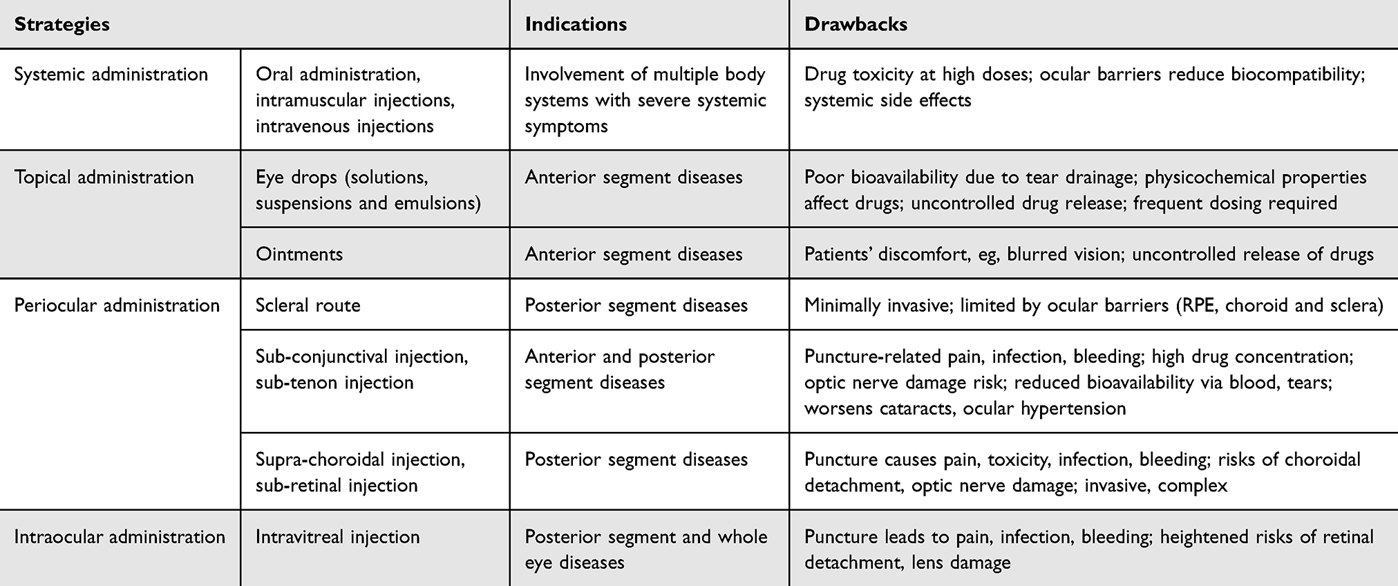 |
Table 1 Overview of Advantages and Limitations of Conventional Drug Delivery Methods for Ophthalmic Disorders41 |
 |
Figure 2 Multiple barriers of the eye. It exhibits various physiological barriers and factors that may influence drug distribution in ophthalmic therapy, encompassing the tear film, the blood-retina barrier, and other critical components. It highlights the complex interplay of these barriers, which can significantly impact the delivery and efficacy of therapeutic agents targeting ocular diseases. Understanding these barriers is crucial for the development of effective drug delivery systems that can navigate these challenges, ensuring optimal treatment outcomes in ophthalmic care. Drawn with Procreate. |
Diverse NIM Platform from Different Materials
The NIM platform leverages its nanoscale dimensions and surface characteristics to effectively overcome ocular barriers and achieve targeted drug delivery (Figure 1). While conventional NPs (eg, liposomes, polymeric NPs) demonstrate some ocular delivery capability, they inherently struggle with the “penetration-retention paradox” - smaller NPs can penetrate ocular barriers but are rapidly cleared, while larger particles with better retention capacity lack penetration ability.42–45 The NIM platform ingeniously addresses this limitation through hierarchical engineering that decouples these functions into two cooperative scales.46
The NIM platform incorporates several groundbreaking innovations that collectively address the challenges of ocular drug delivery. One key advancement is its Dual-Ligand Engineering system, which strategically combines microscale carriers enhanced with mucoadhesive polymers (eg, chitosan) for improved precorneal retention47,48 with nanoscale modules designed for cell-specific targeting, such as the AS1411 aptamer for retinoblastoma therapy.49–51 Another significant innovation is the platform’s Stimuli-Responsive release capability, where drug delivery can be precisely activated by specific biological conditions including pH, ROS levels, or glucose concentrations. This is exemplified by glucose-responsive carboxyphenylboronic acid (CPBA)-functionalized mesoporous silica NPs (MSNs) that enable controlled delivery of 1,25-dihydroxyvitamin D3 for diabetic retinopathy treatment.52 Furthermore, the platform’s Scalable Manufacturing process, utilizing microfluidic production techniques, demonstrates markedly superior batch-to-batch consistency compared to conventional methods, effectively overcoming critical translational challenges in pharmaceutical development.53,54 These integrated innovations position the NIM platform as a transformative approach in ocular therapeutics.
Inorganic Nanomaterials for Ophthalmic Applications (Table 2)
Inorganic NPs (eg, gold, silver, silica, quantum dots) possess controllable dimensions, structures, and unique optoelectromagnetic properties that make them promising candidates for drug delivery and therapeutic applications. However, they face several challenges including susceptibility to degradation, poor biocompatibility, and insufficient stability/dispersibility, which limit their clinical utility.55,56 Surface functionalization with organic/inorganic coatings can significantly improve their dispersion and stability in biological systems.57 These materials have been strategically incorporated into NIM platforms to overcome the limitations of single-component systems58,59 (Figure 3), demonstrating unique advantages in ophthalmic therapies.
 |
Table 2 Representative Applications of Inorganic NPs in Drug Delivery Systems |
 |
Figure 3 Schematic model of a magnetic nanoparticle for drug delivery. It presents a schematic model of a magnetic nanoparticle specifically designed for drug delivery applications. This illustrative example showcases the intricate structure and functionality of such particles, which leverage magnetic properties to enable precise targeting and controlled release of therapeutic agents. By incorporating magnetic cores encapsulated within biodegradable and biocompatible materials, these nanoparticles facilitate enhanced drug delivery efficacy and reduced side effects, marking a significant advancement in the field of targeted therapies. |
Nanozymes exhibit enzyme-mimicking catalytic activities with remarkable antioxidant and anti-inflammatory effects. Their nanoscale dimensions and ease of modification enable effective ocular barrier penetration, making them ideal for targeted treatment of multifactorial eye diseases like dry eye syndrome and AMD.107 A recent study108 developed a novel nanozyme-incorporated hydrogel coating (NHC) through Schiff base reaction between polyaldehyde oligomers (PAO) and amino-functionalized hyaluronic acid (AHA). This system co-loaded voriconazole and copper procyanidin (CuPC) nanozymes for fungal keratitis treatment, where the combined catalase-like and superoxide dismutase-like activities synergistically promoted corneal wound healing, with enhanced efficacy through prolonged retention and improved corneal permeability.
Quantum dots (QDs), particularly carbon-based quantum dots (CDs), have emerged as attractive nanomaterials due to their exceptional fluorescence properties, ultra-small size for cellular/tissue penetration, facile synthesis and surface modification, low cytotoxicity and superior aqueous dispersibility. These characteristics make CDs excellent candidates for ocular imaging, drug delivery, and disease diagnosis.109–112 Notably, spermidine-derived carbon QDs (CQDSpds) synthesized via a one-step dry-heat method demonstrated antibacterial properties and enhanced corneal epithelial permeability through their ultrahigh positive charge-induced tight junction opening.73
Mesoporous silica NPs (MSNs) offer tremendous potential for drug delivery owing to their high stability, large surface area and pore volume, tunable pore sizes and ease of production and functionalization. Hydrophilic MSNs serve as stable, biocompatible carriers that prolong blood circulation, making them a research hotspot.113,114 Surface-functionalized MSNs with NH2 and PEG modifications, combined with nanomolding technology for drug conjugation, have shown improved bioavailability and prolonged therapeutic effects in treating retinal pathological neovascularization.115
Innovative metal nanomaterial designs have demonstrated significant therapeutic enhancements. PEG-coated nanoceria (P/CeO2) developed by Haijie Han’s team exhibited improved biocompatibility, hydrophilicity, and remarkable ocular tolerance while prolonging precorneal retention.116 Urchin-like gold nanostructures with optimized branch length showed 150-fold greater corneal retention compared to smooth gold NPs. In dry eye rabbit models, quercetin-loaded nano-urchins (NU-Q(H)) demonstrated: 30-fold increase in tear production, 49-fold suppression of IL-6 expression, 32-fold reduction in pathological angiogenesis, as well as 18-fold enhancement in nerve regeneration.117 Ion-responsive alginate-capped nanoceria (Ce-ALG) for β-1,3-glucan delivery in corneal abrasion treatment: Alginate coating improved mucoadhesion via hydrogen bonding, Ca2+-mediated “egg-box” structure enhanced drug loading and sustained release, single-dose treatment reduced epithelial defects by 99%. It Showed 45–53 times greater efficacy than conventional treatments.118
Carbon nanomaterials derive their antioxidant, radical-scavenging, and anti-inflammatory properties from: heteroatom doping, sp2 domains, edge structures, and functional groups Notably, sodium alginate/1,8-diaminooctane-derived carbon nanodots (SA/DAO-CNDs) demonstrated >10-fold stronger binding affinity to VEGF-A165 than clinically used inhibitors (aflibercept and ranibizumab).89
Polymeric-Based Materials for the Treatment of Ocular Diseases
Polymers (Table 3) can be categorized into natural and synthetic types, offering high stability and drug-loading capacity. Their mucoadhesive properties make them ideal carriers for ocular drug delivery, prolonging drug retention and reducing clearance.41,119 Some polymers exhibit stimuli responsiveness, allowing smart polymers to function as in situ gelling systems. Biodegradability is a significant advantage, facilitating sustained drug release.120,121 Recent advancements in precisely controlling the molecular weight and sequence of synthetic polymers have enabled effective mucoadhesion and physiological barrier penetration. Controlled radical polymerization techniques allow for the preparation of complex polymer ligands, with homogeneous monomer sequence polymers supporting precise delivery.8,121
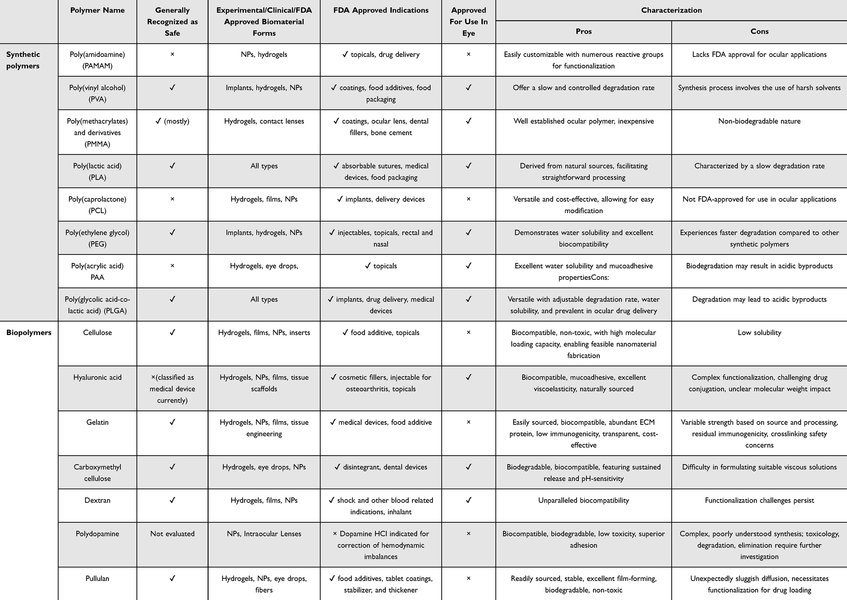 |
Table 3 Condensed Overview of Polymers in Ophthalmic Applications.122 |
Polysaccharide-based biomaterials have emerged as a highly promising class of ocular drug delivery vehicles, demonstrating superior tissue compatibility and outperforming synthetic materials in both drug retention and ocular permeability. Natural polysaccharides like chitosan and hyaluronic acid have been successfully engineered into nanocarriers that combine excellent biocompatibility with enhanced drug bioavailability and favorable safety profiles. Through strategic chemical modifications, researchers have further optimized these systems to improve ocular residence time and drug solubility.123 The therapeutic potential of polysaccharide-based nanocarriers has been extensively documented across various ocular diseases, with numerous studies highlighting their clinical translation potential.124 However, despite these advances, significant knowledge gaps remain regarding their long-term ocular safety, as comprehensive monitoring data are still lacking. Additionally, while polysaccharides are generally biodegradable, their degradation kinetics within the unique ocular microenvironment require further refinement to achieve optimal performance.125 Recent innovations in this field include two particularly noteworthy developments: First, a sophisticated resveratrol-loaded polycaprolactone nanoparticle system (R@PCL NP) that was functionalized with cell-penetrating peptide and metformin through amide bond formation. This design achieved an impressive 15-fold enhancement in retinal permeability following single intravitreal injection.126 Second, an advanced surface-engineered ceria nanocage platform (SRCN) incorporating poly(L-histidine) coatings that enabled multiple therapeutic functions, including enhanced corneal penetration and lesion-specific dual-drug release. This innovative formulation demonstrated remarkable efficacy, showing a 19-fold greater wound reduction than commercial eye drops, 93% suppression of pathological angiogenesis, and nearly complete corneal clarity restoration within just four days.127 These cutting-edge systems exemplify how engineered polysaccharide-based nanocarriers are pushing the boundaries of ocular therapeutics through enhanced delivery efficiency and superior treatment outcomes.
Lipid-Based Materials for the Treatment of Ocular Diseases
Solid lipid NPs (SLNs) and liposomes represent two distinct types of lipid-based drug carriers with different structural and functional characteristics. SLNs are composed of solid lipids (such as glyceryl palmitate) that form a crystalline matrix, enabling high encapsulation efficiency and sustained release of hydrophobic drugs. In contrast, liposomes feature phospholipid bilayers surrounding an aqueous core, making them particularly suitable for hydrophilic payloads and rapid drug release.128–130
Solid lipid NPs (10–1000 nm) offer multiple advantages as ocular delivery vehicles, including enhanced permeability, prolonged retention, improved solubility, reduced toxicity, and targeted delivery capabilities.131 Their self-assembly properties, arising from lipid-aqueous phase interactions, have shown therapeutic potential for various ocular conditions including conjunctivitis, glaucoma, and retinal diseases (Table 4). The therapeutic efficacy of SLNs largely depends on formulation strategies that maximize drug concentration in target tissues. Co-loading of multiple drugs in SLNs enables sophisticated delivery paradigms, significantly increasing therapeutic payloads to specific ocular sites.132,133
 |
Table 4 Representative Lipid-Based Materials for Ophthalmic Application |
Currently, both unilamellar and multilayer vesicular liposomes have been developed to carry hydrophilic and lipophilic substances, quickly absorbed by the reticuloendothelial system. However, SLNs face non-specific uptake by the mononuclear phagocytic system. By attaching different ligands to the surface of SLNs, circulation time and targeted drug delivery to specific sites can be enhanced, thereby overcoming these limitations.177 Selecting surface biomarkers can improve targeting specificity.130 Additionally, widespread clinical application faces challenges related to the reproducibility and reliability of methods. Production requires a multi-component processing line involving centrifugation, filtration, freeze-drying, emulsification, crosslinking, ultrasonication, solvent evaporation, homogenization, and milling, making it difficult to optimize process parameters for stable key quality attributes at a commercial scale, even though small-scale prototypes are relatively easy to obtain.178
Extracellular Vesicles for Ophthalmic Applications
Extracellular vesicles (EVs), including exosomes (50–150 nm), microvesicles, and apoptotic bodies, originate from cellular membrane structures and play roles in biological metabolism, immune responses, cell communication, and disease progression.179 Exosomes, in particular, possess therapeutic potential and value as disease biomarkers due to their low immunogenicity, low toxicity, and membrane marker characteristics.180 However, the high complexity and heterogeneity of EVs, including variations in size, content, function, and source, can significantly influence their effects on recipient cells. Moreover, the super-physiological injection doses and administration frequencies of EVs in different studies contribute to uncertainties regarding their safety. As our understanding of EVs, their cargo, and functional heterogeneity continues to evolve, the demand for precise and accurate characterization of EVs in the context of ocular disease mechanisms and therapies will persist and flourish (Table 5).
 |
Table 5 Representative Exosomes-Associated Nanomaterial Therapeutics |
Nanofibers
Electrospun nanofibers (100–500 nm in diameter) have emerged as a transformative platform for ocular drug delivery by biomimetically replicating key features of the extracellular matrix.200 These advanced systems offer three major therapeutic advantages: First, their sustained release capability has been demonstrated through innovative coaxial electrospun constructs, such as the corn-derived core (zein)-shell (PLA) nanofibers dual-loaded with rutin and celastrol for conjunctival repair.201 This design enables sequential drug release - an initial anti-inflammatory phase from the PLA shell followed by prolonged antifibrotic activity from the zein core. Second, their therapeutic versatility extends to oxidative stress management, as shown by Juan Ye team’s ROS-scavenging dual-network system comprising a poly(PEGMA-co-GMA) hydrogel integrated with electrospun polyurethane membranes for corneal burn treatment.202
The exceptional performance of electrospun nanofibers in ocular applications stems from their unique structural characteristics produced via this established biomedical technique.101,203,204 Their high surface area-to-volume ratio and optimal porosity enable: (1) precise spatiotemporal control of drug release profiles, (2) enhanced biocompatibility and biodegradability matching ocular tissues, (3) significant reduction of administration side effects, and (4) marked improvement in therapeutic outcomes compared to conventional formulations.123,205 These advantages, combined with the ability to incorporate multiple active compounds and functional components in a single system, position electrospun nanofibers as a next-generation platform capable of addressing complex ocular disease pathophysiology through sophisticated, biomimetic drug delivery approaches.
Nucleic Acid-Based Nanomaterials for Ophthalmic Applications
Nucleic acid-based nanomaterials (Table 6) are assembled through hybridization and self-folding processes, encompassing DNA nanomaterials, RNA nanomaterials, RNA-based motifs, and DNA/RNA origami structures. Their notable advantages include high biocompatibility and low immunogenicity. Additionally, these materials can be structurally and functionally programmed for highly selective target binding via aptamers, demonstrating immense potential in biomedical applications such as biosensing, bioimaging, cell reprogramming, gene expression, and targeted delivery.206,207 DNA nanostructures, particularly the tetrahedral framework pioneered by Turberfield in 2005, show promise due to their structural stability and capabilities in scavenging reactive oxygen species, enhancing membrane permeability, and programmable stimuli responsiveness.208 Furthermore, by embedding stimulus-sensitive sequences—such as lysosome-activated, nucleotide-sensitive, or pH-sensitive sequences—dynamic targeting and precise release of cargo are achieved.209 RNA nanostructures also display unique advantages in in vivo isothermal transcription. However, challenges such as susceptibility to degradation, off-target effects, and cross-reactivity still need to be addressed.
 |
Table 6 Representative Nucleic Acid-Associated Nanomaterial Therapeutics |
The strategic selection of materials for NIM platforms plays a pivotal role in determining both drug-loading capacity and biocompatibility, necessitating meticulous optimization to achieve optimal therapeutic outcomes. Different material classes offer distinct advantages and challenges: polymeric matrices (eg, PLGA, chitosan) achieve high drug encapsulation efficiency through their porous architectures and adjustable degradation kinetics,226,227 though their inherent immunogenicity can be effectively addressed through innovative erythrocyte membrane coatings that substantially reduce reticuloendothelial system recognition and clearance.228 Inorganic carriers such as mesoporous silica and gold NPs provide exceptionally large surface areas for small molecule loading,229 yet require biocompatible surface modifications to mitigate potential oxidative stress effects. Lipid-based systems demonstrate superior biocompatibility through their endogenous components that minimize immune clearance, but require stabilization with agents like trehalose to prevent lipid leakage.230–232
The true innovation of NIM platforms lies in their ability to synergistically combine these materials through hierarchical engineering. A prime example is the encapsulation of silica NPs within phosphatidylserine (PS)-modified liposomes, which creates an advanced hybrid system that simultaneously achieves: (1) targeted drug delivery, (2) enhanced cellular uptake, (3) controlled release profiles, (4) excellent biocompatibility, and (5) preserved cell viability. This materials integration approach represents a paradigm shift in ocular therapeutics, as it enables precise balancing of drug payload capacity with biological safety parameters. The modular design philosophy underlying NIM technology allows for rational combination of material advantages while systematically addressing their individual limitations, thereby creating optimized delivery systems that transcend the capabilities of single-component platforms.
Commonality of the Properties of Nano-Micro Particles for Different Administration Approaches (Figure 4)
Nanoparticle interactions and biological behavior in the eye largely depend on their diverse characteristics, particularly size, surface charge, shape, and other physicochemical properties that determine the nano-bio interface interactions in various biological systems.233 For instance, positively charged NPs tend to have longer retention times in the cornea and facilitate penetration through phospholipid membranes.121 Thus, when designing nanoparticle platforms for ocular applications, balancing physicochemical properties (eg, size, surface charge) with biological compatibility is critical to ensure optimal performance.
 |
Figure 4 The application of drug delivery systems in the treatment of various ocular diseases. It illustrates the diverse applications of drug carriers in various ocular diseases. It showcases how these innovative delivery systems are tailored to address a spectrum of eye conditions, each demanding unique therapeutic strategy. It underscores the versatility and significance of drug carriers in ophthalmology, demonstrating how they are pivotal in advancing the treatment of a wide range of eye diseases, ultimately aiming to enhance patient quality of life and visual health. Created in BioRender. Lin, E. (2025) https://BioRender.com/tgyfyi1. |
While various types of nanomaterials exist, they often share common properties under specific delivery routes. In vitreous injections, the nano-biointerface between NPs and the vitreous cavity plays a key role in cellular uptake, particularly regarding targeting.234 The vitreous is mainly composed of 98–99% water, glycosaminoglycans, salts, and various matrix proteins, all of which influence nanoparticle behavior.235 Research indicates that smaller NPs (approximately 100 nm) move more easily within the vitreous matrix, while larger particles encounter significant obstacles. Anionic NPs are observed to penetrate the vitreous more effectively than cationic ones,236 and the shape of NPs also impacts their mobility.237
Topical eye drops are a non-invasive and convenient method for delivering drugs to the anterior eye tissues, widely used for conditions like dry eye, glaucoma, and infections.238 Given that the ocular surface carries a negative charge, positively charged drug carriers generally exhibit better permeability.239 Beyond charge and size, factors like lubricity, muco-adhesiveness, viscosity, and biocompatibility are critical in eye drop formulations. Recent studies have explored novel nanoparticle platforms for local delivery, such as hydrogels, fluid gels, and lipid NPs, significantly enhancing corneal retention time and drug bioavailability in anterior tissues.240 A notable example is the development of rosmarinic acid-conjugated gelatin nanogels co-loaded with diquafosol sodium, which has demonstrated remarkable improvements in ocular surface retention time and therapeutic efficacy for dry eye treatment.241 This dual-functional system combines the anti-inflammatory properties of rosmarinic acid with the mucin secretagogue action of diquafosol, exemplifying the potential of multifunctional nanocarriers in optimizing ocular drug delivery.
Systemic injections face challenges in effectively reaching the retina due to the tight junctions of the blood-retinal barrier (BRB). In this route, NPs may circulate to non-target organs with the bloodstream, necessitating careful evaluation of their biosafety, including biodistribution, excretion, tissue clearance, and potential side effects or toxicity. Insights from blood-brain barrier (BBB) or cancer therapy research suggest that the ideal size for NPs suitable for systemic injection ranges between 2 to 200 nm, providing valuable reference for BRB-related studies.242 For example, 20 nm gold NPs (AuNPs) can successfully penetrate the BRB, while those larger than 100 nm cannot.243 Although smaller NPs facilitate retinal penetration, unmodified conventional NPs struggle to accurately target pathological cells or accumulate at lesions.
Recent advancements in ocular drug delivery have led to the development of sophisticated smart nanoparticle platforms engineered with precision-targeting ligands and aptamers to address the challenges of ocular therapy.244 These next-generation systems employ innovative ligand engineering strategies to overcome existing limitations in drug delivery. A prime example is the PLGA@AST/AXI nanoparticle system,226 which utilizes FDA-approved poly(lactic-co-glycolic acid) to co-encapsulate astaxanthin (a multifunctional carotenoid with antioxidant, anti-inflammatory, and anti-apoptotic properties) and axitinib (a selective VEGF receptor tyrosine kinase inhibitor). This dual-drug platform demonstrates four key advantages: (1) multi-targeted action against wet AMD pathogenesis, (2) sustained release kinetics from a single subconjunctival administration, (3) excellent ocular biocompatibility without tissue damage, and (4) significant therapeutic potential for posterior segment diseases.
The field has further expanded to include several breakthrough platforms: the bioadhesive nanoparticle network system (BNP/CA-PEG) combining cefuroxime axetil with 8-arm polyethylene glycol for enhanced antibiotic delivery,245 and the chondroitin sulfate-cysteine modified nanostructured lipid carriers (Dex-cNLC) that specifically target ocular mucin substructures for efficient dry eye treatment.246 These systems exemplify the growing sophistication in ocular nanomedicine through their targeted delivery mechanisms and improved therapeutic profiles.
However, the translation of these technologies faces substantial challenges, particularly in optimizing ligand-receptor interactions and addressing interspecies variability in ocular biology. Future progress requires a concerted effort to:
- Standardize ligand density and binding parameters
- Elucidate species-specific differences in ocular receptor expression
- Develop scalable, reproducible manufacturing processes
- Establish comprehensive biocompatibility assessment protocols
The integration of these considerations with continued nanoplatform innovation will be crucial for advancing precision ocular therapeutics from laboratory concepts to clinically viable treatments, ultimately enabling more effective management of complex ocular diseases while minimizing systemic side effects. This holistic approach represents the next frontier in ophthalmic drug delivery, combining cutting-edge nanotechnology with rigorous translational science.
Biosafety and Toxicity Profiles of Ocular Nanomaterials: Mechanistic Insights and Interspecies Variability
The NIM platform represents a paradigm shift in ocular drug delivery biosafety, offering transformative advantages over conventional nanocarriers through its innovative hierarchical architecture. This sophisticated design fundamentally addresses the longstanding “toxicity-efficacy paradox” in ocular therapeutics by simultaneously enhancing treatment precision while reducing systemic and local toxicity (Figure 5). The platform’s success stems from three synergistic safety mechanisms: (1) Barrier-shielded delivery exemplified by the PVA/PDA-PBA@MT eye drop system, which combines polydopamine NPs’ exceptional radical scavenging capacity with prolonged ocular retention and controlled melatonin release for dry eye management;247 (2) Spatiotemporal release control demonstrated by pH-responsive hydrogels and transformable microneedle patches that precisely target pathological microenvironments while minimizing off-target effects - particularly the remarkable soft MN patch that delivers antimicrobial NPs to infected corneas before converting to a sustained-release contact lens for wound healing;248 and (3) Enhanced immune evasion through PEGylated microcarriers that significantly reduce dendritic cell activation and promote immune tolerance compared to bare NPs.249
 |
Figure 5 Potential toxicity of nanomaterials. It depicts potential drug toxicities that may arise from the administration of nanomedicines. These toxicities encompass a wide spectrum of adverse effects, including disruptions to the cell cycle, which can lead to abnormal cell proliferation or arrest, as well as DNA damage, a critical concern given its potential to induce mutations and genomic instability. It underscores the importance of rigorous toxicity assessments and the development of safer nanocarriers to minimize these risks, ensuring the safe and effective application of nanomedicines in therapeutic interventions. Created in BioRender. Lin, E. (2025) https://BioRender.com/wcw3s1r. |
These technological breakthroughs are supported by the platform’s unique ability to decouple and optimize two critical safety parameters: microscale components control systemic exposure through regulated retention, while nanoscale elements modulate local cytotoxicity via precision engineering. However, comprehensive safety assessment requires addressing several key challenges: nanoparticle-specific toxicity mechanisms including oxidative stress and DNA damage,35 size- and surface charge-dependent biological interactions,250 species variability in ocular physiology, long-term exposure effects, and dynamic nanoparticle-protein corona formation in ocular fluids. Moving forward, the field must prioritize standardized safety evaluation protocols that bridge preclinical and clinical studies, with particular emphasis on surface engineering strategies to mitigate risks while maintaining therapeutic efficacy. The NIM platform’s success in harmonizing these complex parameters positions it as a groundbreaking approach in ocular therapeutics, offering new hope for treating challenging eye diseases while setting a new standard for drug delivery biosafety.
Factors Affecting the Bio-Performance of Nano/Micro Delivery Systems
The biological performance of nano- and microparticle delivery systems, including biodistribution and bioavailability, is influenced by multiple factors including size, surface charge, solubility, and biodegradability.251 To enhance these performances, researchers aim to prolong bioavailability, broaden biodistribution, and reduce toxicity. The loading capacity of the delivery system is crucial for in vivo performance and primarily depends on the manufacturing process. For chronic disease treatment, matrices with good biodegradability or intelligent responsiveness are more suitable, as they can prevent preloaded drugs from directly interacting with specific tissues, which is essential for intracellular applications like genome editing.252
Release kinetics is another important indicator for assessing the performance of delivery systems.253 Particulate and hydrogel formulations excel in improving drug release profiles. Additionally, enhanced cellular uptake is particularly critical for intracellular applications. While small-sized NPs have advantages, their size must be optimized to avoid rapid clearance or ocular irritation.
The surface charge of NPs affects cellular uptake and intracellular transport.254 Positively charged NPs may disrupt cell membranes or exhibit toxicity, while anionic NPs are internalized via specific endocytic pathways. Therefore, selecting NPs requires a balance between size and charge to maximize cellular uptake and minimize toxicity.
Beyond surface charge, the stiffness, hydrophobicity, and topology of nano/microsystems also influence cell adhesion, thereby affecting biocompatibility and uptake mechanisms.255 To enhance biocompatibility, researchers have explored various coating materials to modify the nanoparticle interface.
In summary, optimizing nano/microparticle delivery systems necessitates a comprehensive consideration of factors such as size, surface charge, solubility, biodegradability, and other surface characteristics. Through meticulous design and appropriate surface modifications, more efficient and safe drug delivery can be achieved, with the introduction of smart release capabilities further enhancing system performance (Figure 6).
 |
Figure 6 The basic parameters involved in the process of drug delivery. It illustrates the fundamental parameters that are integral to the process of drug delivery. Looking ahead, the trajectory of advancement lies in the continued pursuit of smart drug development, which encompasses the realm of nanomedicines engineered to respond to various physiological factors such as pH levels in bodily fluids and blood glucose concentrations. This evolution toward smarter therapeutic solutions aims to enhance the precision and efficacy of drug administration, tailoring. Created in BioRender. Lin, E. (2025) https://BioRender.com/wcw3s1r. |
FDA Approved and Under Clinical Trial Nanomedicine for Ocular Diseases
Nanocarriers, due to their nanoscale size and surface characteristics, hold great promise for penetrating ocular barriers and delivering drugs precisely to target sites. Extensive research on nanoformulations for anterior and posterior segment diseases has yielded positive results in clinical trials (Table 7). Commercially available products like Restasis256 and Durezol257 are used for treating dry eye syndrome and ocular inflammation, respectively. Other marketed nanostructured products include Cequa®258 and Cyclokat®259 (both cyclosporine A nanoemulsions), Lacrisek®260 (liposomal vitamin), and Artelac Rebalance®261 (lubricant). Visudyne262 is indicated for conditions like choroidal neovascularization. InSite Vision’s Durasite® is a novel drug delivery system, with its besifloxacin formulation approved by the FDA.263 Several nanoformulations, such as Ozurdex,264 Iluvien265 and Trivaris266 are used for treating macular edema and uveitis. Currently, various nanoformulations, including TLC399 (ProDex),267 latanoprost-coated liposomes (POLAT-001),268 and SYSTANE®,269 are undergoing clinical trials for conditions like ocular hypertension, glaucoma, AMD, diabetic macular edema, and ocular infections.
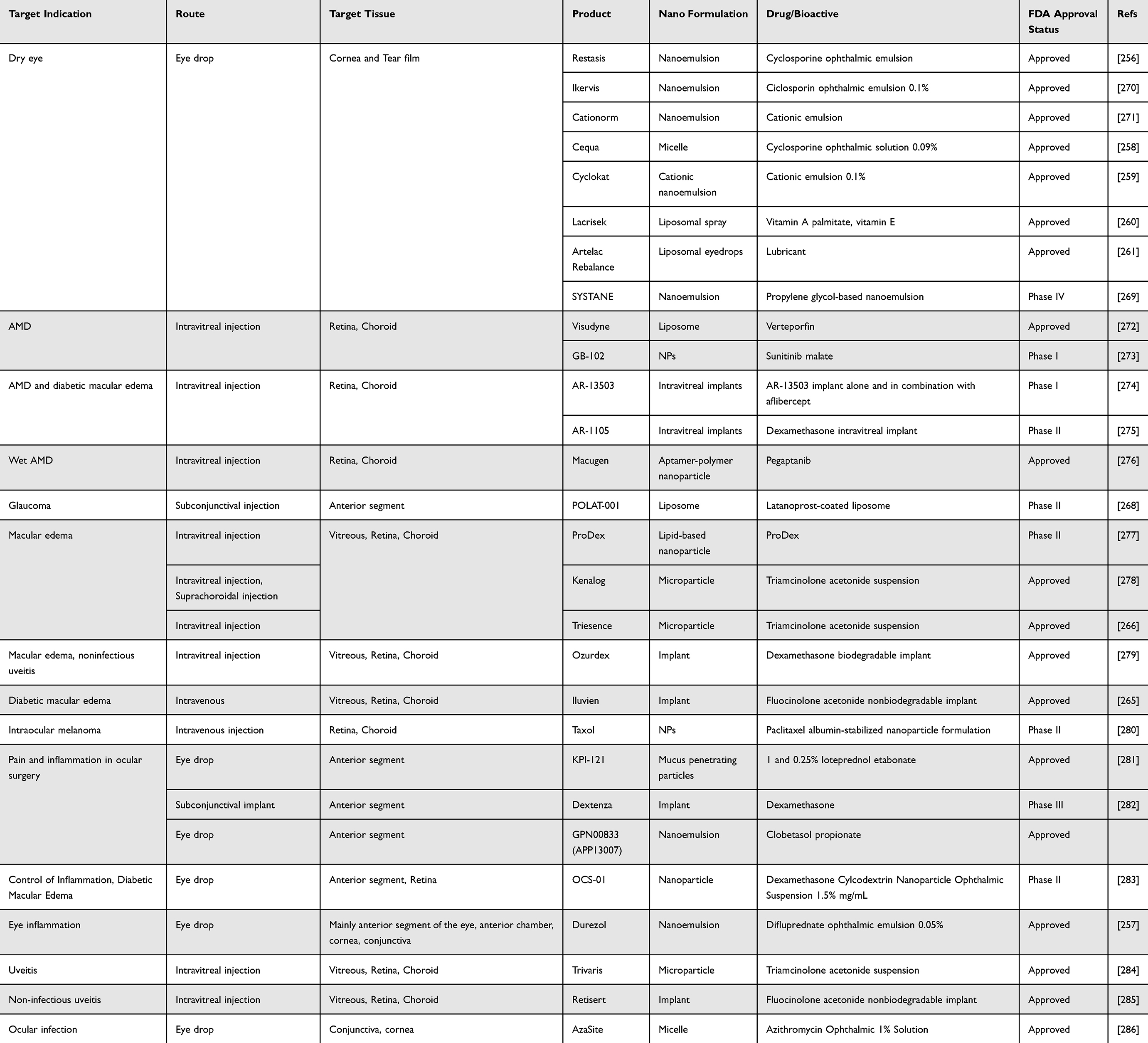 |
Table 7 FDA Approved and Under Clinical Trial Nanomedicine for Ocular Diseases |
The rapid development of nanotechnology and microsystems has integrated NPs with micro-matrices (such as hydrogels, microspheres, and liposomes), combining the advantages of both to provide a larger drug-loading surface area. However, the biocompatibility of inorganic NPs is relatively poor, which can lead to side effects or cytotoxicity. To address this, researchers have combined inorganic NPs with biocompatible polymers to shield encapsulated components from in vivo clearance, thereby enhancing biocompatibility and optimizing drug release profiles.287 In ophthalmic applications, this system demonstrates significant potential for sustained drug delivery, cell encapsulation, and transplantation. Additionally, cell encapsulation technology is being used to create novel therapeutic platforms by encapsulating genetically engineered cells to produce therapeutic factors. Given their longevity, immune privilege characteristics, and ease of gene editing, human retinal pigment epithelial cell lines (ARPE-19) and mesenchymal stem cells are preferred choices. A notable ARPE-19-based delivery system is Neurotech Inc.’s NT-501 device,288 which has been used clinically for the controlled release of ciliary neurotrophic factor.
A number of FDA-approved drugs have completed or are undergoing clinical trials for further validation. Dexamethasone, a cornerstone anti-inflammatory agent, is increasingly delivered via sustained-release implants to address clinical challenges associated with frequent dosing and poor patient compliance. Ozurdex® and DEXTENZA® are exemplary models of sustained-release systems for dexamethasone delivery, with their applications significantly enhancing patient compliance and clinical outcomes. The Ozurdex® system encapsulates dexamethasone within biodegradable poly(lactic-co-glycolic acid) (PLGA) microspheres, enabling prolonged drug release at the target site. In the DME trial (NCT05372562), patients treated with Ozurdex® demonstrated superior outcomes: 22–28% achieved ≥15-letter visual acuity improvement (vs 12% in controls), alongside central macular thickness reductions of 100–150 μm (vs 30 μm in controls). A multicenter Chinese trial (NCT06548568) is currently underway to further validate these findings. The system’s efficacy, lasting up to six months, underscores its potential to reduce treatment burden and improve quality of life. DEXTENZA®, on the other hand, is an FDA-approved intracanalicular insert, employs a polyethylene glycol (PEG)-based plug to deliver dexamethasone nanocrystals for postoperative inflammation/pain management following cataract or glaucoma surgeries. Phase III trial data (NCT02525036) demonstrated significant resolution of inflammation and pain reduction, highlighting its clinical viability. Another example based on biodegradable PLGA microsphere-based platform, is OTX-TIC system, incorporating travoprost nanocrystals for glaucoma management. Clinical trials evaluating its efficacy and safety in patients with open-angle glaucoma (OAG) or ocular hypertension (OHT) aim to validate its potential as a single-application alternative to daily topical therapies.
The NIM platform demonstrates significant translational potential, with emerging clinical adoption in ophthalmic therapeutics. Clinical evaluations have demonstrated superior therapeutic efficacy compared to traditional drug delivery approaches, particularly in targeting chronic ocular pathologies. Nevertheless, the full clinical translation of NIM formulations continues to face persistent challenges.
Challenges in the Clinical Translation
Nanomaterial drug delivery systems face multiple challenges in ophthalmic applications, leading to slow progress and high costs. The primary obstacles involve safety, regulatory approval, scalability, and cost-effectiveness.289 The complexity of new nanotherapeutic products makes the approval process time-consuming and challenging. Although the FDA has released draft guidance documents, final guidelines are still pending, and current regulatory requirements remain aligned with small molecules. These testing standards may not be suitable for nanoproducts, potentially resulting in biased outcomes.
The transition to clinical applications hinges on reproducibility and large-scale production; yet the structural and physicochemical complexity of nanomaterials often leads to poor reproducibility. Issues such as inconsistency, inadequate quality control, low yield, and high manufacturing costs are prevalent in nanodrug production, complicating quality assurance and control. Lastly, patient safety is paramount. An ideal nanoplatform should exhibit good biocompatibility and safety, minimizing adverse effects or ocular accumulation. Although extensive research has explored the toxicity of NPs on ocular tissues, most data are based on animal models, which may not accurately reflect human conditions due to significant differences in retinal immune composition.290 Additionally, the biocompatibility and toxicity of NPs are closely tied to their physicochemical properties, and there remains insufficient evidence to confirm their biocompatibility and toxicity in the human eye.
Conclusion and Prospects
The NIM platform combines nanoscale precision with micron-scale stability to address the ongoing challenges of drug retention, penetration, and biocompatibility in ocular drug delivery, representing a transformative approach in the field of ocular therapy. This innovative technology has shown significant potential, especially in the treatment of genetic retinal diseases. The CRISPR-Cas9 lipid NPs delivered through the NIM platform achieved high gene correction efficiency in a model of retinitis pigmentosa.291–293 However, there are still some key obstacles hindering its clinical translation. The challenges of materials science, especially at the interface of inorganic polymers, require innovative solutions to ensure optimal performance of the system, while regulatory ambiguity significantly limits the progress of NIM preclinical research towards clinical trials. Clinical translation has shown that only a few ocular nanotherapeutic drugs have entered Phase 3 trials, mainly hindered by inconsistent manufacturing and scalability limitations of microfluidic production systems.294,295 Safety considerations remain paramount, as evidenced by the heterogeneity of diseases and the large number of mutations in genes such as RPE65, which directly affect the toxicity threshold of nanomaterials.296,297 These challenges underscore the urgent need to balance efficacy and biocompatibility, especially for long-term treatment regimens. Looking ahead, several key research directions must be prioritized in this field: (1) the development of advanced delivery systems that combine artificial intelligence-guided drug release and sustainable biodegradable matrices; (2) creating enhanced formulations with improved stability and pharmacokinetic characteristics for small molecules and biologics; (3) Expand translational research through comprehensive in vivo studies and optimization of non-invasive delivery methods. When developing solutions for diseases, multidisciplinary collaboration is crucial to addressing these challenges. By successfully overcoming these obstacles, NIM technology has the potential to move beyond incremental improvements and become the foundational platform for the next generation of ophthalmology, effectively bridging the gap between nanoscale innovations and meaningful clinical impact in ophthalmic treatments.
Data Sharing Statement
No datasets were generated or analyzed during the current study.
Funding
This study was supported by grants from the National Natural Science Foundation of China (No. 82271054; No. U20A20363), and the 2024 National Natural Science Foundation Cultivation Program (Youth Fund) (No. gyfynsfc[2024]-05) and Guizhou Provincial Health Commission Science and Technology Fund (No. gzwkj2025-433).
Disclosure
The authors declare that they have no competing interests.
References
1. Abdolalizadeh P, Chaibakhsh S, Falavarjani KG. Global burden of paediatric vision impairment: a trend analysis from 1990 to 2017. Eye. 2021;35(8):2136–2145. doi:10.1038/s41433-021-01598-8
2. Bourne R, Steinmetz JD, Flaxman S, et al. Trends in prevalence of blindness and distance and near vision impairment over 30 years: an analysis for the Global Burden of Disease Study. Lancet Glob Health. 2021;9(2):e130–e143. doi:10.1016/S2214-109X(20)30425-3
3. Tang B, Wang Q, Zhang G, et al. OCTN2- and ATB(0,+)-targeted nanoemulsions for improving ocular drug delivery. J Nanobiotechnology. 2024;22(1):130. doi:10.1186/s12951-024-02402-x
4. Chen L, Wu M-Y, Chen S-L, et al. The guardian of vision: intelligent bacteriophage-based eyedrops for clinical multidrug-resistant ocular surface infections. Adv Mater. 2024;36(38):e2407268. doi:10.1002/adma.202407268
5. Wang X, Zhang M, Li Y, Cong H, Yu B, Shen Y. Research status of dendrimer micelles in tumor therapy for drug delivery. Small. 2023;19(50):e2304006. doi:10.1002/smll.202304006
6. Lyu Q, Peng L, Hong X, et al. Smart nano-micro platforms for ophthalmological applications: the state-of-the-art and future perspectives. Biomaterials. 2021;270:120682. doi:10.1016/j.biomaterials.2021.120682
7. Fathi-Karkan S, Amiri Ramsheh N, Arkaban H, et al. Nanosuspensions in ophthalmology: overcoming challenges and enhancing drug delivery for eye diseases. Int J Pharm. 2024;658:124226. doi:10.1016/j.ijpharm.2024.124226
8. Puricelli C, Gigliotti CL, Stoppa I, et al. Use of poly lactic-co-glycolic acid nano and micro particles in the delivery of drugs modulating different phases of inflammation. Pharmaceutics. 2023;15(6):1772.
9. Huang J, Jiang T, Qie J, et al. Biologically inspired bioactive hydrogels for scarless corneal repair. Sci Adv. 2024;10(51):eadt1643. doi:10.1126/sciadv.adt1643
10. Sheng AA, Lin L, Zhu J, et al. Micro/nanodevices for assessment and treatment in stomatology and ophthalmology. Microsys Nanoeng. 2021;7(1):11. doi:10.1038/s41378-021-00238-1
11. Wang W, Li PF, Xie R, Ju XJ, Liu Z, Chu LY. Designable micro‐/nano‐structured smart polymeric materials. Adv Mater. 2022;34(46):2107877. doi:10.1002/adma.202107877
12. Choi H, Yi J, Cho SH, Hahn SK. Multifunctional micro/nanomotors as an emerging platform for smart healthcare applications. Biomaterials. 2021;279:121201. doi:10.1016/j.biomaterials.2021.121201
13. Ghaderi M, Bi H, Dam-Johansen K. Advanced materials for smart protective coatings: unleashing the potential of metal/covalent organic frameworks, 2D nanomaterials and carbonaceous structures. Adv Colloid Interface Sci. 2024;323:103055. doi:10.1016/j.cis.2023.103055
14. Dludla SB, Mashabela LT, Ng’andwe B, Makoni PA, Witika BA. Current advances in nano-based and polymeric stimuli-responsive drug delivery targeting the ocular microenvironment: a review and envisaged future perspectives. Polymers. 2022;14(17):3580. doi:10.3390/polym14173580
15. Cheng Y, Cai S, Wu H, et al. Revolutionizing eye care: the game-changing applications of nano-antioxidant in ophthalmology. Nanoscale. 2024;16(15):7307–7322. doi:10.1039/D4NR00611A
16. Wu Y, Li X, Fu X, et al. Innovative nanotechnology in drug delivery systems for advanced treatment of posterior segment ocular diseases. Adv Sci. 2024;11(32):2403399. doi:10.1002/advs.202403399
17. Chaudhari P, Birangal S, Mavlankar N, et al. Oil-free eye drops containing Cyclosporine A/cyclodextrin/PVA supramolecular complex as a treatment modality for dry eye disease. Carbohydr Polym. 2022;297:120007. doi:10.1016/j.carbpol.2022.120007
18. Liu L-C, Chen Y-H, Lu D-W. Overview of recent advances in nano-based ocular drug delivery. Int J Mol Sci. 2023;24(20):15352. doi:10.3390/ijms242015352
19. Kahana M, Weizman A, Gabay M, et al. Liposome-based targeting of dopamine to the brain: a novel approach for the treatment of Parkinson’s disease. Mol Psychiatry. 2021;26(6):2626–2632. doi:10.1038/s41380-020-0742-4
20. Barbosa-Alfaro D, Andrés-Guerrero V, Fernandez-Bueno I, et al. Dexamethasone plga microspheres for sub-tenon administration: influence of sterilization and tolerance studies. Pharmaceutics. 2021;13(2):228. doi:10.3390/pharmaceutics13020228
21. Kupikowska-Stobba B, Lewińska D. Polymer microcapsules and microbeads as cell carriers for in vivo biomedical applications. Biomater Sci. 2020;8(6):1536–1574. doi:10.1039/c9bm01337g
22. Mayol L, Silvestri T, Fusco S, Borzacchiello A, De Rosa G, Biondi M. Drug micro-carriers with a hyaluronic acid Corona toward a diffusion-limited aggregation within the vitreous body. Carbohydr Polym. 2019;220:185–190. doi:10.1016/j.carbpol.2019.05.065
23. Joseph RR, Venkatraman SS. Drug delivery to the eye: what benefits do nanocarriers offer? Nanomedicine. 2017;12(6):683–702. doi:10.2217/nnm-2016-0379
24. Zugic A, Tadic V, Savic S. Nano-and microcarriers as drug delivery systems for usnic acid: review of literature. Pharmaceutics. 2020;12(2):156. doi:10.3390/pharmaceutics12020156
25. AlYabhouni SA, Mozumder MS, Hassan N, Mourad A-HI, Issa TM. Nanocarrier-Based, ocular drug delivery: challenges, prospects, and the therapeutic landscape in the United Arab Emirates. Int J Pharm. 2024;124899. doi:10.1016/j.ijpharm.2024.124899
26. Wang T, Yu T, Liu Q, Sung T-C, Higuchi A. Lipid nanoparticle technology-mediated therapeutic gene manipulation in the eyes. Mol Ther Nucleic Acids. 2024;35(3):102236. doi:10.1016/j.omtn.2024.102236
27. Chapa González C, Martínez Saráoz JV, Roacho Pérez JA, Olivas Armendáriz I. Lipid nanoparticles for gene therapy in ocular diseases. DARU J Pharma Sci. 2023;31(1):75–82. doi:10.1007/s40199-023-00455-1
28. Sharmile N, Chowdhury RR, Desai S. A comprehensive review of quality control and reliability research in micro–nano technology. Technologies. 2025;13(3):94. doi:10.3390/technologies13030094
29. Ran R, Sun Q, Baby T, Wibowo D, Middelberg AP, Zhao C-X. Multiphase microfluidic synthesis of micro-and nanostructures for pharmaceutical applications. Chem Eng Sci. 2017;169:78–96. doi:10.1016/j.ces.2017.01.008
30. Carvalho BG, Ceccato BT, Michelon M, Han SW, de la Torre LG. Advanced microfluidic technologies for lipid nano-microsystems from synthesis to biological application. Pharmaceutics. 2022;14(1):141. doi:10.3390/pharmaceutics14010141
31. Chyzy A, Tomczykowa M, Plonska-Brzezinska ME. Hydrogels as potential nano-, micro-and macro-scale systems for controlled drug delivery. Materials. 2020;13(1):188. doi:10.3390/ma13010188
32. Yang S, Wang F, Han H, et al. Fabricated technology of biomedical micro-nano hydrogel. Biomed Technol. 2023;2:31–48. doi:10.1016/j.bmt.2022.11.012
33. Bhujel B, Oh SH, Kim CM, et al. Current advances in regenerative strategies for dry eye diseases: a comprehensive review. Bioengineering. 2023;11(1):39. doi:10.3390/bioengineering11010039
34. Lv Z, Li S, Zeng G, Yao K, Han H. Recent progress of nanomedicine in managing dry eye disease. Adv Ophthalmol Pract Res. 2024;4(1):23–31. doi:10.1016/j.aopr.2024.01.008
35. Xie G, Lin S, Wu F, Liu J. Nanomaterial-based ophthalmic drug delivery. Adv Drug Deliv Rev. 2023;200:115004. doi:10.1016/j.addr.2023.115004
36. Kannan RM, Pitha I, Parikh KS. A new era in posterior segment ocular drug delivery: translation of systemic, cell-targeted, dendrimer-based therapies. Adv Drug Deliv Rev. 2023;200:115005. doi:10.1016/j.addr.2023.115005
37. Fleckenstein M, Schmitz-Valckenberg S, Chakravarthy U. Age-related macular degeneration: a review. JAMA. 2024;331(2):147–157.
38. Van Gelder RN, Chiang MF, Dyer MA, et al. Regenerative and restorative medicine for eye disease. Nat Med. 2022;28(6):1149–1156. doi:10.1038/s41591-022-01862-8
39. Cobrinik D, Longo DL. Retinoblastoma origins and destinations. N Engl J Med. 2024;390(15):1408–1419. doi:10.1056/NEJMra1803083
40. Carvajal RD, Sacco JJ, Jager MJ, et al. Advances in the clinical management of uveal melanoma. Nat Rev Clin Oncol. 2023;20(2):99–115. doi:10.1038/s41571-022-00714-1
41. Wong KY, Nie Z, Wong MS, Wang Y, Liu J. Metal-drug coordination nanoparticles and hydrogels for enhanced delivery. Adv Mater. 2024;36(26):e2404053. doi:10.1002/adma.202404053
42. Jo DH, Kim JH, Lee TG, Kim JH. Size, surface charge, and shape determine therapeutic effects of nanoparticles on brain and retinal diseases. Nanomed Nanotechnol Biol Med. 2015;11(7):1603–1611. doi:10.1016/j.nano.2015.04.015
43. Schneider-Futschik EK, Reyes-Ortega F. Advantages and disadvantages of using magnetic nanoparticles for the treatment of complicated ocular disorders. Pharmaceutics. 2021;13(8):1157. doi:10.3390/pharmaceutics13081157
44. Vaneev A, Tikhomirova V, Chesnokova N, et al. Nanotechnology for topical drug delivery to the anterior segment of the eye. Int J Mol Sci. 2021;22(22):12368. doi:10.3390/ijms222212368
45. Pagels RF, Prud’Homme RK. Polymeric nanoparticles and microparticles for the delivery of peptides, biologics, and soluble therapeutics. J Control Release. 2015;219:519–535. doi:10.1016/j.jconrel.2015.09.001
46. Lee H, Noh H. Advancements in nanogels for enhanced ocular drug delivery: cutting-edge strategies to overcome eye barriers. Gels. 2023;9(9):718. doi:10.3390/gels9090718
47. Arabpour Z, Salehi M, An S, et al. Exploring hydrogel nanoparticle systems for enhanced ocular drug delivery. Gels. 2024;10(9):589. doi:10.3390/gels10090589
48. Wu KY, Ashkar S, Jain S, Marchand M, Tran SD. Breaking barriers in eye treatment: polymeric nano-based drug-delivery system for anterior segment diseases and glaucoma. Polymers. 2023;15(6):1373. doi:10.3390/polym15061373
49. Cao J, Zhang F, Xiong W. Discovery of aptamers and the acceleration of the development of targeting research in ophthalmology. Int J Nanomed. 2023;4421–4430. doi:10.2147/IJN.S418115
50. Eivazzadeh-Keihan R, Noruzi EB, Mehrban SF, et al. The latest advances in biomedical applications of chitosan hydrogel as a powerful natural structure with eye-catching biological properties. J Mater Sci;2022. 1–37. doi:10.1007/s10856-022-06709-9
51. Zhang Z, Ai S, Yang Z, Li X. Peptide-based supramolecular hydrogels for local drug delivery. Adv Drug Delivery Rev. 2021;174:482–503. doi:10.1016/j.addr.2021.05.010
52. Sarkar S, Osman N, Thrimawithana T, Wann SB, Kalita J, Manna P. Alleviation of diabetic retinopathy by glucose-triggered delivery of Vitamin D via dextran-gated functionalized mesoporous silica nanoparticles. ACS Appl Bio Mater. 2024;7(2):1260–1270. doi:10.1021/acsabm.3c01200
53. Zhang L, Zhu H, Ye P, Zhu L, Ren Y, Lei J. Controlled production of liposomes with novel microfluidic membrane emulsification for application of entrapping hydrophilic and lipophilic drugs. J Ind Eng Chem. 2024;131:470–480. doi:10.1016/j.jiec.2023.10.051
54. Gökçe O, Castonguay S, Temiz Y, Gervais T, Delamarche E. Self-coalescing flows in microfluidics for pulse-shaped delivery of reagents. Nature. 2019;574(7777):228–232. doi:10.1038/s41586-019-1635-z
55. Shasha C, Krishnan KM. Nonequilibrium dynamics of magnetic nanoparticles with applications in biomedicine. Adv Mater. 2021;33(23):e1904131. doi:10.1002/adma.201904131
56. Gavilán H, Avugadda SK, Fernández-Cabada T, et al. Magnetic nanoparticles and clusters for magnetic hyperthermia: optimizing their heat performance and developing combinatorial therapies to tackle cancer. Chem Soc Rev. 2021;50(20):11614–11667. doi:10.1039/d1cs00427a
57. Araújo EV, Carneiro SV, Neto DMA, et al. Advances in surface design and biomedical applications of magnetic nanoparticles. Adv Colloid Interface Sci. 2024;328:103166. doi:10.1016/j.cis.2024.103166
58. Xu B, Li S, Shi R, Liu H. Multifunctional mesoporous silica nanoparticles for biomedical applications. Signal Transduct Target Ther. 2023;8(1):435. doi:10.1038/s41392-023-01654-7
59. Pei Z, Lei H, Cheng L. Bioactive inorganic nanomaterials for cancer theranostics. Chem Soc Rev. 2023;52(6):2031–2081. doi:10.1039/D2CS00352J
60. Yanai A, Häfeli UO, Metcalfe AL, et al. Focused magnetic stem cell targeting to the retina using superparamagnetic iron oxide nanoparticles. Cell Transpl. 2012;21(6):1137–1148. doi:10.3727/096368911X627435
61. Demirci H, Slimani N, Pawar M, Kumon RE, Vaishnava P, Besirli CG. Magnetic hyperthermia in Y79 retinoblastoma and ARPE-19 retinal epithelial cells: tumor selective apoptotic activity of iron oxide nanoparticle. Trans Vision Sci Technol. 2019;8(5). doi:10.1167/tvst.8.5.18
62. Zargarzadeh M, MadaahHosseini HR, Delavari H, Irajirad R, Aghaie E. Synthesis of magnetite (Fe3O4)-Avastin nanocomposite as a potential drug for AMD treatment. Micro Nano Lett. 2018;13(8):1141–1145. doi:10.1049/mnl.2017.0820
63. Mousavikhamene Z, Abdekhodaie MJ, Ahmadieh H. Facilitation of transscleral drug delivery by drug loaded magnetic polymeric particles. Mater Sci Eng C-Mater Biol Appl. 2017;79:812–820. doi:10.1016/j.msec.2017.05.015
64. Giannaccini M, Pedicini L, De Matienzo G, Chiellini F, Dente L, Raffa V. Magnetic nanoparticles: a strategy to target the choroidal layer in the posterior segment of the eye. Sci Rep. 2017;7. doi:10.1038/srep43092
65. Krause M, Kwong KK, Xiong J, Gragoudas ES, Young LHY. MRI of blood volume and cellular uptake of superparamagnetic iron in an animal model of choroidal melanoma. Ophthal Res. 2002;34(4):241–250. doi:10.1159/000063883
66. Krause MHJ, Kwong KK, Gragoudas ES, Young LHY. MRI of blood volume with superparamagnetic iron in choroidal melanoma treated with thermotherapy. Mag Reson Imag. 2004;22(6):779–787. doi:10.1016/j.mri.2004.01.052
67. Giannaccini M, Usai A, Chiellini F, et al. Neurotrophin-conjugated nanoparticles prevent retina damage induced by oxidative stress. Cell Mol Life Sci. 2018;75(7):1255–1267. doi:10.1007/s00018-017-2691-x
68. Giannaccini M, Giannini M, Calatayud MP, et al. Magnetic nanoparticles as intraocular drug delivery system to target retinal pigmented epithelium (RPE). Int J Mol Sci. 2014;15(1):1590–1605. doi:10.3390/ijms15011590
69. Bassetto M, Ajoy D, Poulhes F, et al. Magnetically assisted drug delivery of topical eye drops maintains retinal function in vivo in mice. Pharmaceutics. 2021;13(10). doi:10.3390/pharmaceutics13101650.
70. Hatamie S, Shih PJ, Chen BW, Wang IJ, Young TH, Yao DJ. Synergic effect of novel WS2 carriers holding spherical Cobalt Ferrite @cubic Fe3O4 (WS2/s-CoFe2O4@c-Fe3O4) nanocomposites in magnetic resonance imaging and photothermal therapy for ocular treatments and investigation of corneal endothelial cell migration. Nanomaterials. 2020;10(12):2555. doi:10.3390/nano10122555
71. Czugala M, Mykhaylyk O, Böhler P, et al. Efficient and safe gene delivery to human corneal endothelium using magnetic nanoparticles. Nanomedicine. 2016;11(14):1787–1800. doi:10.2217/nnm-2016-0144
72. Noh S, Hong HK, Kim DG, et al. Magnetically controlled intraocular delivery of dexamethasone using silica-coated magnetic nanoparticles. Acs Omega. 2024;9(26):27888–27897. doi:10.1021/acsomega.3c07033
73. Jian HJ, Wu RS, Lin TY, et al. Super-cationic carbon quantum dots synthesized from spermidine as an eye drop formulation for topical treatment of bacterial keratitis. Acs Nano. 2017;11(7):6703–6716. doi:10.1021/acsnano.7b01023
74. Wu ZX, Xia WB, Ou LL, et al. Utilization of nitrogen-doped graphene quantum dots to neutralize ROS and modulate intracellular antioxidant pathways to improve dry eye disease therapy. Int J Nanomed. 2024;19:2691–2708. doi:10.2147/IJN.S445398
75. Tam ALC, Gupta N, Zhang Z, Yücel YH. Quantum dots trace lymphatic drainage from the mouse eye. Nanotechnology. 2011;22(42):425101. doi:10.1088/0957-4484/22/42/425101
76. Barras A, Sauvage F, de Hoon I. Carbon quantum dots as a dual platform for the inhibition and light-based destruction of collagen fibers: implications for the treatment of eye floaters. Nanoscale Horiz. 2021;6(6):449–461. doi:10.1039/D1NH00157D
77. Wei W, Cao HL, Shen D, Sun XY, Jia ZZ, Zhang MZ. Antioxidant carbon dots nanozyme loaded in thermosensitive in situ hydrogel system for efficient dry eye disease treatment. Int J Nanomed. 2024;19:4045–4060. doi:10.2147/IJN.S456613
78. Fang YR, Zhuo L, Yuan H, Zhao H, Zhang LS. Construction of graphene quantum dot-based dissolving microneedle patches for the treatment of bacterial keratitis. Int J Pharm. 2023;639:122945. doi:10.1016/j.ijpharm.2023.122945
79. Mao YY, Yu SR, Kang YY, et al. CuInS/ZnS quantum dots modified intraocular lens for photothermal therapy of posterior capsule opacification. Exp Eye Res. 2021;202:108282. doi:10.1016/j.exer.2020.108282
80. Fang YR, Cheng P, Yuan H, Zhao H, Zhang LS. Nitrogen-doped reduced graphene oxide/black phosphorus quantum dot composites for electrocatalytic treatment of choroidal melanoma. Journal of Alloys and Compounds. 2024;971:172692. doi:10.1016/j.jallcom.2023.172692
81. Zhang HK, Li XS, You P, et al. Highly tumoricidal efficiency of non-oxidized MXene-Ti3C2Tx quantum dots on human uveal melanoma. Front Bioeng Biotechnol. 2022;10:1028470.doi:10.3389/fbioe.2022.1028470
82. Wang G, Zeng L, Gong C, et al. Extracellular vesicles derived from mouse adipose-derived mesenchymal stem cells promote diabetic corneal epithelial wound healing through NGF/TrkA pathway activation involving dendritic cells. Exp Eye Res. 2023;231:109484. doi:10.1016/j.exer.2023.109484
83. Jian HJ, Anand A, Lai JY, et al. In situ hybridization of polymeric curcumin to arginine-derived carbon quantum dots for synergistic treatment of bacterial infections. ACS Appl Mater Interfaces. 2023;15(22):26457–26471. doi:10.1021/acsami.3c04316
84. Yang M, Chen TT, Chen X, et al. Development of graphitic carbon nitride quantum dots-based oxygen self-sufficient platforms for enhanced corneal crosslinking. Nat Commun. 2024;15(1):5508. doi:10.1038/s41467-024-49645-8
85. Shoval A, Markus A, Zhou ZX, et al. Anti-VEGF-Aptamer modified C-dots-a hybrid nanocomposite for topical treatment of ocular vascular disorders. Small. 2019;15(40). doi:10.1002/smll.201902776.
86. Lin PH, Jian HJ, Li YJ, et al. Alleviation of dry eye syndrome with one dose of antioxidant, anti-inflammatory, and mucoadhesive lysine-carbonized nanogels. Acta Biomater. 2022;141:140–150. doi:10.1016/j.actbio.2022.01.044
87. Jian HJ, Chiou YR, Anand A, et al. Carbon-in-carbon: hybrid carbonized nanomaterials with multifunctional activities for the treatment of endophthalmitis. Chem Eng J. 2024;491. doi:10.1016/j.cej.2024.151997
88. Tang MM, Ji XY, Xu H, et al. Photostable and biocompatible fluorescent silicon nanoparticles-based theranostic probes for simultaneous imaging and treatment of ocular neovascularization. Anal Chem. 2018;90(13):8188–8195. doi:10.1021/acs.analchem.8b01580
89. Jian HJ, Anand A, Lai JY, et al. Ultrahigh‐efficacy VEGF neutralization using carbonized nanodonuts: implications for intraocular anti‐angiogenic therapy. Adv Healthcare Mater. 2024;13(7):2302881. doi:10.1002/adhm.202302881
90. Xu HY, Mao BX, Ni SL, et al. Engineering matrix-free drug protein nanoparticles with promising penetration through biobarriers for treating corneal neovascularization. Acs Nano. 2024;18(11):8209–8228. doi:10.1021/acsnano.3c12203
91. Nguyen DD, Luo LJ, Lai JY. Effects of shell thickness of hollow poly(lactic acid) nanoparticles on sustained drug delivery for pharmacological treatment of glaucoma. Acta Biomater. 2020;111:302–315. doi:10.1016/j.actbio.2020.04.055
92. Kim H, Nguyen VP, Manivasagan P, et al. Doxorubicin-fucoidan-gold nanoparticles composite for dual-chemo-photothermal treatment on eye tumors. Oncotarget. 2017;8(69):113719–113733. doi:10.18632/oncotarget.23092
93. Ma F, Feng J, Liu X, et al. A synergistic therapeutic nano-eyedrop for dry eye disease based on ascorbic acid-coupled exosomes. Nanoscale. 2023;15(4):1890–1899. doi:10.1039/D2NR05178H
94. Posch C, Latorre A, Crosby MB, et al. Detection of GNAQ mutations and reduction of cell viability in uveal melanoma cells with functionalized gold nanoparticles. Biomed. Microdevices. 2015;17(1). doi:10.1007/s10544-014-9908-7.
95. Yang M, Zhu B, Zhang YP, Yang QH, Amraii SA. Protective properties of AgNPs green-synthesized by Camellia sinensis on ovalbumin-induced allergic conjunctivitis. J Eng Res. 2024;12(1):17–24. doi:10.1016/j.jer.2023.07.013
96. Bai YJ, Ma L, Huang YC, Lang SY, Fan WJ, Liu GY. Zwitterionic silver nanoparticle based antibacterial eye drops for efficient therapy of bacterial keratitis. Biomater Sci. 2023;11(22):7397–7407. doi:10.1039/D3BM01346D
97. Tang F, Mahdavi B. Protective properties of AgNPs green-synthesized by Abelmoschus esculentus on retinal damage on the virtue of its anti-inflammatory and antioxidant effects in diabetic rat. Open Chem. 2023;21(1). doi:10.1515/chem-2023-0138
98. Lachmapure M, Paralikar P, Palanisamy M, Alves M, Rai M. Efficacy of biogenic silver nanoparticles against clinical isolates of fungi causing mycotic keratitis in humans. IET Nanobiotechnol. 2017;11(7):809–814. doi:10.1049/iet-nbt.2017.0003
99. Stati G, Rossi F, Trakoolwilaiwan T, et al. Development and characterization of curcumin-silver nanoparticles as a promising formulation to test on human pterygium-derived keratinocytes. Molecules. 2022;27(1). doi:10.3390/molecules27010282
100. Hendiger EB, Padzik M, Sifaoui I, et al. Silver nanoparticles as a novel potential preventive agent against acanthamoeba keratitis. Pathogens. 2020;9(5). doi:10.3390/pathogens9050350.
101. Yan D, Yao Q, Yu F, et al. Surface modified electrospun poly(lactic acid) fibrous scaffold with cellulose nanofibrils and Ag nanoparticles for ocular cell proliferation and antimicrobial application. Mater Sci Eng C Mater Biol Appl. 2020;111:110767. doi:10.1016/j.msec.2020.110767
102. Badia A, Duarri A, Salas A, et al. Repeated topical administration of 3 nm cerium oxide nanoparticles reverts disease atrophic phenotype and arrests neovascular degeneration in AMD mouse models. Acs Nano. 2023;17(2):910–926. doi:10.1021/acsnano.2c05447
103. Ling X, Jarubula R. Synthesis of negatively charged CeO2 NPs and in vitro cytotoxicity human lens epithelial (HLE) cell lines-investigation for new therapy for cataract treatment. J Inorg Organomet Polym Mater. 2021;31(3):1373–1380. doi:10.1007/s10904-020-01793-2
104. Lin Y, Li Q, Wang L, et al. Advances in regenerative medicine applications of tetrahedral framework nucleic acid-based nanomaterials: an expert consensus recommendation. Int J Oral Sci. 2022;14(1):51. doi:10.1038/s41368-022-00199-9
105. Zhou YR, Li L, Li SH, et al. Autoregenerative redox nanoparticles as an antioxidant and glycation inhibitor for palliation of diabetic cataracts. Nanoscale. 2019;11(27):13126–13138. doi:10.1039/C9NR02350J
106. Mitra RN, Merwin MJ, Han ZC, Conley SM, Al-Ubaidi MR, Naash MI. Yttrium oxide nanoparticles prevent photoreceptor death in a light-damage model of retinal degeneration. Free Radic Biol Med. 2014;75:140–148. doi:10.1016/j.freeradbiomed.2014.07.013
107. Wong KY, Wong MS, Liu J. Nanozymes for Treating Ocular Diseases. Adv Healthc Mater. 2024;e2401309. doi:10.1002/adhm.202401309
108. Shi D, Qi X, Ma L, et al. Fabrication of nanozyme-thixotropic anionic hydrogel coating with multi-enzyme-mimicking activity for the treatment of fungal keratitis. Chem Eng J. 2024;486:150264. doi:10.1016/j.cej.2024.150264
109. Kumar VB, Sher I, Rencus-Lazar S, Rotenstreich Y, Gazit E. Functional carbon quantum dots for ocular imaging and therapeutic applications. Small. 2023;19(7):e2205754. doi:10.1002/smll.202205754
110. Liao Z, Mallem K, Prodanov MF, et al. Ultralow roll-off quantum dot light-emitting diodes using engineered carrier injection layer. Adv Mater. 2023;35(47):e2303950. doi:10.1002/adma.202303950
111. Ren M, Xu J. Quantum dot nanocomposites for flexible retina. Nat Nanotechnol. 2022;17(8):819–820. doi:10.1038/s41565-022-01190-5
112. Reilly CE, Keller S, Nakamura S, DenBaars SP. Metalorganic chemical vapor deposition of InN quantum dots and nanostructures. Light Sci Appl. 2021;10(1):150. doi:10.1038/s41377-021-00593-8
113. Wang Y, Wang X, Xie R, Burger JC, Tong Y, Gong S. Overcoming the blood-brain barrier for gene therapy via systemic administration of GSH-responsive silica nanocapsules. Adv Mater. 2023;35(6):e2208018. doi:10.1002/adma.202208018
114. Yue Q, Sun J, Kang Y, Deng Y. Advances in the interfacial assembly of mesoporous silica on magnetite particles. Angew Chem Int Ed Engl. 2020;59(37):15804–15817. doi:10.1002/anie.201911690
115. Sun J, Nie H, Pan P, et al. Combined anti-angiogenic and anti-inflammatory nanoformulation for effective treatment of ocular vascular diseases. Int J Nanomed. 2023;18:437–453. doi:10.2147/IJN.S387428
116. Cui W, Chen S, Hu T, et al. Nanoceria-mediated Cyclosporin A delivery for dry eye disease management through modulating immune–epithelial crosstalk. ACS nano. 2024;18(17):11084–11102. doi:10.1021/acsnano.3c11514
117. Ghosh S, Su YH, Yang CJ, Lai JY. Design of highly adhesive urchin‐like gold nanostructures for effective topical drug administration and symptomatic relief of corneal dryness. Small Struct. 2025;6(2):2400484. doi:10.1002/sstr.202400484
118. Ger T-Y, Yang C-J, Bui HL, Lue SJ, Yao C-H, Lai J-Y. Alginate-functionalized nanoceria as ion-responsive eye drop formulation to treat corneal abrasion. Carbohydr Polym. 2025;352:123164. doi:10.1016/j.carbpol.2024.123164
119. Das P, Marvi PK, Ganguly S, et al. MXene-based elastomer mimetic stretchable sensors: design, properties, and applications. Nanomicro Lett. 2024;16(1):135. doi:10.1007/s40820-024-01349-w
120. Balhaddad AA, Kansara AA, Hidan D, Weir MD, Xu HHK, Melo MAS. Toward dental caries: exploring nanoparticle-based platforms and calcium phosphate compounds for dental restorative materials. Bioact Mater. 2019;4(1):43–55. doi:10.1016/j.bioactmat.2018.12.002
121. Imperiale JC, Acosta GB, Sosnik A. Polymer-based carriers for ophthalmic drug delivery. J Control Release. 2018;285:106–141. doi:10.1016/j.jconrel.2018.06.031
122. Allyn MM, Luo RH, Hellwarth EB, Swindle-Reilly KE. Considerations for polymers used in ocular drug delivery. Front Med Lausanne. 2021;8:787644. doi:10.3389/fmed.2021.787644
123. Wang TJ, Rethi L, Ku MY, Nguyen HT, Chuang AE. A review on revolutionizing ophthalmic therapy: unveiling the potential of chitosan, hyaluronic acid, cellulose, cyclodextrin, and poloxamer in eye disease treatments. Int J Biol Macromol. 2024;273(Pt 2):132700. doi:10.1016/j.ijbiomac.2024.132700
124. Dmour I. Absorption enhancement strategies in chitosan-based nanosystems and hydrogels intended for ocular delivery: latest advances for optimization of drug permeation. Carbohydr Polym. 2024;343:122486. doi:10.1016/j.carbpol.2024.122486
125. Karim A, Rehman A, Feng J, et al. Alginate-based nanocarriers for the delivery and controlled-release of bioactive compounds. Adv Colloid Interface Sci. 2022;307:102744. doi:10.1016/j.cis.2022.102744
126. Nguyen DD, Luo L-J, Yang C-J, Lai J-Y. Highly retina-permeating and long-acting resveratrol/metformin nanotherapeutics for enhanced treatment of macular degeneration. ACS nano. 2022;17(1):168–183. doi:10.1021/acsnano.2c05824
127. Yang CJ, Nguyen DD, Lai JY. Poly (l‐Histidine)‐mediated on‐demand therapeutic delivery of roughened ceria nanocages for treatment of chemical eye injury. Adv Sci. 2023;10(26):2302174. doi:10.1002/advs.202302174
128. Yadav M, Schiavone N, Guzman-Aranguez A, et al. Atorvastatin-loaded solid lipid nanoparticles as eye drops: proposed treatment option for age-related macular degeneration (AMD. Drug Delivery Transl Res. 2020;10:919–944. doi:10.1007/s13346-020-00733-4
129. Bonilla L, Espina M, Severino P, et al. Lipid nanoparticles for the posterior eye segment. Pharmaceutics. 2021;14(1):90. doi:10.3390/pharmaceutics14010090
130. Gugleva V, Andonova V. Recent progress of solid lipid nanoparticles and nanostructured lipid carriers as ocular drug delivery platforms. Pharmaceuticals. 2023;16(3):474. doi:10.3390/ph16030474
131. Han H, Li S, Xu M, et al. Polymer-and lipid-based nanocarriers for ocular drug delivery: current status and future perspectives. Adv Drug Delivery Rev. 2023;196:114770. doi:10.1016/j.addr.2023.114770
132. Wu S, Lin L, Shi L, Liu S. An overview of lipid constituents in lipid nanoparticle mRNA delivery systems. Wiley Interdiscip Rev Nanomed Nanobiotechnol. 2024;16(4):e1978. doi:10.1002/wnan.1978
133. Chen S, Deng Z, Ji D. Advances in the development of lipid nanoparticles for ophthalmic therapeutics. Biomed Pharmacother. 2024;178:117108. doi:10.1016/j.biopha.2024.117108
134. Yadav M, Schiavone N, Guzman-Aranguez A, et al. Atorvastatin-loaded solid lipid nanoparticles as eye drops: proposed treatment option for age-related macular degeneration (AMD. Drug Delivery Transl Res. 2020;10(4):919–944.
135. Kumari S, Dandamudi M, Rani S, et al. Dexamethasone-loaded nanostructured lipid carriers for the treatment of dry eye disease. Pharmaceutics. 2021;13(6). doi:10.3390/pharmaceutics13060905
136. González-Fernández FM, Delledonne A, Nicoli S, et al. Nanostructured lipid carriers for enhanced transscleral delivery of dexamethasone acetate: development, ex vivo characterization and multiphoton microscopy studies. Pharmaceutics. 2023;15(2):20. doi:10.3390/pharmaceutics15020407
137. Ozdemir S, Uner B. Prolonged release niosomes for ocular delivery of loteprednol: ocular distribution assessment on dry eye disease induced rabbit model. AAPS Pharm Sci Tech. 2024;25(5):14. doi:10.1208/s12249-024-02838-2
138. Garrós N, Mallandrich M, Beirampour N, et al. Baricitinib liposomes as a new approach for the treatment of Sjogren’s syndrome. Pharmaceutics. 2022;14(9):20. doi:10.3390/pharmaceutics14091895
139. Chen LB, Wu F, Pang Y, et al. Therapeutic nanocoating of ocular surface. Nano Today. 2021;41:101309. doi:10.1016/j.nantod.2021.101309
140. López-Machado A, Díaz-Garrido N, Cano A, et al. Development of lactoferrin-loaded liposomes for the management of dry eye disease and ocular inflammation. Pharmaceutics. 2021;13(10):19. doi:10.3390/pharmaceutics13101698
141. Bonilla-Vidal L, Espina M, García ML, et al. Novel nanostructured lipid carriers loading Apigenin for anterior segment ocular pathologies. Int J Pharm. 2024;658:14. doi:10.1016/j.ijpharm.2024.124222
142. Elmotasem H, Awad GEA. A stepwise optimization strategy to formulate in situ gelling formulations comprising fluconazole-hydroxypropyl-beta-cyclodextrin complex loaded niosomal vesicles and Eudragit nanoparticles for enhanced antifungal activity and prolonged ocular delivery. Asian J Pharm Sci. 2020;15(5):617–636. doi:10.1016/j.ajps.2019.09.003
143. Yang JJ, Liang Z, Lu P, et al. Development of a luliconazole nanoemulsion as a prospective ophthalmic delivery system for the treatment of fungal keratitis: in vitro and in vivo evaluation. Pharmaceutics. 2022;14(10):2052. doi:10.3390/pharmaceutics14102052
144. Gómez-Aguado I, Rodríguez-Castejón J, Beraza-Millor M, et al. mRNA-based nanomedicinal products to address corneal inflammation by Interleukin-10 supplementation. Pharmaceutics. 2021;13(9):1472. doi:10.3390/pharmaceutics13091472
145. Youssef AAA, Dudhipala N, Majumdar S. Dual drug loaded lipid nanocarrier formulations for topical ocular applications. Int J Nanomed. 2022;17:2283–2299. doi:10.2147/IJN.S360740
146. Youssef A, Dudhipala N, Majumdar S. Ciprofloxacin loaded nanostructured lipid carriers incorporated into in-situ gels to improve management of bacterial endophthalmitis. Pharmaceutics. 2020;12(6):19. doi:10.3390/pharmaceutics12060572
147. Asasutjarit R, Managit C, Phanaksri T, Treesuppharat W, Fuongfuchat A. Formulation development and in vitro evaluation of transferrin-conjugated liposomes as a carrier of ganciclovir targeting the retina. Int J Pharm. 2020;577:12. doi:10.1016/j.ijpharm.2020.119084
148. Varela-Fernandez R, Garcia-Otero X, Diaz-Tome V, et al. Lactoferrin-loaded nanostructured lipid carriers (NLCs) as a new formulation for optimized ocular drug delivery. Eur J Pharm Biopharm. 2022;172:144–156. doi:10.1016/j.ejpb.2022.02.010
149. Aytekin E, Öztürk N, Vurla I, et al. Design of ocular drug delivery platforms and in vitro - in vivo evaluation of riboflavin to the cornea by non-interventional (epi-on) technique for keratoconus treatment. J Control Release. 2020;324:238–249. doi:10.1016/j.jconrel.2020.05.017
150. Razavi MS, Ebrahimnejad P, Javar HA, et al. Development of dual-functional core-shell electrospun mats with controlled release of anti-inflammatory and anti-bacterial agents for the treatment of corneal alkali burn injuries. Biomater Adv. 2023;154:213648. doi:10.1016/j.bioadv.2023.213648
151. El-Emam GA, Girgis GNS, Hamed MF, Soliman OAA, Gawad A. Formulation and pathohistological study of mizolastine-solid lipid nanoparticles-loaded ocular hydrogels. Int J Nanomed. 2021;16:7775–7799. doi:10.2147/IJN.S335482
152. Sun KX, Hu K. Preparation and characterization of tacrolimus-loaded SLNs in situ gel for ocular drug delivery for the treatment of immune conjunctivitis. Drug Des Dev Ther. 2021;15:141–150. doi:10.2147/DDDT.S287721
153. Shi JR, Yang JJ, Xu HH, et al. Preparation of a Sunitinib loaded microemulsion for ocular delivery and evaluation for the treatment of corneal neovascularization in vitro and in vivo. Front Pharmacol. 2023;14. doi:10.3389/fphar.2023.1157084
154. Luo Q, Yang JJ, Xu HH, et al. Sorafenib-loaded nanostructured lipid carriers for topical ocular therapy of corneal neovascularization: development, in-vitro and in vivo study. Drug Deliv. 2022;29(1):837–855. doi:10.1080/10717544.2022.2048134
155. Wang K, Jiang L, Zhong YY, et al. Ferrostatin-1-loaded liposome for treatment of corneal alkali burn via targeting ferroptosis. Bioeng Transl Med. 2022;7(2):16.
156. Wadetwar RN, Agrawal AR, Kanojiya PS. In situ gel containing Bimatoprost solid lipid nanoparticles for ocular delivery: in-vitro and ex-vivo evaluation. J Drug Deliv Sci Technol. 2020;56:14.
157. Hu J, Li HH, Zhao YS, et al. Critical evaluation of multifunctional betaxolol hydrochloride nanoformulations for effective sustained intraocular pressure reduction. Int J Nanomed. 2022;17:5915–5931. doi:10.2147/IJN.S382968
158. Liu S, Han XY, Liu HY, et al. Incorporation of ion exchange functionalized-montmorillonite into solid lipid nanoparticles with low irritation enhances drug bioavailability for glaucoma treatment. Drug Deliv. 2020;27(1):652–661. doi:10.1080/10717544.2020.1756984
159. Chen LP, Wu RX. Brinzolamide- and latanoprost-loaded nano lipid carrier prevents synergistic retinal damage in glaucoma. Acta Biochim Pol. 2022;69(2):423–428. doi:10.18388/abp.2020_5885
160. Ding Y, Chow SH, Chen JY, et al. Targeted delivery of LM22A-4 by cubosomes protects retinal ganglion cells in an experimental glaucoma model. Acta Biomater. 2021;126:433–444. doi:10.1016/j.actbio.2021.03.043
161. Sakr MG, El-Zahaby SA, Al-Mahallawi AM, Ghorab DM. Fabrication of betaxolol hydrochloride-loaded highly permeable ocular bilosomes (HPOBs) to combat glaucoma: in vitro, ex vivo & in vivo characterizations. J Drug Deliv Sci Technol. 2023;82:15.
162. Nemr AA, El-Mahrouk GM, Badie HA. Hyaluronic acid-enriched bilosomes: an approach to enhance ocular delivery of agomelatine via D-optimal design: formulation, in vitro characterization, and in vivo pharmacodynamic evaluation in rabbits. Drug Deliv. 2022;29(1):2343–2356. doi:10.1080/10717544.2022.2100513
163. Lin S, Gao W, Zhu CC, et al. Efficiently suppress of ferroptosis using deferoxamine nanoparticles as a new method for retinal ganglion cell protection after traumatic optic neuropathy. Biomater Adv. 2022;138:12. doi:10.1016/j.bioadv.2022.212936
164. Nirbhavane P, Sharma G, Singh B, et al. Triamcinolone acetonide loaded-cationic nano-lipoidal formulation for uveitis: evidences of improved biopharmaceutical performance and anti-inflammatory activity. Colloid Surf B-Biointerfaces. 2020;190:9. doi:10.1016/j.colsurfb.2020.110902
165. Nirbhavane P, Moksha L, Sharma G, Velpandian T, Singh B, Katare OP. Cationic nano-lipidic carrier mediated ocular delivery of triamcinolone acetonide: a preclinical investigation in the management of uveitis. Life-Basel. 2023;13(4):13.
166. Bonaccorso A, Pepe V, Zappulla C, et al. Sorafenib repurposing for ophthalmic delivery by lipid nanoparticles: a preliminary study. Pharmaceutics. 2021;13(11):16. doi:10.3390/pharmaceutics13111956
167. Wang QY, He J, Qi YC, et al. Ultrasound-enhanced nano catalyst with ferroptosis-apoptosis combined anticancer strategy for metastatic uveal melanoma. Biomaterials. 2024;305:14. doi:10.1016/j.biomaterials.2023.122458
168. Cimino C, López ES, Bonaccorso A, et al. In vitro and in vivo studies of ocular topically administered NLC for the treatment of uveal melanoma. Int J Pharm. 2024;660:124300. doi:10.1016/j.ijpharm.2024.124300
169. Bonilla-Vidal L, Espina M, García ML, et al. Melatonin loaded nanostructured lipid carriers for the treatment of uveal melanoma. J Drug Deliv Sci Technol. 2024;100:12.
170. Sun R, Zhang AA, Ge Y, et al. Ultra-small-size Astragaloside-IV loaded lipid nanocapsules eye drops for the effective management of dry age-related macular degeneration. Expert Opin Drug Delivery. 2020;17(9):1305–1319. doi:10.1080/17425247.2020.1783236
171. Cao XW, Su LL, Chen H. A potent bioreducible ionizable lipid nanoparticle enables siRNA delivery for retinal neovascularization inhibition. Eur J Pharm Biopharm. 2024;199:10. doi:10.1016/j.ejpb.2024.114296
172. Eygeris Y, Henderson MI, Curtis AG, et al. Preformed vesicle approach to LNP manufacturing enhances retinal mRNA delivery. Small. 2024;20(37):16. doi:10.1002/smll.202400815
173. Schnichels S, Simmang D, Loescher M, et al. Lipid-DNA nanoparticles as drug-delivery vehicles for the treatment of retinal diseases. Pharmaceutics. 2023;15(2). doi:10.3390/pharmaceutics15020532
174. Patel S, Ryals RC, Weller KK, Pennesi ME, Sahay G. Lipid nanoparticles for delivery of messenger RNA to the back of the eye. J Control Release. 2019;303:91–100. doi:10.1016/j.jconrel.2019.04.015
175. Christensen G, Urimi D, Lorenzo-Soler L, Schipper N, Paquet-Durand F. Ocular permeability, intraocular biodistribution of lipid nanocapsule formulation intended for retinal drug delivery. Eur J Pharm Biopharm. 2023;187:175–183. doi:10.1016/j.ejpb.2023.04.012
176. Sun D, Sun WY, Gao SQ, et al. Intravitreal delivery of PEGylated-ECO plasmid DNA nanoparticles for gene therapy of Stargardt disease. Pharm Res. 2024;41(4):807–817. doi:10.1007/s11095-024-03679-1
177. Lv Y, Zhai C, Sun G, He Y. Chitosan as a promising materials for the construction of nanocarriers for diabetic retinopathy: an updated review. J Biol Eng. 2024;18(1):18. doi:10.1186/s13036-024-00414-7
178. Wang R, Gao Y, Liu A, Zhai G. A review of nanocarrier-mediated drug delivery systems for posterior segment eye disease: challenges analysis and recent advances. J Drug Target. 2021;29(7):687–702. doi:10.1080/1061186X.2021.1878366
179. Su Y, Chen M, Xu W, Gu P, Fan X. Advances in extracellular-vesicles-based diagnostic and therapeutic approaches for ocular diseases. ACS Nano. 2024;18(34):22793–22828. doi:10.1021/acsnano.4c08486
180. Tian Y, Zhang T, Li J, Tao Y. Advances in development of exosomes for ophthalmic therapeutics. Adv Drug Deliv Rev. 2023;199:114899. doi:10.1016/j.addr.2023.114899
181. Yang C, Gao Q, Liu J, et al. M2 macrophage-derived extracellular vesicles ameliorate Benzalkonium Chloride-induced dry eye. Exp Eye Res. 2024;247. doi:10.1016/j.exer.2024.110041
182. Zhao DD, Ji H, Zhao HX, et al. BMSC-derived exosomes regulate the Treg/Th17 balance through the miR-21-5p/TLR4/MyD88/NF-κB pathway to alleviate dry eye symptoms in mice. In Vitro Cell Dev Biol-Anim. 2024;60(6):644–656. doi:10.1007/s11626-024-00910-6
183. Zhao Q, Bae EH, Zhang Y, et al. Inhibitory effects of extracellular vesicles from iPS-Cell-Derived mesenchymal stem cells on the onset of Sialadenitis in Sjögren’s syndrome are mediated by immunomodulatory splenocytes and improved by inhibiting miR-125b. Int J Mol Sci. 2023;24(6):5258. doi: 10.3390/ijms24065258
184. Jia Z, Lv Y, Zhang W, et al. Mesenchymal stem cell derived exosomes-based immunological signature in a rat model of corneal allograft rejection therapy. FBL. 2022;27(3):86. doi:10.31083/j.fbl2703086
185. Zhou T, He C, Lai P, et al. miR-204–containing exosomes ameliorate GVHD-associated dry eye disease. Sci Adv. 2022;8(2):eabj9617. doi:10.1126/sciadv.abj9617
186. Zou M, Gong L, Ke Q, et al. Heterochromatin inhibits cGAS and STING during oxidative stress-induced retinal pigment epithelium and retina degeneration. Free Radic Biol Med. 2022;178:147–160. doi:10.1016/j.freeradbiomed.2021.11.040
187. Wang L, Wang X, Chen Q, et al. MicroRNAs of extracellular vesicles derived from mesenchymal stromal cells alleviate inflammation in dry eye disease by targeting the IRAK1/TAB2/NF-κB pathway. Ocul Surf. 2023;28:131–140. doi:10.1016/j.jtos.2023.03.002
188. Li T, Zhang WM, Wang J, et al. Circulating small extracellular vesicles involved in systemic regulation respond to RGC degeneration in glaucoma. Adv Sci. 2024;11(32):e2309307. doi:10.1002/advs.202309307
189. Ji SL, Peng YF, Liu J, Xu P, Tang SB. Human adipose tissue-derived stem cell extracellular vesicles attenuate ocular hypertension-induced retinal ganglion cell damage by inhibiting microglia-TLR4/MAPK/NF-κB proinflammatory cascade signaling. Acta Neuropathol Commun. 2024;12(1). doi:10.1186/s40478-024-01753-8
190. Duan YN, Chen XT, Shao H, et al. Enhanced immunosuppressive capability of mesenchymal stem cell-derived small extracellular vesicles with high expression of CD73 in experimental autoimmune uveitis. Stem Cell Res Ther. 2024;15(1). doi:10.1186/s13287-024-03764-7
191. Li BY, Zhang M, Chen S, Zhao C, Li XR, Zhang XM. Small extracellular vesicle-based delivery of interleukin-10 improves treatment of experimental autoimmune uveitis. Exp Eye Res. 2024;244. doi:10.1016/j.exer.2024.109936
192. Li B, Sun N, Yang FH, et al. Plasma-derived small extracellular vesicles from VKH patients suppress T cell proliferation via microRNA-410-3p modulation of CXCL5 Axis. Invest Ophthalmol Visual Sci. 2023;64(12). doi:10.1167/iovs.64.12.11
193. Li H, Zhang ZH, Li YT, et al. Therapeutic effect of rapamycin-loaded small extracellular vesicles derived from mesenchymal stem cells on experimental autoimmune uveitis. Front Immunol. 2022;13:864956. doi:10.3389/fimmu.2022.864956
194. Fan RY, Su L, Zhang H, et al. Enhanced therapeutic effect of PEDF-loaded mesenchymal stem cell-derived small extracellular vesicles against oxygen-induced retinopathy through increased stability and penetrability of PEDF. J Nanobiotechnol. 2023;21(1):21. doi:10.1186/s12951-023-02066-z
195. Tang Y, Kang YY, Zhang XR, Cheng CY. Mesenchymal stem cell exosomes as nanotherapeutics for dry age-related macular degeneration. J Control Release. 2023;357:356–370. doi:10.1016/j.jconrel.2023.04.003
196. Hadady H, Karamali F, Ejeian F, Soroushzadeh S, Nasr-Esfahani MH. Potential neuroprotective effect of stem cells from apical papilla derived extracellular vesicles enriched by lab-on-chip approach during retinal degeneration. Cell Mol Life Sci. 2022;79(7):350. doi:10.1007/s00018-022-04375-2
197. Chen YQ, Tong J, Liu C, et al. MSC-derived small extracellular vesicles mitigate diabetic retinopathy by stabilizing Nrf2 through miR-143-3p-mediated inhibition of neddylation. Free Radic Biol Med. 2024;219:76–87. doi:10.1016/j.freeradbiomed.2024.04.216
198. Sun FT, Sun YT, Wang XL, et al. Engineered mesenchymal stem cell-derived small extracellular vesicles for diabetic retinopathy therapy through HIF-1α/EZH2/PGC-1α pathway. Bioact Mater. 2024;33:444–459. doi:10.1016/j.bioactmat.2023.11.008
199. Reddy SK, Ballal AR, Shailaja S, et al. Small extracellular vesicle-loaded bevacizumab reduces the frequency of intravitreal injection required for diabetic retinopathy. Theranostics. 2023;13(7):2241–2255. doi:10.7150/thno.78426
200. Thakkar S, Misra M. Electrospun polymeric nanofibers: new horizons in drug delivery. Eur J Pharm Sci. 2017;107:148–167. doi:10.1016/j.ejps.2017.07.001
201. Ning Q, Sun X, Cui H, et al. Dual-corn-derived nanofiber membrane for subconjunctival injury: sequential release of dual-natural products for programmed anti-inflammation and anti-fibrosis. J Control Release. 2025;381:113577. doi:10.1016/j.jconrel.2025.02.073
202. Shi X, Zhou T, Huang S, et al. An electrospun scaffold functionalized with a ROS-scavenging hydrogel stimulates ocular wound healing. Acta Biomater. 2023;158:266–280. doi:10.1016/j.actbio.2023.01.016
203. Jiang F, Yan D, Lin J, Kong H, Yao Q. Implantation of multiscale silk fibers on poly (lactic acid) fibrous membrane for biomedical applications. Mater Today Chem. 2021;21:100494. doi:10.1016/j.mtchem.2021.100494
204. Yan D, Yao QK, Yu F, et al. Surface modified electrospun poly(lactic acid) fibrous scaffold with cellulose nanofibrils and Ag nanoparticles for ocular cell proliferation and antimicrobial application. Mater Sci Eng C. 2020;111:110767.
205. Datta D, Priyanka Bandi S, Colaco V, Dhas N, Siva Reddy DV, Vora LK. Fostering the unleashing potential of nanocarriers-mediated delivery of ocular therapeutics. Int J Pharm. 2024;658:124192. doi:10.1016/j.ijpharm.2024.124192
206. Huayamares SG, Loughrey D, Kim H, Dahlman JE, Sorscher EJ. Nucleic acid-based drugs for patients with solid tumours. Nat Rev Clin Oncol. 2024;21(6):407–427. doi:10.1038/s41571-024-00883-1
207. Hu Q, Li H, Wang L, Gu H, Fan C. DNA nanotechnology-enabled drug delivery systems. Chem Rev. 2019;119(10):6459–6506. doi:10.1021/acs.chemrev.7b00663
208. Yan D, Huang C, Ouyang W, Hu J, Liu Z. Unleashing novel therapeutic strategies for dry eye: targeting ROS and the cGAS-STING signaling pathway with tetrahedral framework nucleic acids. Adv Healthc Mater. 2024;13(30):e2400198. doi:10.1002/adhm.202400198
209. Zhang T, Tian T, Lin Y. Functionalizing framework nucleic-acid-based nanostructures for biomedical application. Adv Mater. 2022;34(46):e2107820. doi:10.1002/adma.202107820
210. Xie M, Wu YQ, Zhang YL, et al. Membrane fusion-mediated loading of therapeutic siRNA into exosome for tissue-specific application. Adv Mater. 2024;36(33). doi:10.1002/adma.202403935.
211. Faber K, Zorzi GK, Brazil NT, Rott MB, Teixeira HF. siRNA-loaded liposomes: inhibition of encystment of Acanthamoeba and toxicity on the eye surface. Chem Biol Drug Des. 2017;90(3):406–416. doi:10.1111/cbdd.12958
212. Li W, Chen L, Gu Z, et al. Co-delivery of microRNA-150 and quercetin by lipid nanoparticles (LNPs) for the targeted treatment of age-related macular degeneration (AMD. J Control Release. 2023;355:358–370. doi:10.1016/j.jconrel.2023.01.080
213. Ryals RC, Patel S, Acosta C, et al. The effects of PEGylation on LNP based mRNA delivery to the eye. PLoS One. 2020;15(10):e0241006. doi:10.1371/journal.pone.0241006
214. Laitinen J. ICG, Liposome and Ionizable Lipid Nanoparticle Mediated mRNA Delivery–Option in Ocular Drug Treatment. Itä-Suomen yliopisto; 2022.
215. Xia X-B, Xiong S-Q, Xu H-Z, Jiang J, Li Y. Suppression of retinal neovascularization by shRNA targeting HIF-1α. Curr Eye Res. 2008;33(10):892–902. doi:10.1080/02713680802416670
216. Jiang M, Gan L, Zhu C, Dong Y, Liu J, Gan Y. Cationic core–shell liponanoparticles for ocular gene delivery. Biomaterials. 2012;33(30):7621–7630. doi:10.1016/j.biomaterials.2012.06.079
217. Rajala A, Wang Y, Zhu Y, et al. Nanoparticle-assisted targeted delivery of eye-specific genes to eyes significantly improves the vision of blind mice in vivo. Nano Lett. 2014;14(9):5257–5263. doi:10.1021/nl502275s
218. Alanazi JS, Alqahtani FY, Aleanizy FS, et al. MicroRNA-539-5p-Loaded PLGA nanoparticles grafted with iRGD as a targeting treatment for choroidal neovascularization. Pharmaceutics. 2022;14(2):243. doi:10.3390/pharmaceutics14020243
219. Chaharband F, Daftarian N, Kanavi MR, et al. Trimethyl chitosan-hyaluronic acid nano-polyplexes for intravitreal VEGFR-2 siRNA delivery: formulation and in vivo efficacy evaluation. Nanomed Nanotechnol Biol Med. 2020;26:102181. doi:10.1016/j.nano.2020.102181
220. Marano R, Toth I, Wimmer N, Brankov M, Rakoczy P. Dendrimer delivery of an anti-VEGF oligonucleotide into the eye: a long-term study into inhibition of laser-induced CNV, distribution, uptake and toxicity. Genet Ther. 2005;12(21):1544–1550. doi:10.1038/sj.gt.3302579
221. Lee J, Ryoo N-K, Han H, et al. Anti-VEGF polysiRNA polyplex for the treatment of choroidal neovascularization. Mol Pharmaceut. 2016;13(6):1988–1995. doi:10.1021/acs.molpharmaceut.6b00148
222. Tong Y-C, Chang S-F, Kao WW-Y, Liu C-Y, Liaw J. Polymeric micelle gene delivery of bcl-xL via eye drop reduced corneal apoptosis following epithelial debridement. J Control Release. 2010;147(1):76–83. doi:10.1016/j.jconrel.2010.06.006
223. Shi Z, Li SK, Charoenputtakun P, Liu C-Y, Jasinski D, Guo P. RNA nanoparticle distribution and clearance in the eye after subconjunctival injection with and without thermosensitive hydrogels. J Control Release. 2018;270:14–22. doi:10.1016/j.jconrel.2017.11.028
224. Yan D, Huang C, Ouyang W, Hu J, Liu Z. Unleashing novel therapeutic strategies for dry eye: targeting ROS and the cGAS-STING signaling pathway with tetrahedral framework nucleic acids. Adv Healthc Mater. 2024;13(30):e2400198. doi:10.1002/adhm.202400198
225. Schnichels S, Hurst J, de Vries JW, et al. Self-assembled DNA nanoparticles loaded with travoprost for glaucoma-treatment. Nanomed Nanotechnol Biol Med. 2020;29:102260. doi:10.1016/j.nano.2020.102260
226. Chang W, Lv X, Zhu J, et al. Multifunctional nanotherapeutics with long-acting release against macular degeneration by minimally invasive administration. ACS Nano. 2024. Epub ahead of print. doi:10.1021/acsnano.4c04494
227. Long L, Ge Z, Zhang F, et al. Development of injectable hyaluronic acid-based hydrogels with antioxidant activity for the treatment of corneal neovascularization. Chem Eng J. 2023;478:147147. doi:10.1016/j.cej.2023.147147
228. Nguyen PHD, Jayasinghe MK, Le AH, Peng B, Le MT. Advances in drug delivery systems based on red blood cells and their membrane-derived nanoparticles. ACS nano. 2023;17(6):5187–5210. doi:10.1021/acsnano.2c11965
229. Sun A, Qian D, Wang Z, et al. Protective effect of lipoic acid modification on brain dysfunctions of mice induced by mesoporous silica nanoparticles. Chem Eng J. 2021;415:128957. doi:10.1016/j.cej.2021.128957
230. Allen TM, Cullis PR. Liposomal drug delivery systems: from concept to clinical applications. Adv Drug Delivery Rev. 2013;65(1):36–48. doi:10.1016/j.addr.2012.09.037
231. Lila ASA, Ishida T. Liposomal delivery systems: design optimization and current applications. Biol Pharm Bull. 2017;40(1):1–10. doi:10.1248/bpb.b16-00624
232. Chee SH. Trehalose Glycolipids as Immunomodulators and Their Incorporation Into Liposomes for Drug Delivery, Open Access Te Herenga Waka-Victoria. University of Wellington; 2015.
233. Han H, Santos HA. Nano- and micro-platforms in therapeutic proteins delivery for cancer therapy: materials and strategies. Adv Mater. 2024;36(45):e2409522. doi:10.1002/adma.202409522
234. Huang X, Chau Y. Intravitreal nanoparticles for retinal delivery. Drug Discov Today. 2019;24(8):1510–1523. doi:10.1016/j.drudis.2019.05.005
235. López-Contreras AK, Martínez-Ruiz MG, Olvera-Montaño C, et al. Importance of the use of oxidative stress biomarkers and inflammatory profile in aqueous and vitreous humor in diabetic retinopathy. Antioxidants. 2020;9(9):891. doi:10.3390/antiox9090891
236. Kwon K, Hwang Y, Jung J, Tae G. Enhanced transport and permeation of a polymeric nanocarrier across the retina by mixing with ATP upon intravitreal injection. Pharmaceutics. 2021;13(4):463. doi:10.3390/pharmaceutics13040463
237. Del Amo EM, Rimpelä AK, Heikkinen E, et al. Pharmacokinetic aspects of retinal drug delivery. Prog Retin Eye Res. 2017;57:134–185. doi:10.1016/j.preteyeres.2016.12.001
238. Wang C, Pang Y. Nano-based eye drop: topical and noninvasive therapy for ocular diseases. Adv Drug Deliv Rev. 2023;194:114721. doi:10.1016/j.addr.2023.114721
239. Liang Z, Zhang Z, Lu P, et al. The effect of charges on the corneal penetration of solid lipid nanoparticles loaded Econazole after topical administration in rabbits. Eur J Pharm Sci. 2023;187:106494. doi:10.1016/j.ejps.2023.106494
240. Meza-Rios A, Navarro-Partida J, Armendariz-Borunda J, Santos A. Therapies based on nanoparticles for eye drug delivery. Ophthalmol Ther. 2020;9(3):1–14. doi:10.1007/s40123-020-00257-7
241. Zhou T, Li S, Zhu J, et al. Rosmarinic acid-grafted gelatin nanogels for efficient diquafosol delivery in dry eye disease therapy. J Control Release. 2024;373:306–318. doi:10.1016/j.jconrel.2024.07.026
242. Tang L, Yang X, Yin Q, et al. Investigating the optimal size of anticancer nanomedicine. Proc Natl Acad Sci U S A. 2014;111(43):15344–15349. doi:10.1073/pnas.1411499111
243. Kim JH, Kim JH, Kim KW, Kim MH, Yu YS. Intravenously administered gold nanoparticles pass through the blood-retinal barrier depending on the particle size, and induce no retinal toxicity. Nanotechnology. 2009;20(50):505101. doi:10.1088/0957-4484/20/50/505101
244. Chehelgerdi M, Chehelgerdi M, Allela OQB, et al. Progressing nanotechnology to improve targeted cancer treatment: overcoming hurdles in its clinical implementation. Mol Cancer. 2023;22(1):169. doi:10.1186/s12943-023-01865-0
245. Kong X, Jia Y, Wang H, et al. Effective treatment of Haemophilus influenzae-induced bacterial conjunctivitis by a bioadhesive nanoparticle reticulate structure. ACS Appl Mater Interfaces. 2023;15(19):22892–22902. doi:10.1021/acsami.3c01308
246. Zhu R, Chen W, Gu D, Wang T, Li J, Pan H. Chondroitin sulfate and L-Cysteine conjugate modified cationic nanostructured lipid carriers: pre-corneal retention, permeability, and related studies for dry eye treatment. Int J Biol Macromol. 2023;228:624–637. doi:10.1016/j.ijbiomac.2022.12.238
247. Yang X, Wang B, Zeng H, et al. A modified polydopamine nanoparticle loaded with melatonin for synergistic ROS scavenging and anti-inflammatory effects in the treatment of dry eye disease. Adv Healthc Mater. 2025;14(7):e2404372. doi:10.1002/adhm.202404372
248. Jiang X, Liu S, Chen J, et al. A transformative wearable corneal microneedle patch for efficient therapy of ocular injury and infection. Adv Sci. 2025;12(12):e2414548. doi:10.1002/advs.202414548
249. Li PY, Bearoff F, Zhu P, et al. PEGylation enables subcutaneously administered nanoparticles to induce antigen-specific immune tolerance. J Control Release. 2021;331:164–175. doi:10.1016/j.jconrel.2021.01.013
250. Zheng Y, Luo S, Xu M, et al. Transepithelial transport of nanoparticles in oral drug delivery: from the perspective of surface and holistic property modulation. Acta Pharm Sin B. 2024;14(9):3876–3900. doi:10.1016/j.apsb.2024.06.015
251. Alshawwa SZ, Kassem AA, Farid RM, Mostafa SK, Labib GS. Nanocarrier drug delivery systems: characterization, limitations, future perspectives and implementation of artificial intelligence. Pharmaceutics. 2022;14(4):883. doi:10.3390/pharmaceutics14040883
252. Li XY, Li LL, Zhang ZL, Chen H. An overview on pharmacokinetics, disposition, and safety of nanoparticles in ocular applications. Curr Drug Metab. 2013;14(8):857–862. doi:10.2174/138920021131400107
253. Adepu S, Ramakrishna S. Controlled drug delivery systems: current status and future directions. Molecules. 2021;26(19):5905. doi:10.3390/molecules26195905
254. Behzadi S, Serpooshan V, Tao W, et al. Cellular uptake of nanoparticles: journey inside the cell. Chem Soc Rev. 2017;46(14):4218–4244. doi:10.1039/C6CS00636A
255. Luo J, Walker M, Xiao Y, Donnelly H, Dalby MJ, Salmeron-Sanchez M. The influence of nanotopography on cell behaviour through interactions with the extracellular matrix - A review. Bioact Mater. 2022;15:145–159. doi:10.1016/j.bioactmat.2021.11.024
256. Schwartz LM, Woloshin S. A clear-eyed view of restasis and chronic dry eye disease. JAMA Intern Med. 2018;178(2):181–182. doi:10.1001/jamainternmed.2017.7904
257. Foster CS, DaVanzo R, Flynn TE, et al. Durezol ® (Difluprednate ophthalmic emulsion 0.05%) compared with Pred Forte ® 1% ophthalmic suspension in the treatment of endogenous anterior uveitis. J Ocul Pharmacol Ther. 2010;26(5):475–483. doi:10.1089/jop.2010.0059
258. Mandal A, Gote V, Pal D, et al. Ocular pharmacokinetics of a topical ophthalmic nanomicellar solution of cyclosporine (Cequa®) for dry eye disease. Pharm Res. 2019;36(2):1–21. doi:10.1007/s11095-018-2556-5
259. Buggage RR, Amrane M, Ismail D, et al. The effect of cyclokat®(preservative-free cyclosporine 0.1% cationic emulsion) on dry eye disease signs and symptoms in sjogren and non-sjogren patients with moderate to severe DED in a phase III randomized clinical trial. Invest Ophthalmol Visual Sci. 2012;53(14):576.
260. Modugno R, Feuerman O, La Gloria Valerio A, Scalora T, Salami E. A novel liposome-based, aminoacid-and vitamin-containing tear substitute in patients with evaporative dry eye disease. a pilot prospective study. J Comm Med and Pub Health Rep. 2021;2(2).
261. Versura P, Profazio V, Giannaccare G, Fresina M, Campos EC. Discomfort symptoms reduction and ocular surface parameters recovery with Artelac rebalance treatment in mild–moderate dry eye. Eur J Ophthalmol. 2013;23(4):488–495. doi:10.5301/ejo.5000267
262. Renaud RC, Xuereb H. Vision impairment therapies. Nat Rev Drug Discov. 2003;2(6):425. doi:10.1038/nrd1113
263. Tótoli EG, Salgado HRN. Besifloxacin: a critical review of its characteristics, properties, and analytical methods. Crit Rev Anal Chem. 2018;48(2):132–142. doi:10.1080/10408347.2018.1429885
264. Garweg JG, Zandi S. Retinal vein occlusion and the use of a dexamethasone intravitreal implant (Ozurdex®) in its treatment. Graefes Arch Clin Exp Ophthalmol. 2016;254:1257–1265. doi:10.1007/s00417-016-3350-x
265. Kane FE, Burdan J, Cutino A, Green KE. Iluvien™: a new sustained delivery technology for posterior eye disease. Expert Opin Drug Delivery. 2008;5(9):1039–1046. doi:10.1517/17425247.5.9.1039
266. Chang AA, Li H, Broadhead GK, Luo K, Zhu M. Safety and efficacy of intravitreal preservative-free triamcinolone acetonide (Triesence) for macular edema. J Ocul Pharmacol Ther. 2015;31(9):563–569. doi:10.1089/jop.2015.0021
267. Agarwal P, Huang D, Thakur SS, Rupenthal ID. Nanotechnology for Ocular Drug Delivery, Design of Nanostructures for Versatile Therapeutic Applications. Elsevier; 2018:137–188.
268. Shouchane-Blum K, Geffen N, Zahavi A, Bhartiya S. Sustained drug delivery platforms–A new era for glaucoma treatment. Clin Exp Vis Eye Res. 2019;2(1):22. doi:10.15713/ins.clever.24
269. Benelli U. Systane® lubricant eye drops in the management of ocular dryness. Clin Ophthalmol. 2011;783–790. doi:10.2147/OPTH.S13773
270. Hind J, Macdonald E, Lockington D. Real-world experience at a Scottish university teaching hospital regarding the tolerability and persistence with topical Ciclosporin 0.1%(Ikervis) treatment in patients with dry eye disease. Eye. 2019;33(4):685–686. doi:10.1038/s41433-018-0289-7
271. Lyseng-Williamson KA. Cationorm®(cationic emulsion eye drops) in dry eye disease: a guide to its use. Drugs Therapy Perspect. 2016;32:317–322. doi:10.1007/s40267-016-0319-0
272. Bressler NM, Bressler SB. Photodynamic therapy with verteporfin (Visudyne): impact on ophthalmology and visual sciences. Invest Ophthalmol Visual Sci. 2000;41(3):624–628.
273. Yang M, Peterson WM, Yu Y, et al. GB-102 for wet AMD: a novel injectable formulation that safely delivers active levels of sunitinib to the retina and RPE/choroid for over four months. Invest Ophthalmol Visual Sci. 2016;57(12):5037.
274. Levy B, Ramirez N, Novack GD, Kopczynski C. Ocular hypotensive safety and systemic absorption of AR-13324 ophthalmic solution in normal volunteers. Am J Ophthalmol. 2015;159(5):980–985.e1. doi:10.1016/j.ajo.2015.01.026
275. Singer MA, Boyer DS, Williams S, et al. Phase 2 randomized study (ORION-1) of a novel, biodegradable dexamethasone implant (AR-1105) for the treatment of macular edema due to central or branch retinal vein occlusion. Retina. 2023;43(1):25–33. doi:10.1097/IAE.0000000000003632
276. Starita C, Patel M, Katz B, Adamis AP. Vascular endothelial growth factor and the potential therapeutic use of pegaptanib (Macugen®) in diabetic retinopathy. Diab Retinop. 2007;39:122–148.
277. Chacun S, Kodjikian L, Ricard C, et al. Dexamethasone implant under a proactive treatment regimen in clinical setting: the ProDEX study. Ophthalmol Retina. 2024. doi:10.1016/j.oret.2024.03.019
278. Burk SE, Da Mata AP, Snyder ME, Schneider S, Osher RH, Cionni RJ. Visualizing vitreous using Kenalog suspension. J Cataract Refract Surg. 2003;29(4):645–651. doi:10.1016/S0886-3350(03)00016-6
279. Bansal R, Bansal P, Kulkarni P, et al. Wandering ozurdex® implant. J Ophthal Inflam Infect. 2012;2(1):1–5. doi:10.1007/s12348-011-0042-x
280. Wang F, Cao Y, Zhao W, Liu H, Fu Z, Han R. Taxol inhibits melanoma metastases through apoptosis induction, angiogenesis inhibition, and restoration of E-cadherin and nm23 expression. J Pharmacol Sci. 2003;93(2):197–203. doi:10.1254/jphs.93.197
281. Beckman KA, Katz JA, Majmudar PA, Rips AG, Vaidya NS, Rostov AT. KPI-121 1% for pain and inflammation in ocular surgery. Pain Manag. 2022;12(1):17–23. doi:10.2217/pmt-2021-0023
282. Talamo JH, Tyson SL, Bafna S, et al. Results of a phase 3, randomized, double-masked, vehicle controlled study (Phase 3c) evaluating the safety and efficacy of DEXTENZATM (dexamethasone insert) 0.4 mg for the treatment of ocular inflammation and pain after cataract surgery. Invest Ophthalmol Visual Sci. 2017;58(8):1825.
283. Awh CC, Kaiser PK. OCULIS OCS-01.
284. Menke AM. Triamcinolone Acetonide (Kenalog TM Triesence TM Trivaris TM): recommendations.
285. Jaffe GJ, Martin D, Callanan D, Pearson PA, Levy B, Comstock T, F.A.U.S. Group. Fluocinolone acetonide implant (Retisert) for noninfectious posterior uveitis: thirty-four–week results of a multicenter randomized clinical study. Ophthalmology. 2006;113(6):1020–1027. doi:10.1016/j.ophtha.2006.02.021
286. Opitz DL, Harthan JS. Review of azithromycin ophthalmic 1% solution (AzaSite®) for the treatment of ocular infections. Ophthalmol Eye Dis. 2012;4:
287. Jiao M, Zhang P, Meng J, et al. Recent advancements in biocompatible inorganic nanoparticles towards biomedical applications. Biomater Sci. 2018;6(4):726–745. doi:10.1039/C7BM01020F
288. Emerich DF, Thanos CG. NT-501: an ophthalmic implant of polymer-encapsulated ciliary neurotrophic factor-producing cells. CurR Opin Mol Ther. 2008;10(5):506–515.
289. Ezike TC, Okpala US, Onoja UL, et al. Advances in drug delivery systems, challenges and future directions. Heliyon. 2023;9(6):e17488. doi:10.1016/j.heliyon.2023.e17488
290. Zhu S, Gong L, Li Y, Xu H, Gu Z, Zhao Y. Safety assessment of nanomaterials to eyes: an important but neglected issue. Adv Sci. 2019;6(16):1802289. doi:10.1002/advs.201802289
291. Chien Y, Hsiao Y-J, Chou S-J, et al. Nanoparticles-mediated CRISPR-Cas9 gene therapy in inherited retinal diseases: applications, challenges, and emerging opportunities. J Nanobiotechnol. 2022;20(1):511. doi:10.1186/s12951-022-01717-x
292. Zeng S, Chen Y, Zhou F, et al. Recent advances and prospects for lipid-based nanoparticles as drug carriers in the treatment of human retinal diseases. Adv Drug Delivery Rev. 2023;199:114965. doi:10.1016/j.addr.2023.114965
293. Mehta NJ, Mehta SN. Nanotechnology in retinal disease: current concepts and future directions. J Ocul Pharmacol Ther. 2024;40(1):3–12. doi:10.1089/jop.2023.0083
294. Bezelya A, Küçüktürkmen B, Bozkır A. Microfluidic Devices for Precision Nanoparticle Production. Micro, MDPI; 2023:822–866. doi:10.3390/micro.3040058
295. Shegokar R, Nakach M. Large-scale manufacturing of nanoparticles—An industrial outlook. Drug Del Aspects. 2020;2:57–77.
296. Leinonen H, Zhang J, Occelli LM, et al. A combination treatment based on drug repurposing demonstrates mutation-agnostic efficacy in pre-clinical retinopathy models. Nat Commun. 2024;15(1):5943. doi:10.1038/s41467-024-50033-5
297. Botto C, Rucli M, Tekinsoy MD, Pulman J, Sahel J-A, Dalkara D. Early and late stage gene therapy interventions for inherited retinal degenerations. Prog Retinal Eye Res. 2022;86:100975. doi:10.1016/j.preteyeres.2021.100975
 © 2025 The Author(s). This work is published and licensed by Dove Medical Press Limited. The
full terms of this license are available at https://www.dovepress.com/terms.php
and incorporate the Creative Commons Attribution
- Non Commercial (unported, 4.0) License.
By accessing the work you hereby accept the Terms. Non-commercial uses of the work are permitted
without any further permission from Dove Medical Press Limited, provided the work is properly
attributed. For permission for commercial use of this work, please see paragraphs 4.2 and 5 of our Terms.
© 2025 The Author(s). This work is published and licensed by Dove Medical Press Limited. The
full terms of this license are available at https://www.dovepress.com/terms.php
and incorporate the Creative Commons Attribution
- Non Commercial (unported, 4.0) License.
By accessing the work you hereby accept the Terms. Non-commercial uses of the work are permitted
without any further permission from Dove Medical Press Limited, provided the work is properly
attributed. For permission for commercial use of this work, please see paragraphs 4.2 and 5 of our Terms.